Non-Operative Success Rate High | Radial Nerve at Risk | Functional Bracing Gold Standard
AO/OTA CLASSIFICATION (12-A/B/C)
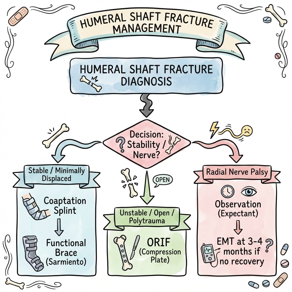
Critical Must-Knows
- Functional bracing achieves over 90% union with acceptable alignment in most cases
- Radial nerve spirals around posterior humerus - vulnerable at junction of middle and distal thirds
- Holstein-Lewis fracture: Distal third spiral fracture with high radial nerve palsy risk
- Acceptable alignment: under 20° anterior angulation, under 30° varus/valgus, less than 3cm shortening, under 15° rotation
- Primary radial nerve palsy (at injury): observe 3-4 months before exploration
Clinical Pearls
- "70% of radial nerve palsies recover spontaneously without exploration
- "Secondary palsy after manipulation = urgent exploration
- "Pendulum exercises begin immediately with functional brace
- "Antegrade IMN avoids radial nerve but risks rotator cuff injury
Clinical Imaging
Imaging Gallery


Critical Humeral Shaft Fracture Exam Points
Radial Nerve Assessment
Document wrist/finger extension and sensation before any manipulation. Primary palsy = observe, Secondary palsy post-manipulation = explore. Nerve crosses posterior humerus at spiral groove.
Functional Bracing
Gold standard non-operative management. Humeral brace with adjustable straps. Allows elbow and shoulder motion. Gravity alignment maintains reduction.
Surgical Indications
Polytrauma, open fractures, vascular injury, floating elbow, bilateral fractures, pathological fractures. Also consider for unacceptable alignment and patient factors (obesity).
Operative Options
ORIF with plate vs IMN. Plates preferred for distal fractures (avoid rotator cuff). IMN for proximal/middle shaft. MIPO technique reduces iatrogenic nerve injury.
At a Glance Treatment Decision Guide
Quick Decision Guide
| Fracture Pattern | Nerve Status | Key Factor | Treatment |
|---|---|---|---|
| Simple, acceptable alignment | Intact | Compliant patient | Functional brace - gold standard |
| Simple fracture | Primary radial palsy | Closed injury | Functional brace + observe nerve 3-4 months |
| Any pattern | Secondary palsy post-manipulation | New deficit after reduction | Urgent ORIF + nerve exploration |
| Holstein-Lewis (distal spiral) | With or without palsy | High nerve risk location | Strong consideration for ORIF |
| Open/Polytrauma/Floating elbow | Any status | Associated injuries | ORIF or IMN mandatory |
Key Mnemonics
WESTRadial Nerve Motor Testing
| W | Wrist extension ECRL, ECRB - radial nerve proper |
| E | Elbow extension (triceps) May be spared if injury distal to branches |
| S | Supination (of forearm) Supinator muscle - posterior interosseous nerve |
| T | Thumb and finger extension EPL, EDC - posterior interosseous nerve |
| W | Wrist extension ECRL, ECRB - radial nerve proper | S | Supination (of forearm) Supinator muscle - posterior interosseous nerve |
| E | Elbow extension (triceps) May be spared if injury distal to branches | T | Thumb and finger extension EPL, EDC - posterior interosseous nerve |
Hook:WEST - Wrist drop is the classic finding! Test wrist extension against gravity, if absent = radial nerve palsy.
STOP BRACESurgical Indications for Humeral Shaft
| S | Segmental fractures Multiple fracture levels = poor bracing |
| T | Transverse pattern at isthmus Narrow canal, poor contact |
| O | Open fractures Require debridement and stable fixation |
| P | Polytrauma/Pathological Need early mobilization, impending fractures |
| B | Bilateral fractures Cannot use crutches with both arms braced |
| R | Radial nerve secondary palsy After manipulation = nerve exploration |
| A | Arterial injury (vascular) Brachial artery injury requires fixation |
| C | Cannot tolerate brace Obesity, skin issues, non-compliance |
| E | Extended joints (floating elbow) Ipsilateral forearm fracture |
| S | Segmental fractures Multiple fracture levels = poor bracing | P | Polytrauma/Pathological Need early mobilization, impending fractures | A | Arterial injury (vascular) Brachial artery injury requires fixation |
| T | Transverse pattern at isthmus Narrow canal, poor contact | B | Bilateral fractures Cannot use crutches with both arms braced | C | Cannot tolerate brace Obesity, skin issues, non-compliance |
| O | Open fractures Require debridement and stable fixation | R | Radial nerve secondary palsy After manipulation = nerve exploration | E | Extended joints (floating elbow) Ipsilateral forearm fracture |
Hook:STOP the BRACE when surgery is needed - these are your absolute and relative surgical indications!
20-30-3-15Acceptable Angulation (Rule of 20-30-3-15)
| 20 | 20 degrees sagittal angulation Anterior/posterior bowing acceptable |
| 30 | 30 degrees coronal angulation Varus/valgus acceptable (hidden by shoulder) |
| 3 | 3cm shortening Well tolerated functionally |
| 15 | 15 degrees rotation Acceptable rotational malunion |
| 20 | 20 degrees sagittal angulation Anterior/posterior bowing acceptable | 3 | 3cm shortening Well tolerated functionally |
| 30 | 30 degrees coronal angulation Varus/valgus acceptable (hidden by shoulder) | 15 | 15 degrees rotation Acceptable rotational malunion |
Hook:20-30-3-15: The shoulder hides angulation well, making humeral shaft fractures forgiving for non-operative management!
HoLLyHolstein-Lewis Fracture Features
| Ho | Holstein-Lewis Named after Holstein and Lewis (1963) |
| L | Lateral spiral distal third Distal third spiral oblique fracture |
| L | Lateral intermuscular septum Nerve tethered here at spiral groove |
| y | High radial palsy risk 22-32% nerve palsy rate at this level |
| Ho | Holstein-Lewis Named after Holstein and Lewis (1963) | L | Lateral intermuscular septum Nerve tethered here at spiral groove |
| L | Lateral spiral distal third Distal third spiral oblique fracture | y | High radial palsy risk 22-32% nerve palsy rate at this level |
Hook:HoLLy Hurts the Radial nerve! Distal third spiral = high risk for nerve injury = consider ORIF.
Overview and Epidemiology
Clinical Significance
Humeral shaft fractures are unique in that non-operative management achieves excellent outcomes in the majority of cases. The shoulder and elbow joints compensate well for residual angulation and shortening, making functional bracing the gold standard treatment.
Demographics
- Bimodal distribution: Young males (high-energy), elderly females (low-energy falls)
- Male predominance in working age (sports, MVA)
- Female predominance in elderly (osteoporotic)
- Mean age: 45-55 years
Mechanism
- Direct blow: Transverse fractures (50%)
- Indirect/Torsion: Spiral/oblique fractures (50%)
- Falls (most common overall)
- Motor vehicle accidents
- Sports (throwing, arm wrestling)
Fracture Distribution by Location
| Location | Frequency | Common Pattern | Nerve at Risk |
|---|---|---|---|
| Proximal third | 30% | Spiral/Oblique | Axillary nerve (rare) |
| Middle third | 60% | Transverse/Spiral | Radial nerve (spiral groove) |
| Distal third | 10% | Spiral (Holstein-Lewis) | Radial nerve (highest risk) |
Anatomy and Pathophysiology
The Radial Nerve Course
The radial nerve is the most commonly injured nerve in humeral shaft fractures. It enters the posterior compartment from medial to lateral, wrapping around the posterior humerus in the spiral groove at the junction of the middle and distal thirds. Here it is tethered by the lateral intermuscular septum.

Muscular Deforming Forces by Fracture Level
| Fracture Level | Proximal Fragment | Distal Fragment | Resulting Deformity |
|---|---|---|---|
| Above pectoralis major insertion | Abduction, ER (rotator cuff) | Adduction (pec major, deltoid) | Apex lateral angulation |
| Between pec major and deltoid | Adduction (pec major) | Abduction (deltoid) | Apex medial angulation |
| Below deltoid insertion | Abduction (deltoid) | Proximal pull (biceps, triceps) | Shortening, variable angulation |
Key Anatomical Landmarks
- Surgical neck: Transition to shaft (axillary nerve)
- Deltoid insertion: V-shaped, mid-shaft lateral
- Spiral groove: Posterior, middle-distal junction
- Supracondylar ridge: Transition to distal humerus
Radial Nerve Anatomy
- Enters arm in axilla (posterior cord)
- Supplies triceps proximally
- Spiral groove: 14-20cm from lateral epicondyle
- Pierces lateral intermuscular septum at distal third
- Divides into PIN and superficial radial at elbow
Exam Trap: Radial Nerve Branches
The radial nerve supplies the triceps before entering the spiral groove. Therefore, triceps function may be preserved despite radial nerve injury at the spiral groove level. Always test wrist and finger extension - these are the key clinical findings for radial nerve palsy.
Classification Systems
AO/OTA Classification (12-A/B/C)
| Type | Description | Subgroups | Treatment Tendency |
|---|---|---|---|
| 12-A (Simple) | Two fragments, over 90% cortical contact | A1: Spiral, A2: Oblique (over 30°), A3: Transverse (under 30°) | Functional bracing |
| 12-B (Wedge) | Three fragments, contact between main fragments possible | B1: Spiral wedge, B2: Bending wedge, B3: Fragmentary wedge | Bracing or surgery |
| 12-C (Complex) | Multiple fragments, no contact between main fragments | C1: Spiral, C2: Segmental, C3: Irregular comminuted | Usually surgical |
Key Point
AO/OTA classification determines complexity and comminution. Simple fractures (A) have excellent outcomes with functional bracing. Complex fractures (C) often require surgical stabilization.
Clinical Assessment
History
- Mechanism: Direct blow vs torsion vs fall
- Hand dominance: Functional implications
- Occupation: Manual vs sedentary work
- Comorbidities: Diabetes, smoking, osteoporosis
- Previous injury: Pathological fracture concern
Examination
- Look: Deformity, swelling, bruising, skin condition
- Feel: Point tenderness, crepitus, neurovascular status
- Move: Limited by pain, test shoulder and elbow
- Neurovascular: Radial nerve critical, brachial artery
Radial Nerve Examination is MANDATORY
Document radial nerve function before and after any intervention. Test: wrist extension against gravity, finger MCP extension, thumb extension (hitchhiker), sensation first dorsal web space. Record in notes clearly.
Radial Nerve Assessment
| Test | Technique | Finding | Interpretation |
|---|---|---|---|
| Wrist extension | Extend wrist against resistance | Wrist drop if absent | Radial nerve palsy - most reliable sign |
| Finger MCP extension | Extend fingers at MCP against resistance | Cannot extend MCPs | PIN involvement |
| Thumb extension | Extend thumb (hitchhiker sign) | Cannot extend thumb | EPL - PIN involvement |
| Sensation first web space | Light touch dorsal first web | Numbness/decreased | Superficial radial nerve |
| Triceps function | Extend elbow against resistance | Usually preserved | Branches given before spiral groove |
Primary vs Secondary Palsy
Primary palsy (present at injury): Usually neurapraxia from stretch/contusion. Observe for 3-4 months - 70% recover spontaneously. Secondary palsy (develops after manipulation/reduction): Suggests nerve entrapment in fracture site - warrants urgent exploration.
Differential Diagnosis
| Diagnosis | Distinguishing Features | Key Investigation | Pitfall to Avoid |
|---|---|---|---|
| Humeral shaft fracture | Mid-arm deformity, crepitus, abnormal mobility; possible wrist drop | AP/lateral humerus including shoulder and elbow | Missing associated radial nerve palsy by not testing wrist extension |
| Pathological fracture | Low-energy mechanism, antecedent arm pain, known malignancy, lytic lesion | Radiographs +/- CT/MRI, bloods, staging if primary unknown | Treating as simple trauma and missing underlying tumour or myeloma |
| Proximal humerus fracture | Pain and deformity localised to shoulder, not mid-arm; axillary nerve at risk | Shoulder AP and axillary/scapular-Y views | Mislabelling a surgical-neck fracture as a shaft fracture |
| Distal humerus / intercondylar fracture | Elbow-centred pain, articular involvement, possible ulnar nerve signs | CT for articular extension | Underestimating articular involvement on plain films |
| Isolated traumatic radial nerve palsy (no fracture) | Wrist drop without bony injury (e.g. Saturday-night palsy, penetrating wound) | Nerve conduction studies, ultrasound/MRI of nerve | Attributing wrist drop to a fracture that is not present |
| Septic process of arm/humerus | Fever, systemic upset, atraumatic or trivial trauma, raised inflammatory markers | Bloods (CRP, WCC), MRI, aspiration | Plating through occult infection |
Investigations
Imaging Protocol
AP and lateral of entire humerus. Must include shoulder and elbow joints. Assess fracture pattern, displacement, angulation, and any articular involvement.
Include shoulder and elbow joints on separate views if not fully visualized. Rule out associated injuries (floating elbow, shoulder dislocation).
Indicated for: articular extension, preoperative planning for comminuted fractures, pathological fracture workup.
MRI: pathological fracture evaluation, soft tissue injury. Angiography: if vascular injury suspected (rare).
Radiographic Assessment
| Parameter | What to Assess | Normal/Acceptable | Action if Abnormal |
|---|---|---|---|
| Fracture pattern | Simple vs comminuted | Simple preferred for bracing | Comminuted may need surgery |
| Sagittal angulation | AP view | less than 20 degrees | Reduction needed if over 20° |
| Coronal angulation | Lateral view | less than 30 degrees | Reduction needed if over 30° |
| Shortening | Compare with normal | less than 3cm | Usually acceptable |
| Joint involvement | Shoulder and elbow | None | CT if suspected |
Holstein-Lewis Recognition
Look specifically for distal third spiral oblique fracture pattern on radiographs. This is the Holstein-Lewis fracture with high radial nerve palsy risk. Even with intact nerve function, consider lower threshold for surgical intervention.

Management Algorithm
Non-Operative Management - Functional Bracing
Gold standard for most humeral shaft fractures. Pioneered by Sarmiento, achieves over 90% union rate.
Functional Bracing Protocol
Coaptation splint (sugar tong) or U-slab. Sling for comfort. Ice, elevation. Begin pendulum exercises immediately if pain allows.
Convert to prefabricated humeral brace once swelling subsides. Circumferential compression with adjustable straps. Allows elbow and shoulder motion.
Active shoulder and elbow ROM. Pendulum exercises 4-6 times daily. Gravity alignment maintains reduction. Wean sling.
Weekly X-rays initially to ensure maintained alignment. Then every 2-4 weeks until union (usually 8-12 weeks).
Wean brace once clinical and radiographic union. Progressive strengthening. Full activity by 4-6 months.
Functional Bracing Mechanism
The brace provides hydraulic compression via soft tissue containment. This, combined with gravity, aligns the fracture. The key is early motion - muscle contraction maintains alignment and promotes healing.
Success Factors
- Compliant patient
- Simple fracture pattern
- Acceptable initial alignment
- Early active motion
- Weekly X-ray follow-up initially
Poor Brace Candidates
- Obesity (poor soft tissue compression)
- Skin problems (burns, dermatitis)
- Non-compliance
- Transverse fractures at narrow isthmus
- Segmental fractures
Surgical Technique
Open Reduction Internal Fixation - Plating

Patient Positioning:
- Supine with arm on arm board (anterolateral approach)
- Lateral decubitus or prone for posterior approach
- Beach chair for proximal fractures
Anterolateral Approach:
- Incision along lateral border of biceps
- Develop interval between brachialis (musculocutaneous) and brachioradialis (radial)
- Radial nerve runs between these muscles - identify and protect
- Plate applied anterolaterally on flat humerus
Posterior Approach:
- Midline posterior incision
- Split triceps in midline or elevate medial and lateral heads
- Radial nerve identified in spiral groove, protected
- Plate applied to flat posterior surface
Plate Choice:
- 4.5mm narrow LCP or broad LCP
- Minimum 3-4 screws (6-8 cortices) each side of fracture
- Compression plate for simple patterns
- Bridge plating for comminution
MIPO Technique
Minimally Invasive Plate Osteosynthesis uses small incisions proximally and distally with submuscular plate passage. Reduces soft tissue stripping and theoretical nerve injury risk. Requires fluoroscopy.

Complications
Complications and Management
| Complication | Incidence | Risk Factors | Management |
|---|---|---|---|
| Radial nerve palsy | 12-18% (primary) | Distal third, spiral, Holstein-Lewis | Observe primary, explore secondary |
| Nonunion | 2-10% | Transverse pattern, distraction, non-compliance | ORIF + bone graft |
| Malunion | Variable | Non-operative with poor follow-up | Usually well tolerated, osteotomy if symptomatic |
| Shoulder stiffness | Common initially | Prolonged immobilization, poor compliance | Early motion, physiotherapy |
| Infection (surgical) | 1-3% | Open fractures, poor soft tissues | Debridement, antibiotics, revision |
| Hardware failure | 2-5% | Early loading, inadequate fixation | Revision fixation |
| Iatrogenic nerve injury | 2-5% (surgical) | Posterior approach, ORIF | Careful dissection, MIPO technique |
Radial Nerve Palsy Management
Management Algorithm
Primary palsy (at injury): 70% recover spontaneously. Observe for 3-4 months with clinical and EMG monitoring. If no recovery by 4 months, explore. Secondary palsy (post-manipulation): Urgent exploration - nerve likely trapped in fracture site.
Primary vs Secondary Radial Nerve Palsy
| Feature | Primary Palsy | Secondary Palsy |
|---|---|---|
| Timing | Present at injury | Develops after manipulation/surgery |
| Mechanism | Stretch/contusion (neurapraxia) | Entrapment in fracture site |
| Spontaneous recovery | 70% within 3-4 months | Unlikely without exploration |
| Management | Observe, wrist splint, EMG at 3-4 weeks | Urgent surgical exploration |
Management of Primary Radial Nerve Palsy
Document findings clearly. Wrist cock-up splint to maintain function. Proceed with non-operative management if otherwise indicated.
Baseline EMG/NCS to confirm nerve injury type and level. Monitor clinically for Tinel sign progression (sign of regeneration).
Repeat EMG if no clinical recovery. Look for reinnervation potentials. Continue monitoring.
If no clinical or EMG recovery, plan surgical exploration. Nerve grafting may be required if gap identified.
If nerve recovery incomplete, consider tendon transfers (PT to ECRB, FCR to EDC, PL to EPL).
Tendon Transfer Options
If radial nerve does not recover: PT to ECRB (wrist extension), FCR to EDC (finger extension), PL to EPL (thumb extension). These restore functional wrist and finger extension.
Nonunion Risk Factors
Transverse fracture pattern has higher nonunion risk with functional bracing (2-10% vs 5% or less for oblique/spiral). The narrow isthmus and transverse orientation provide poor cortical contact. Consider primary ORIF for transverse fractures at the narrow diaphyseal portion.

Shoulder Stiffness Prevention
Pendulum exercises from day one are critical. The key to successful functional bracing is EARLY MOTION. Shoulder stiffness is a preventable complication with appropriate rehabilitation.
Postoperative Care and Rehabilitation
Post-ORIF Rehabilitation
Sling for comfort. Wound care. Gentle pendulum exercises. No active shoulder elevation against gravity.
Active-assisted shoulder ROM. Active elbow ROM. Gentle strengthening begins at 4 weeks if stable fixation.
Progressive resistive exercises. Wean sling. Radiographic union usually evident by 8-12 weeks.
Full activity when clinically and radiographically healed. Sport-specific training as appropriate.
Outcomes and Prognosis
Prognostic Factors
| Factor | Impact | Notes |
|---|---|---|
| Fracture pattern | Significant | Simple patterns do well with bracing; transverse at risk |
| Patient compliance | Critical for bracing | Non-compliance leads to malunion/nonunion |
| Radial nerve status | Important | Primary palsy usually recovers; secondary needs surgery |
| Associated injuries | Variable | Polytrauma patients may need surgical fixation |
| Smoking | Negative | Higher nonunion rates |
| Diabetes | Negative | Delayed healing, higher complications |
Plate vs Nail Outcomes
Meta-analyses show similar union rates for plate vs nail (both over 95%). However, plating has lower reoperation rates and less shoulder problems. Nailing is faster but associated with more shoulder pain (antegrade) or elbow issues (retrograde).
Evidence Base
Sarmiento Functional Bracing Series
- 922 humeral diaphyseal fractures treated with a prefabricated functional brace; 620 (67%) available for follow-up
- Nonunion in 16 of 620 followed patients - 9 of 155 open fractures (6%) versus 7 of 465 closed fractures (less than 2%)
- Coronal angulation healed at less than 16° varus in 87% and anterior angulation less than 16° in 81%
- 98% retained shoulder motion loss of 25° or less at brace removal
Plate vs Nail Meta-Analysis (Heineman)
- Meta-analysis of 4 RCTs (203 patients) comparing intramedullary nails and plates
- No statistically significant difference in total complication rate, non-union, infection, nerve palsy, or reoperation
- Included trials were small with methodological limitations
- Authors called for a definitive large RCT
Plate vs Nail Meta-Analysis (Bhandari)
- Meta-analysis of 3 RCTs (155 patients) of compression plating versus intramedullary nailing
- Plating reduced relative risk of reoperation versus nailing (RR 0.26, 95% CI 0.007-0.9, p=0.03)
- Plating reduced shoulder problems versus nailing (RR 0.10, 95% CI 0.03-0.4, p=0.002)
- Roughly one reoperation prevented for every 10 patients treated with a plate
Radial Nerve Palsy Natural History (Shao Systematic Review)
- Systematic review of 35 studies; overall palsy prevalence 11.8% (532 palsies in 4517 fractures)
- Overall recovery 88.1%; spontaneous recovery 70.7% in conservatively managed patients
- Middle and middle-distal shaft fractures had the highest association with palsy
- No significant difference in final outcome between early exploration and expectant management
Holstein-Lewis Fracture (Original Description)
- Original description of the distal-third spiral oblique humeral fracture associated with radial-nerve paralysis
- Sharp proximal fragment displaces the radial nerve where it pierces the lateral intermuscular septum
- Established the pattern as a recognised cause of fracture-associated radial nerve injury
Bridge Plate vs Functional Brace RCT (Matsunaga)
- RCT of 110 patients - minimally invasive bridge-plate osteosynthesis versus functional brace
- DASH score modestly better with surgery at 6 months only (10.9 vs 16.9, p=0.046)
- Lower nonunion rate with surgery (0% vs 15%)
- No difference in SF-36, pain, or Constant-Murley score; coronal angulation lower with plating (2.0° vs 10.5°)
Population Epidemiology - Swedish Fracture Register
- 2,011 adult humeral fractures from the Swedish Fracture Register (2011-2013); 13% were shaft fractures
- Humeral shaft fracture incidence 13.4 per 100,000 person-years
- Female:male ratio 2.4:1; mean age 66.8 years, with most fractures from low-energy falls in patients over 50
- Only 1.2% open and 1.3% pathological
Exam Viva Scenarios
Use these scenarios to practise clinical reasoning and management decisions
Scenario 1: Initial Assessment and Classification
"A 35-year-old man presents after falling off a ladder onto his outstretched arm. He has obvious deformity of his right upper arm and cannot extend his wrist or fingers. X-rays show a spiral fracture of the distal third of the humerus. How would you assess and manage this patient?"
Scenario 2: Surgical Decision-Making
"A 55-year-old woman sustains a closed transverse humeral shaft fracture at the mid-diaphysis in a motorcycle accident. She also has a closed tibial shaft fracture. Her radial nerve is intact. How would you manage this patient?"
Scenario 3: Complication Management
"You are called to see a 40-year-old man in ED who had a closed humeral shaft fracture reduced in a backslab by the junior doctor. He now cannot extend his wrist or fingers - he had normal function before the reduction. What is your management?"
MCQ Practice Points
Anatomy Question
Q: At what level does the radial nerve cross the posterior humerus in the spiral groove? A: The junction of the middle and distal thirds, approximately 14-20cm from the lateral epicondyle. Here it is tethered by the lateral intermuscular septum.
Classification Question
Q: What is a Holstein-Lewis fracture? A: A distal third spiral oblique fracture of the humerus with high radial nerve palsy rate (22-32%). The sharp proximal spike can trap or lacerate the radial nerve at the lateral intermuscular septum.
Management Question
Q: What are the acceptable angulation limits for humeral shaft fractures treated non-operatively? A: Less than 20° sagittal (AP) angulation, less than 30° coronal (varus/valgus), less than 3cm shortening, less than 15° rotation. The shoulder compensates well for these deformities.
Nerve Question
Q: A patient develops wrist drop after manipulation of a humeral shaft fracture. What is your management? A: This is secondary radial nerve palsy - the nerve is likely trapped in the fracture. Urgent exploration is required. Do not observe as you would for primary palsy.
Treatment Question
Q: What is the expected spontaneous recovery rate for primary radial nerve palsy in closed humeral shaft fractures? A: 70% recover spontaneously within 3-4 months. Observe with wrist splint and EMG monitoring. Explore if no recovery by 4 months.
Guidelines, Registries & Global Practice
Global Epidemiology
Population-Based Incidence (per 100,000 person-years)
| Source / Setting | Population | Humeral Shaft Incidence | Key Pattern |
|---|---|---|---|
| Swedish Fracture Register (Bergdahl 2016) | Sweden, adults | 13.4 | 13% of all humeral fractures; F:M 2.4:1; mean age 66.8 |
| Edinburgh / UK series (widely cited) | Western Europe | Approximately 13-20 | Bimodal: young males high-energy, elderly females low-energy falls |
| Composite literature estimate | Global | Approximately 1-3% of all fractures | Most are closed, low-energy, isolated injuries |
Epidemiology Anchor
The Swedish Fracture Register (Bergdahl et al, BMC Musculoskelet Disord 2016) provides the cleanest contemporary denominator: humeral shaft fractures occur at 13.4 per 100,000 person-years, are predominantly low-energy falls in older women (F:M 2.4:1), and make up about 13% of all humeral fractures. Only ~1% are open.
Guideline & Registry Landscape
International Guidance and Registry Position
| Body / Region | Position on Humeral Shaft Fractures | Evidence Basis |
|---|---|---|
| AAOS / AO Foundation (USA, global) | Functional bracing remains a reasonable default for most closed isolated fractures; surgery for absolute indications and selected relative ones. AO principles guide implant choice (plate for distal/articular, nail or plate for mid-shaft) | Synthesis of RCTs and meta-analyses (Level I-II) |
| NICE / BOAST (UK) | Emphasis on documented neurovascular assessment, early senior decision-making, and shared decision-making between bracing and fixation; no mandate for routine surgery | Consensus standards informed by RCT evidence |
| EFORT / AO (Europe) | Bracing and operative fixation both endorsed; growing interest in primary fixation to reduce nonunion and shorten disability in selected adults | Level I RCT and meta-analysis data |
| RACS / AOA (Australia/NZ) | Competency expected in both non-operative and operative management; mandatory documentation of radial nerve status; informed consent must include nerve-injury discussion | Training and examination standards |
Practice Variation
There is genuine international practice variation: the historical Sarmiento data and Shao systematic review underpin a non-operative-first culture in many UK/European and Australian units, whereas the Matsunaga RCT and lower nonunion rates with fixation have driven a trend toward earlier operative management in some centres. No registry or guideline currently mandates routine surgery for the typical closed isolated fracture.
Australian Context
In Australia, humeral shaft fractures present commonly to emergency departments with the same bimodal pattern seen internationally - high-energy injuries in younger working-age males and low-energy osteoporotic falls in older women. Functional bracing remains the mainstay of treatment across most Australian units, with plating preferred over nailing when an operative indication exists, consistent with the meta-analytic evidence favouring plate fixation for reoperation and shoulder outcomes. Access to orthopaedic care is generally timely through the public hospital trauma system, and smoking cessation support (Quitline 13 7848) is relevant given the adverse effect of smoking on union. RACS/AOA training and the FRACS/FRCS examinations consistently test radial nerve assessment, the primary-versus-secondary palsy distinction, and the documentation and consent standards around nerve injury.
Medicolegal Considerations
Key documentation requirements:
- Document radial nerve function before AND after any manipulation
- Record complete motor (wrist, finger, thumb extension) and sensory (dorsal first web) exam
- Document discussion of treatment options including functional bracing vs surgery
- Informed consent for surgery must include: infection, nonunion, nerve injury (especially iatrogenic), shoulder stiffness (IMN), need for hardware removal
- If radial palsy present: document as primary vs secondary and management plan
Consent for Humeral Shaft ORIF
Specific risks to discuss: Radial nerve injury (iatrogenic 2-5%), infection, nonunion, hardware failure/irritation, need for bone grafting, shoulder stiffness. For IMN: add rotator cuff injury and shoulder pain (15-30%).
HUMERAL SHAFT FRACTURES
Clinical summary
Key Anatomy
- •Radial nerve in spiral groove at middle-distal third junction
- •Nerve tethered by lateral intermuscular septum
- •Triceps branches given BEFORE spiral groove (usually spared)
- •Muscular deforming forces vary by fracture level
Classification
- •AO/OTA: 12-A (simple), B (wedge), C (complex)
- •Holstein-Lewis: distal third spiral = high radial palsy risk
- •Location: proximal (30%), middle (60%), distal (10%)
- •Pattern: transverse (direct blow), spiral (torsion)
Acceptable Alignment (20-30-3-15)
- •under 20° sagittal (AP) angulation
- •under 30° coronal (varus/valgus) angulation
- •less than 3cm shortening
- •under 15° rotation
Treatment Algorithm
- •Most fractures: Functional bracing (over 90% union)
- •STOP BRACE indications: Segmental, Transverse isthmus, Open, Polytrauma, Bilateral, Radial 2° palsy, Arterial injury, Cannot tolerate, Extended joints (floating elbow)
- •Plate preferred over nail (less shoulder pain, lower reop rate)
- •MIPO technique reduces iatrogenic nerve injury
Radial Nerve Palsy
- •Primary palsy: observe 3-4 months, 70% recover
- •Secondary palsy (post-manipulation): URGENT exploration
- •Test: wrist extension, finger MCP extension, thumb extension
- •Tendon transfers if no recovery: PT-ECRB, FCR-EDC, PL-EPL