Clubbing, Periostosis & Arthralgia (incl. Pachydermoperiostosis)
- Hypertrophic osteoarthropathy (HOA) is a clinical syndrome comprising the TRIAD of DIGITAL CLUBBING, PERIOSTOSIS (periosteal new-bone formation along the long bones) and ARTHRALGIA with joint effusions, often with painful swelling of the distal limbs; recognising the triad - rather than any single feature - is the key.
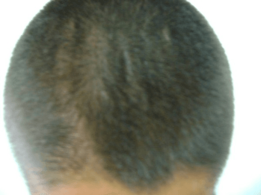
- The PRIMARY (hereditary) form is PACHYDERMOPERIOSTOSIS (Touraine-Solente-Gole syndrome), a GENETIC disorder of the PROSTAGLANDIN E2 degradation/transport pathway (HPGD or SLCO2A1 mutations) that, in addition to clubbing and periostosis, produces PACHYDERMA (thickened, furrowed facial/scalp skin) and CUTIS VERTICIS GYRATA; it typically presents in young men and may be incomplete (clubbing and periostosis without florid skin changes).
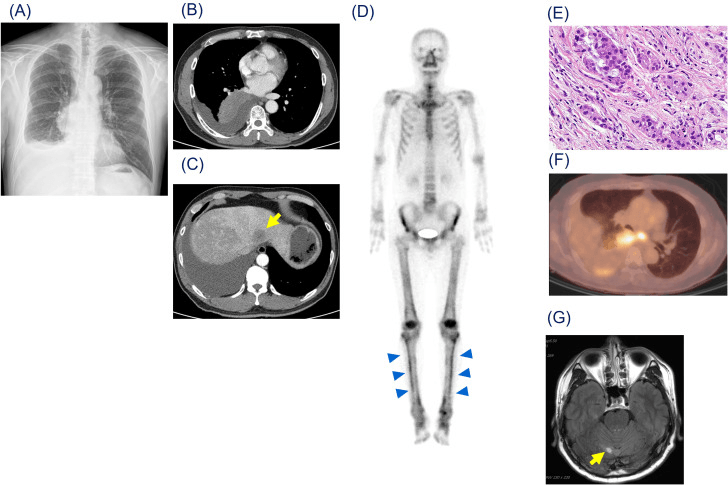
- The SECONDARY form is far COMMONER and clinically the more important to recognise, because it is a PARANEOPLASTIC/systemic MARKER: it is classically associated with INTRATHORACIC MALIGNANCY (especially non-small-cell lung cancer and mesothelioma) and with suppurative lung disease (bronchiectasis, lung abscess, empyema, cystic fibrosis), and also with cyanotic congenital heart disease, inflammatory bowel disease and chronic liver disease - so NEW-ONSET HOA (and especially new clubbing) in an adult mandates a search for an underlying, often pulmonary, cause.
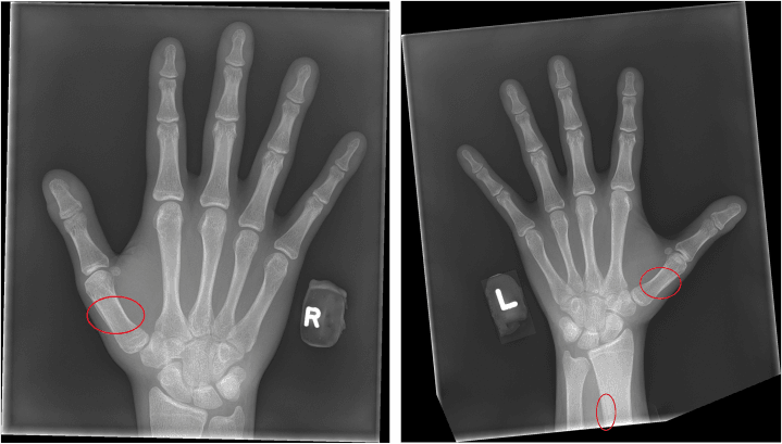
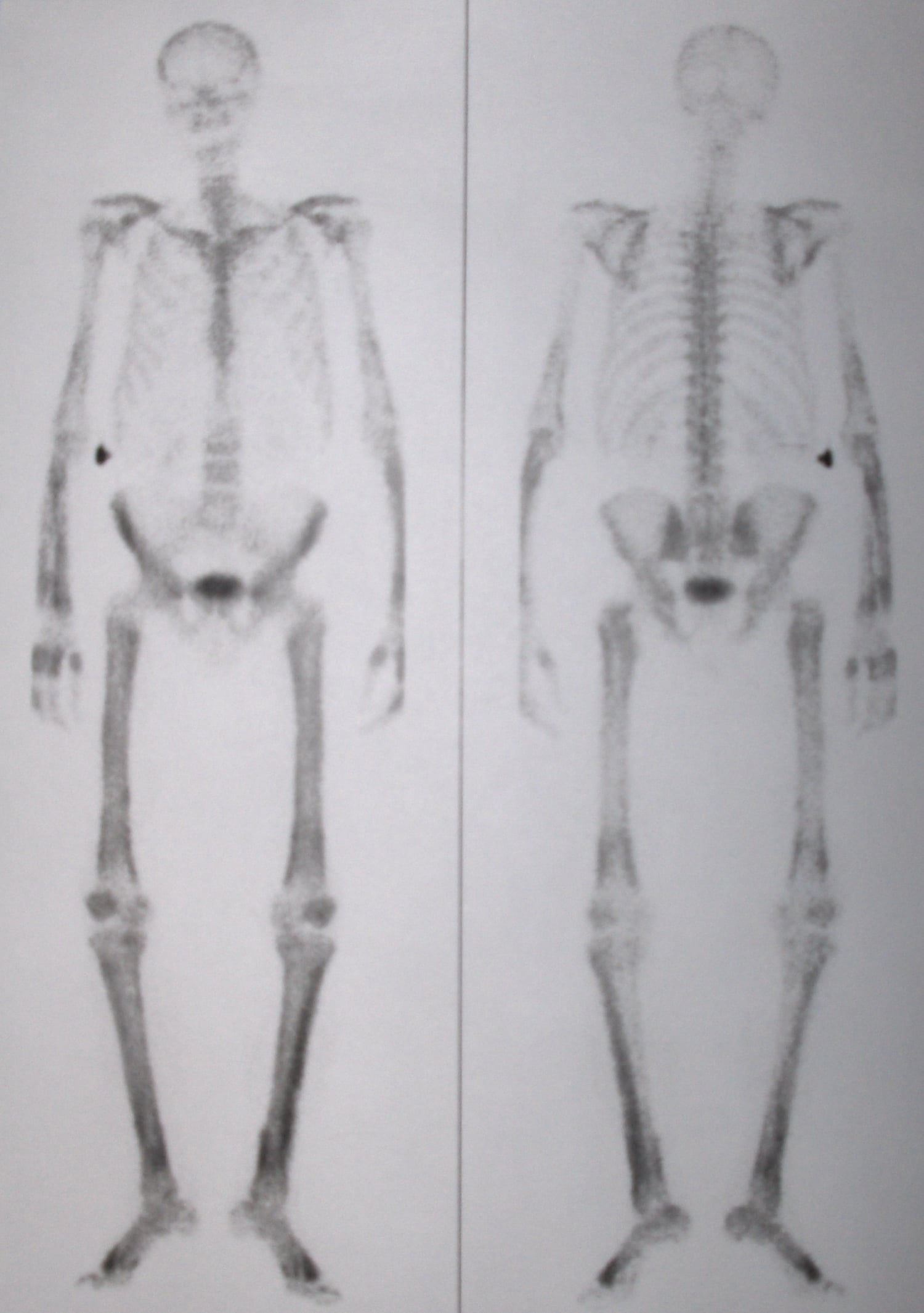
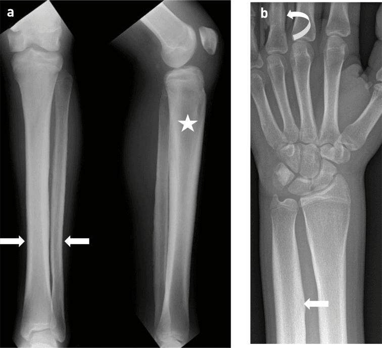
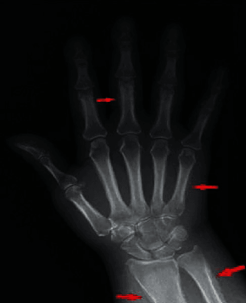
- The IMAGING hallmark is SYMMETRIC PERIOSTEAL NEW BONE along the diaphyses and metaphyses of the long bones (tibia, fibula, radius, ulna), and a radionuclide BONE SCAN shows symmetric 'TRAMLINE' (double-stripe) cortical uptake along the long bones, which is sensitive for early or radiographically occult disease; the Schamroth (window) sign is a simple clinical test for clubbing.
- The DIFFERENTIAL of periosteal reaction and limb pain includes thyroid acropachy, chronic venous stasis/periostitis, vitamin-A toxicity, and (in children) physiological periostosis - but the combination of clubbing, symmetric long-bone periostosis and arthralgia is characteristic of HOA, and the orthopaedic role is often to recognise the syndrome from a periosteal reaction noted on imaging.
- MANAGEMENT depends on the form: SECONDARY HOA is treated by addressing the UNDERLYING CAUSE - resection/treatment of the lung tumour or control of the suppurative/cardiac disease often improves or resolves the HOA - while SYMPTOMATIC relief (non-steroidal anti-inflammatories, and agents acting on the prostaglandin pathway such as bisphosphonates or, in primary disease, COX inhibitors) helps the arthralgia/bone pain; the single most important action in new-onset secondary HOA is to USE it as the clue to find and treat the occult disease.
- “HOA = triad of CLUBBING + PERIOSTOSIS (symmetric long-bone periosteal new bone) + ARTHRALGIA. Primary = pachydermoperiostosis (genetic PGE2 pathway, + pachyderma/cutis verticis gyrata, young men).
- “Secondary HOA is far commoner and is a PARANEOPLASTIC marker - classically intrathoracic malignancy (lung cancer/mesothelioma) + suppurative lung disease. New clubbing/HOA in an adult = SEARCH for the cause (chest!).
- “Imaging: symmetric periosteal new bone of long bones; bone scan 'tramline' cortical uptake. Treat the underlying cause; NSAIDs/bisphosphonates for symptoms.
Clubbing + periostosis (symmetric long-bone periosteal new bone) + arthralgia. Bone scan shows 'tramline' cortical uptake; the Schamroth sign screens for clubbing.
Secondary HOA is a paraneoplastic marker - classically intrathoracic malignancy and suppurative lung disease. New-onset HOA in an adult mandates a search for the underlying (lung) cause.
The Syndrome & Its Two Forms
HOA is the triad of digital clubbing, periostosis (periosteal new bone along the long bones) and arthralgia/effusions. The primary form is pachydermoperiostosis (Touraine-Solente-Gole), a genetic disorder of the prostaglandin E2 pathway (HPGD/SLCO2A1), which adds pachyderma and cutis verticis gyrata and presents in young men (sometimes incomplete). The secondary form is far commoner and is a paraneoplastic/systemic marker - classically intrathoracic malignancy (lung cancer, mesothelioma) and suppurative lung disease (bronchiectasis, abscess, cystic fibrosis), also cyanotic heart disease, IBD and liver disease - so new-onset HOA in an adult should trigger a search for the cause.
- Primary (pachydermoperiostosis)
- Genetic (HPGD/SLCO2A1; PGE2 pathway)
- Secondary
- Paraneoplastic/systemic (lung cancer, suppurative lung disease, etc.)
- Primary (pachydermoperiostosis)
- Pachyderma + cutis verticis gyrata
- Secondary
- Usually absent
- Primary (pachydermoperiostosis)
- Young men; family history
- Secondary
- Adult with underlying disease
- Primary (pachydermoperiostosis)
- Rare
- Secondary
- Far commoner
- Primary (pachydermoperiostosis)
- Symptomatic; genetic counselling
- Secondary
- Find and treat the underlying cause (chest!)
Why Clubbing and Periostosis Happen: The Pathophysiology
The prostaglandin E2 (PGE2) pathway of primary disease and the paraneoplastic secondary form are linked by a single mechanism. Both converge on excess PGE2 and a fibrovascular/growth-factor drive acting on the distal soft tissues and the periosteum - they simply reach it by different routes.
Pachydermoperiostosis is a loss of PGE2 clearance. The two culprit genes both handle PGE2 breakdown: HPGD encodes 15-hydroxyprostaglandin dehydrogenase, the enzyme that degrades prostaglandins, and SLCO2A1 encodes the prostaglandin transporter that carries PGE2 into cells for that degradation. Lose either and PGE2 accumulates systemically, acting on fibroblasts, osteoblasts and vessels to produce the clubbing, periostosis and the skin changes (pachyderma, cutis verticis gyrata).
Normally the lung filters out megakaryocytes and large platelet aggregates. With an intrathoracic tumour, a right-to-left shunt (cyanotic heart disease) or chronic suppurative lung disease, these clumps bypass the pulmonary filter and impact in the distal digital capillaries, releasing platelet-derived growth factor (PDGF) and vascular endothelial growth factor (VEGF). This drives fibrovascular proliferation, oedema and periosteal new bone - with COX-2/PGE2 again the shared mediator.
The unifying theme is PGE2 plus a VEGF/PDGF fibrovascular drive: failed clearance in primary disease, raised platelet-and-growth-factor production in secondary disease. This is why COX inhibitors/NSAIDs ease the bone pain, why the bone scan lights up the cortices ("tramline"), and - crucially - why removing the tumour or treating the lung disease can reverse the HOA.
Defining and Assessing Clubbing
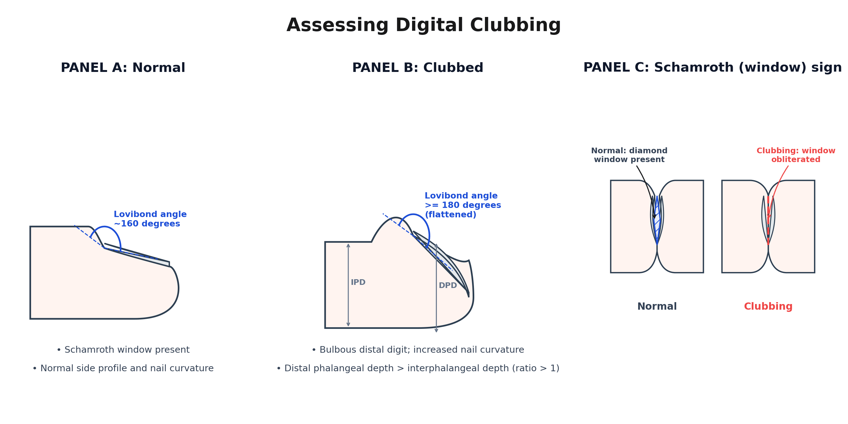
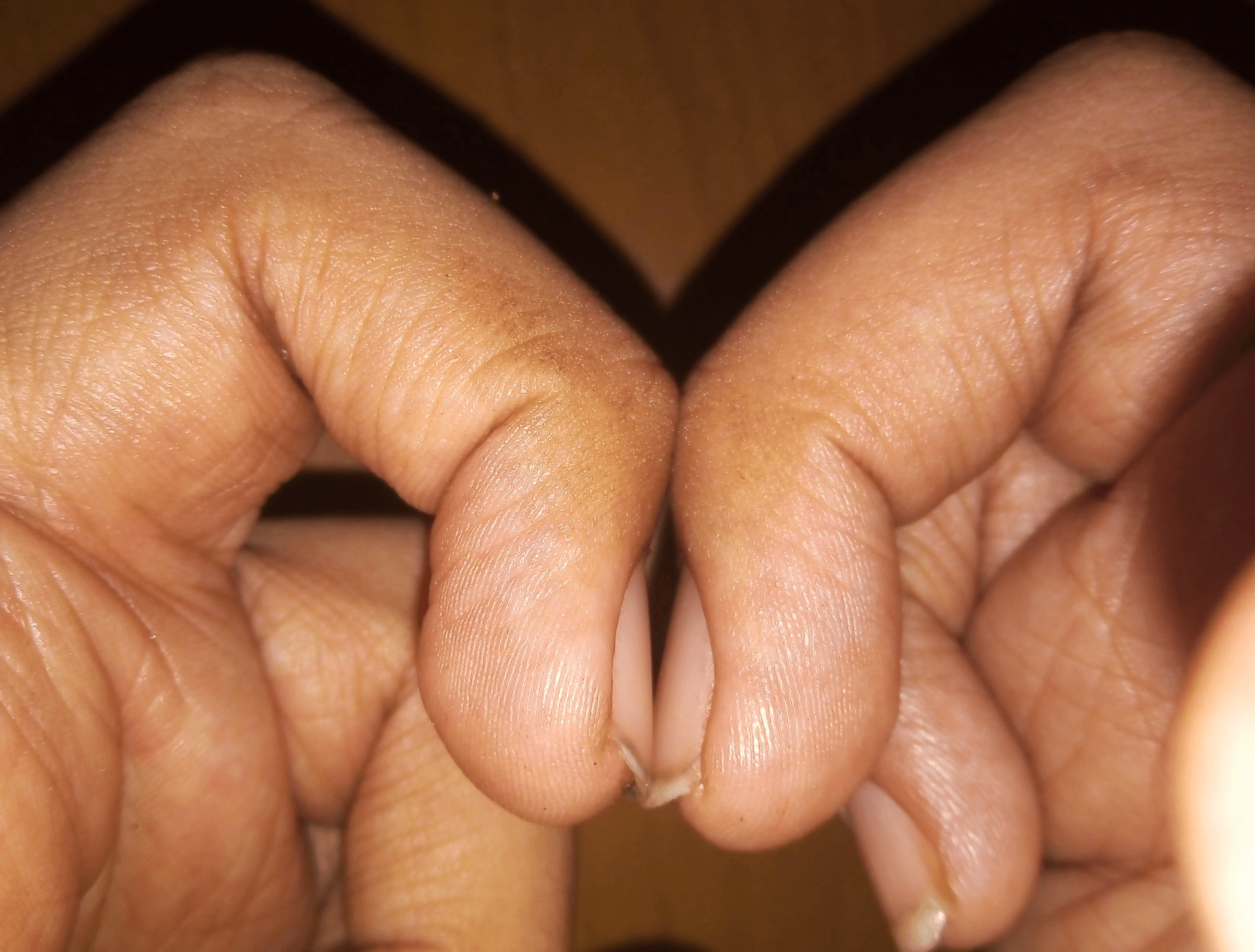
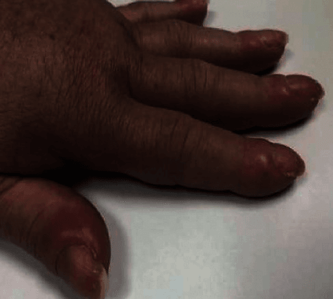
Clubbing is named throughout and screened for with the Schamroth sign, but it has a precise definition and several objective bedside signs the examiner expects. Clubbing is a bulbous enlargement of the distal digit from increased soft tissue at the nail bed, with loss of the normal nail-fold angle and increased nail-bed fluctuation. It evolves through recognised stages: increased nail-bed fluctuation (a "floating nail") - then loss of the nail-fold angle - then increased nail curvature - and finally the drumstick digit.
- Lovibond's angle (the profile nail-fold angle): normally around 160 degrees, it flattens to 180 degrees or more in clubbing.
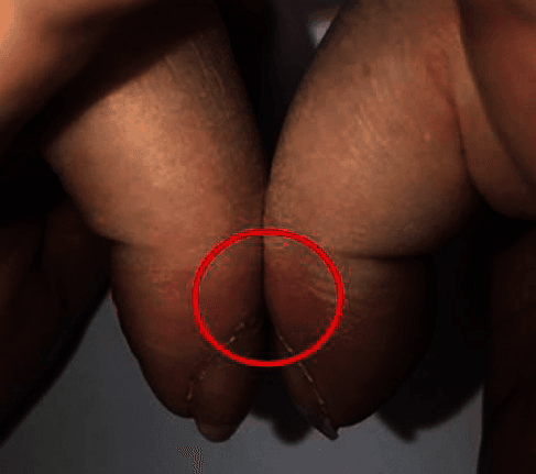
- Schamroth (window) sign: apposing the dorsal surfaces of opposing terminal phalanges normally leaves a diamond-shaped gap; in clubbing this window is obliterated.
- Phalangeal depth ratio: the distal phalangeal depth exceeds the interphalangeal depth (ratio greater than one) in clubbing.
Clubbing can be the earliest and only feature before florid periostosis. New clubbing in an adult is the trigger to investigate for the underlying (especially intrathoracic) cause of secondary HOA - so assessing it objectively, not just noting "looks clubbed", changes management.
Imaging & Management
- Imaging: symmetric periosteal new bone along the long-bone diaphyses/metaphyses (tibia, fibula, radius, ulna) on radiographs; bone scan shows symmetric 'tramline' cortical uptake (sensitive for early/occult disease). The Schamroth (window) sign screens for clubbing.
- Secondary HOA - treat the underlying cause: resection/treatment of the lung tumour or control of the suppurative/cardiac disease often improves or resolves the HOA.
- Symptomatic relief: NSAIDs for arthralgia/bone pain; agents acting on the prostaglandin pathway (bisphosphonates; COX inhibitors in primary disease) may help.
- The clue: in new-onset secondary HOA the most important action is to USE the syndrome to find and treat the occult disease.
The clinically critical point about hypertrophic osteoarthropathy is that, in its common secondary form, it is a messenger for an underlying disease - most importantly an intrathoracic malignancy such as lung cancer or mesothelioma, but also suppurative lung disease and certain cardiac, bowel and liver conditions. New-onset clubbing and symmetric long-bone periostosis with arthralgia in an adult should therefore never be treated as an isolated musculoskeletal complaint; the appropriate response is to investigate for, and treat, the underlying cause - which often improves or resolves the HOA itself - while providing symptomatic relief. The primary (hereditary) form, pachydermoperiostosis, is a genetic prostaglandin-pathway disorder with the additional skin features of pachyderma and cutis verticis gyrata, and is managed symptomatically with genetic counselling; the exam (and clinical) error is to overlook the secondary form's role as a sign of occult, potentially serious, disease.
Working It Up: What to Order, and What if the Chest Is Clear
"Search for the underlying cause" is the right instruction, but the orthopaedic surgeon who has just found symmetric periostitis needs to know what to actually request - and what to do when the obvious test comes back normal.
- 1Confirm the syndrome
Symmetric periosteal new bone on long-bone radiographs, plus clubbing assessed objectively (Lovibond angle, Schamroth window, phalangeal depth ratio), plus arthralgia.
Do not stop at 'periosteal reaction' on a report - look at the hands.
- 2Image the chest - CT, not just a radiograph
A chest radiograph is the first test but a normal film does NOT exclude the cause: a small peripheral adenocarcinoma or an early mesothelioma can be radiographically occult while producing florid HOA.
Most secondary HOA is explained here.
- 3If the chest is genuinely clear, widen the search
Echocardiography for cyanotic congenital heart disease and infective endocarditis; liver function and screening for chronic liver disease; symptom-directed assessment for inflammatory bowel disease and coeliac disease; thyroid function, since thyroid acropachy is the classic mimic.
These are the non-pulmonary causes the classic 'pulmonary osteoarthropathy' name obscures.
- 4Reconsider primary disease
A young man, an onset around adolescence, a family history, thickened furrowed facial skin or cutis verticis gyrata, and no underlying illness point to pachydermoperiostosis rather than a missed malignancy.
Genetic testing (HPGD, SLCO2A1) confirms it and stops a fruitless cancer hunt.
- 5If everything is negative
Do not simply discharge. HOA can PRECEDE the diagnosis of the underlying tumour, sometimes by months.
Arrange interval clinical review and repeat chest imaging rather than closing the episode.
That final step is the one that matters most and is easily missed: the syndrome is sometimes the first manifestation of the malignancy, so a negative initial screen buys surveillance, not reassurance.
If you aspirate the joint
Effusions are part of the triad and are a useful discriminator when the diagnosis is uncertain. The fluid in HOA is non-inflammatory - clear, viscous, with a low white cell count - unlike the turbid, high-cell-count fluid of an inflammatory or septic arthritis. A knee effusion in a patient with clubbing and periostitis that returns a bland aspirate supports HOA rather than pointing away from it.
Telling the Periosteal Reactions Apart
Symmetric periostitis has a short differential, and the discriminators are worth holding because the radiologist's report will often say only "periosteal reaction".
- Distribution and character
- Symmetric, smooth then layered periostitis of long-bone DIAPHYSES and metaphyses (tibia, fibula, radius, ulna); spares epiphyses
- The discriminator
- CLUBBING - and an underlying cause, usually intrathoracic
- Distribution and character
- HANDS and FEET - metacarpals, metatarsals and proximal phalanges; fluffy, spiculated, 'bubbly' periostitis
- The discriminator
- Graves disease with exophthalmos and pretibial myxoedema; typically painless
- Distribution and character
- Distal tibia and fibula, undulating
- The discriminator
- Chronic venous insufficiency, oedema, skin changes; no clubbing
- Distribution and character
- Undulating periostitis, often ulna and metatarsals; children
- The discriminator
- Excess intake history; no clubbing
- Distribution and character
- Mandible, clavicle, ulna in INFANTS under 6 months
- The discriminator
- Age and mandibular involvement are decisive
- Distribution and character
- Symmetric DIAPHYSEAL cortical thickening from both endosteal and periosteal surfaces
- The discriminator
- Progressive diaphyseal dysplasia with waddling gait, in childhood
- Distribution and character
- Symmetric long-bone periostitis
- The discriminator
- PGE1 infusion to maintain a patent ductus - the pharmacological proof of the mechanism
The last row is worth pausing on. Neonates given a prostaglandin infusion develop exactly this periostitis, which is as close to a human experiment as the field offers and is the strongest single argument that PGE2 drives the periosteal reaction described above. The paediatric hyperostoses are covered in Caffey Disease, Camurati-Engelmann Disease and Melorheostosis.
Mnemonics & Memory Aids
CPA
Hook:CPA = the HOA triad: Clubbing (check the Chest), Periostosis, Arthralgia. Secondary = paraneoplastic - find the cause.
Clinical Decision Scenarios
Practise clinical reasoning and management decisions out loud
“An adult is referred with bilateral leg pain; radiographs show symmetric periosteal new bone along the tibiae, and you notice finger clubbing. What is the diagnosis and what must you do?”
The syndrome
- Triad: digital clubbing + periostosis + arthralgia/effusions
- Schamroth (window) sign screens for clubbing
- Recognise the triad, not just one feature
Primary vs secondary
- Primary = pachydermoperiostosis (genetic PGE2 pathway; pachyderma + cutis verticis gyrata; young men)
- Secondary (far commoner) = paraneoplastic/systemic marker
- Secondary causes: lung cancer/mesothelioma, suppurative lung disease, cyanotic heart disease, IBD, liver disease
Imaging
- Symmetric periosteal new bone of long-bone diaphyses/metaphyses
- Bone scan: symmetric 'tramline' cortical uptake (early/occult disease)
- Differential: thyroid acropachy, venous stasis periostitis, vitamin-A toxicity
Management
- Secondary: treat the underlying cause (often improves/resolves HOA) - SEARCH the chest
- Symptomatic: NSAIDs; bisphosphonates / prostaglandin-pathway agents
- Primary: symptomatic + genetic counselling
Evidence & Key Studies
Acquired digital clubbing as the incomplete form of pachydermoperiostosis (primary HOA)
- Reports two healthy young men with acquired digital clubbing as the incomplete form of pachydermoperiostosis (primary hypertrophic osteoarthropathy).
- Illustrates the clinical features relevant to recognition - clubbing, periostosis (periosteal thickening), the Schamroth sign, and the cutaneous features of pachydermoperiostosis (pachyderma/cutis verticis gyrata).
- Reinforces that primary HOA (pachydermoperiostosis) can present incompletely, primarily with clubbing and periostosis, in young men.
The recognition of primary hypertrophic osteoarthropathy as the incomplete form of pachydermoperiostosis (acquired clubbing, periostosis, the Schamroth sign and the cutaneous pachyderma/cutis verticis gyrata features) in young men comes from the cited Staunton and Chang report. The HOA triad, the prostaglandin-pathway genetics (HPGD/SLCO2A1), and - most importantly - the paraneoplastic nature of secondary HOA (its association with intrathoracic malignancy and suppurative lung disease, with management directed at the underlying cause) are standard, well-established teaching, as are the work-up sequence, the non-inflammatory effusion and the periosteal-reaction differential. (See also Caffey Disease, Camurati-Engelmann Disease, Melorheostosis and Plain Radiography Principles.)