Systematic radiographic and MRI evaluation of bone and soft-tissue tumours
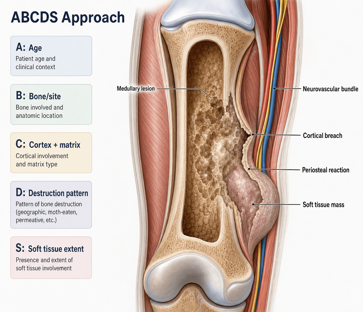
- ABCDS: Age, Border, Cortex, Depth, Soft tissue
- Zone of transition: narrow (benign) vs wide (aggressive)
- Lodwick classification grades aggressiveness (I-III)
- Whole bone MRI mandatory before biopsy for staging
- Biopsy track must be planned with definitive surgeon
- “Age is single most important factor in differential
- “Periosteal reaction: solid (benign) vs interrupted (aggressive)
- “Skip lesions: separate tumour foci in same bone
- “Enneking staging: compartmental vs extracompartmental
- “Don't biopsy before discussing with tumour surgeon
Overview and Principles
Musculoskeletal tumours run from trivial "do-not-touch" lesions to lethal high-grade sarcomas. The candidate's task is not to memorise every entity but to apply a reproducible systematic framework to any radiograph and answer one question: is the lesion non-aggressive, which can be observed or treated by a generalist, or aggressive, which must be referred to a specialist sarcoma centre before any intervention? Age, zone of transition, cortical integrity, periosteal reaction and a soft-tissue mass answer that question faster than trying to name the tumour.
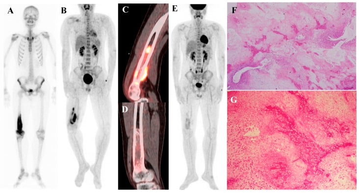
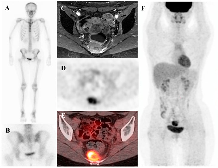
Why the radiograph comes first. Matrix mineralisation, periosteal reaction and the zone of transition are best appreciated on plain film, and they frequently allow a confident diagnosis or a tight differential. Cross-sectional imaging then answers specific questions: MRI for marrow and soft-tissue extent and local staging, CT for cortical detail and pulmonary metastases, and FDG-PET/CT for whole-body staging and response.
Do-not-touch lesions. Non-ossifying fibroma, bone island, fibrous dysplasia, osteochondroma and simple bone cyst have such typical features that biopsy is unnecessary and may cause harm. Recognising them avoids over-investigation.
The danger of acting before staging. A poorly planned biopsy or an unwary excision of an unsuspected sarcoma can convert a limb-salvageable tumour into an amputation or compromise survival. The candidate who states early that any suspicious lesion is referred intact to a sarcoma multidisciplinary team, fully staged, and biopsied along a planned track demonstrates safe practice.
Systematic ABCDS Approach
A-B-C-D-SABCDS - Tumour Imaging Assessment
Hook:Work through each element systematically in viva - this demonstrates methodical approach and covers all key features
Age. Age is the single most important factor in the differential. In patients over 40, metastases are the most common malignant bone lesion by far; in patients under 30, primary bone tumours are more common than metastases. The table gives the entities to consider in each decade.
- Benign
- Eosinophilic granuloma, Simple bone cyst, Osteochondroma
- Malignant
- Ewing sarcoma, Neuroblastoma mets
- Benign
- Osteoid osteoma, Chondroblastoma, Aneurysmal bone cyst
- Malignant
- Osteosarcoma, Ewing sarcoma
- Benign
- Giant cell tumour, Enchondroma
- Malignant
- Primary lymphoma, Parosteal osteosarcoma
- Benign
- Fibrous dysplasia (monostotic), Haemangioma
- Malignant
- Metastases, Myeloma, Chondrosarcoma
- Benign
- Paget's disease
- Malignant
- Metastases (most common), Myeloma
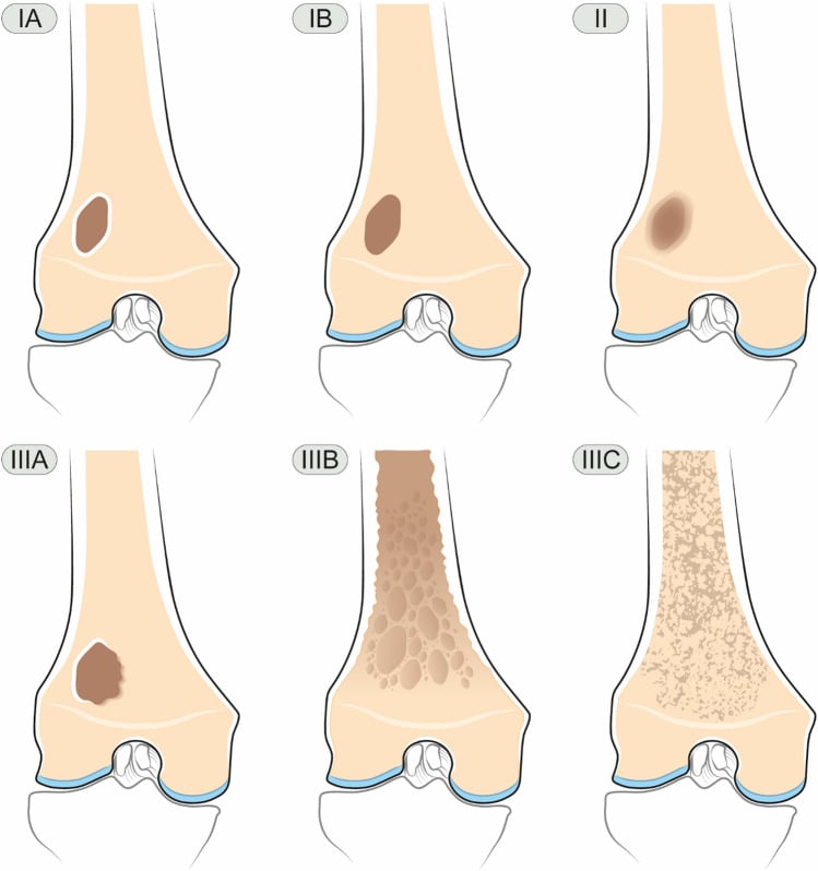
Lodwick Classification
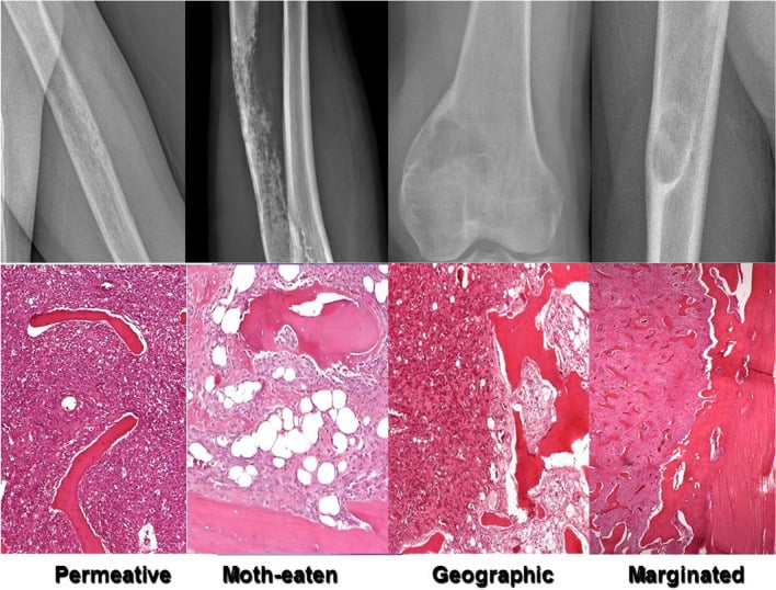
What Lodwick grades. The classification grades the pattern of bone destruction, and with it the growth rate and likely behaviour, from a geographic lesion with a sclerotic rim to permeative destruction. Type I lesions (A, B, C) can often be observed or treated conservatively; Type II and III lesions require urgent investigation and often indicate malignancy or aggressive infection.
- Pattern
- Geographic with sclerotic rim
- Zone of Transition
- Very narrow
- Growth Rate
- Very slow
- Typical Lesions
- NOF, Enchondroma, SBC
- Pattern
- Geographic without sclerosis
- Zone of Transition
- Narrow
- Growth Rate
- Slow
- Typical Lesions
- GCT, ABC, Eosinophilic granuloma
- Pattern
- Geographic with ill-defined margin
- Zone of Transition
- Wide
- Growth Rate
- Moderate
- Typical Lesions
- Low-grade malignancy, Infection
- Pattern
- Moth-eaten
- Zone of Transition
- Wide
- Growth Rate
- Fast
- Typical Lesions
- Aggressive malignancy, Osteomyelitis
- Pattern
- Permeative
- Zone of Transition
- Very wide
- Growth Rate
- Very fast
- Typical Lesions
- Round cell tumours, Ewing, Lymphoma
The modified scheme. The Lodwick-Madewell modification in the figure below numbers the patterns differently from the table above: ill-defined geographic destruction becomes grade II, a margin that changes on serial radiographs is IIIA, and moth-eaten and permeative destruction are IIIB and IIIC. Serial change and the zone of transition are biological observations, not merely labels.
Periosteal Reaction Patterns
Reading the periosteum. A solid, continuous reaction means a slow, benign process; an interrupted one means a rapid, aggressive one. Lamellated onion-skin layers record intermittent growth, the Codman triangle is the periosteum lifted at the edge of the lesion, and sunburst spiculation is the tumour outgrowing its containment. Infection and healing can mimic the periosteal reaction of an aggressive tumour, so always correlate with the clinical features.
- Appearance
- Smooth, uninterrupted
- Implication
- Slow-growing, benign
- Common Causes
- Stress fracture, Osteoid osteoma
- Appearance
- Multiple parallel layers
- Implication
- Intermittent growth
- Common Causes
- Ewing sarcoma, Osteomyelitis
- Appearance
- Perpendicular striations
- Implication
- Aggressive, rapid growth
- Common Causes
- Osteosarcoma
- Appearance
- Fine perpendicular spicules
- Implication
- Aggressive
- Common Causes
- Ewing, Marrow expansion
- Appearance
- Interrupted periosteum lifted at edge
- Implication
- Very aggressive
- Common Causes
- Osteosarcoma, Ewing, Infection
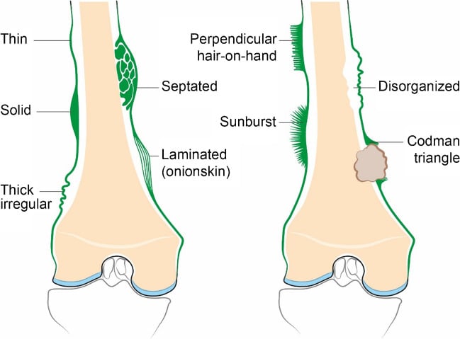
Matrix Mineralisation
What the tumour makes. Osteoid matrix is dense, cloud-like and amorphous, and means bone formation by the tumour; seen with aggressive features it is osteosarcoma until proven otherwise. Chondroid matrix, the rings-and-arcs or popcorn calcification, is pathognomonic of cartilage tumours, but it is seen in both the benign enchondroma and the malignant chondrosarcoma, so the matrix must be interpreted with margins, cortical integrity and clinical symptoms. Fibrous matrix is hazy ground-glass, and mixed patterns belong to dedifferentiated tumours.
- Appearance
- Dense, cloud-like, amorphous
- Tumours
- Osteosarcoma, Osteoid osteoma, Osteoblastoma
- Appearance
- Rings and arcs, popcorn
- Tumours
- Enchondroma, Chondrosarcoma, Chondroblastoma
- Appearance
- Ground-glass, hazy
- Tumours
- Fibrous dysplasia, NOF
- Appearance
- Combination patterns
- Tumours
- Dedifferentiated tumours
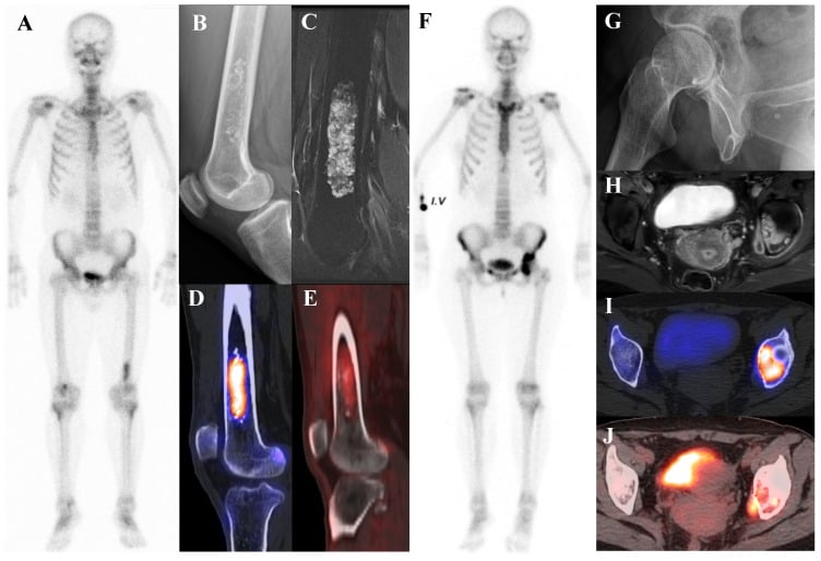
Enneking Surgical Staging
Benign lesions are staged by behaviour: latent lesions are inactive and self-limiting and stay within their capsule, active lesions grow and deform but remain contained, and aggressive lesions breach the capsule and the cortex.
- Behaviour
- Inactive, self-limiting, stays within its capsule
- Examples
- Non-ossifying fibroma, bone island
- Behaviour
- Grows and deforms but remains contained
- Examples
- Aneurysmal bone cyst, active simple bone cyst
- Behaviour
- Locally aggressive, breaches the capsule and cortex
- Examples
- Giant cell tumour
Malignant lesions are staged by combining histological grade (G1 low, G2 high), anatomic site (T1 intracompartmental, T2 extracompartmental) and metastasis (M0 / M1). The T is an imaging decision: MRI shows whether the tumour has breached its compartment, which, with grade and metastasis, sets the stage and the required margin. The Enneking system is increasingly supplemented or superseded by AJCC/UICC TNM, which adds tumour size and depth, for soft-tissue sarcoma staging.
- Grade
- G1 (low)
- Site
- T1 (intracompartmental)
- Metastasis
- M0
- Grade
- G1 (low)
- Site
- T2 (extracompartmental)
- Metastasis
- M0
- Grade
- G2 (high)
- Site
- T1 (intracompartmental)
- Metastasis
- M0
- Grade
- G2 (high)
- Site
- T2 (extracompartmental)
- Metastasis
- M0
- Grade
- Any grade
- Site
- Any site
- Metastasis
- M1 (regional or distant)
MRI in Tumour Imaging
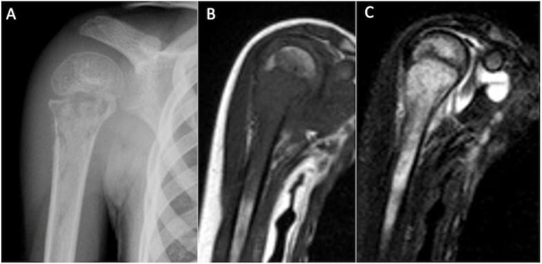
What MRI is for. MRI is the local staging study. It shows intramedullary extent on T1 and STIR, where tumour replacing marrow is low on T1; the soft-tissue mass and its relationship to neurovascular structures on T2 and gadolinium-enhanced sequences; joint involvement, which affects surgical planning; and neurovascular encasement, for resectability assessment.
Whole-bone MRI. It is mandatory before biopsy to identify skip lesions: separate tumour foci in the same bone, which affect staging, prognosis and surgical planning. Skip lesions are found in 3-25% of osteosarcomas; the whole-bone MRI cohort in the evidence base put the true incidence at 6.5% of high-grade long-bone sarcomas once cases with distant disease were excluded.
- Sequence
- T1, STIR
- Key Information
- Low T1 = tumour replacing marrow
- Sequence
- T2, Gd-enhanced
- Key Information
- Size, relationship to NV structures
- Sequence
- Whole bone T1/STIR
- Key Information
- Separate tumour foci (staging, prognosis)
- Sequence
- All sequences
- Key Information
- Affects surgical planning
- Sequence
- Gd-enhanced, MRA
- Key Information
- Resectability assessment
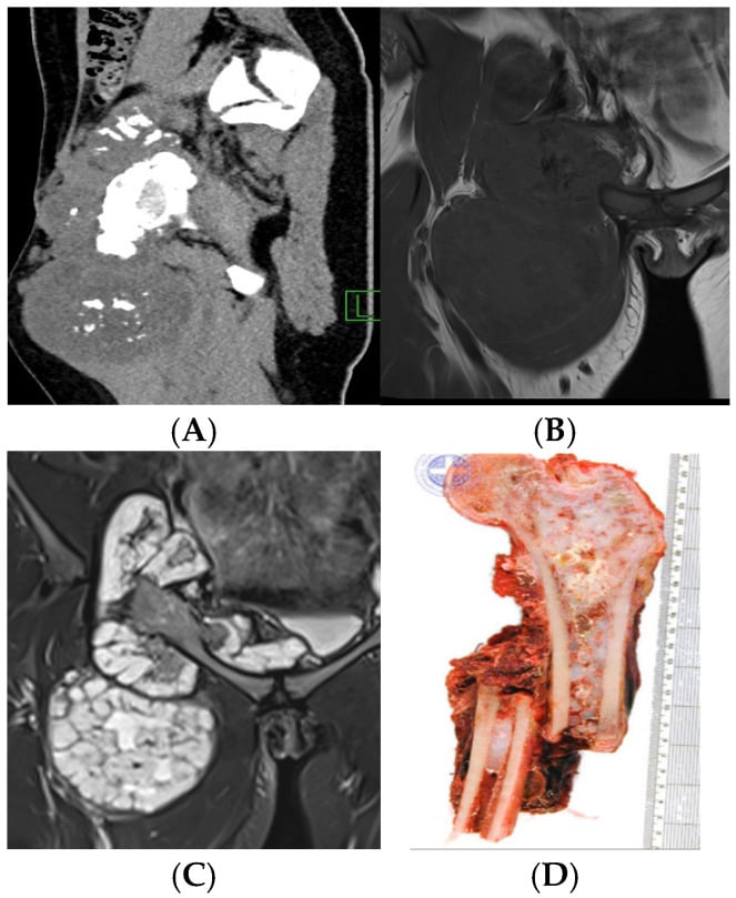
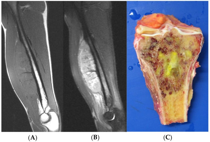
Classic Tumour Appearances
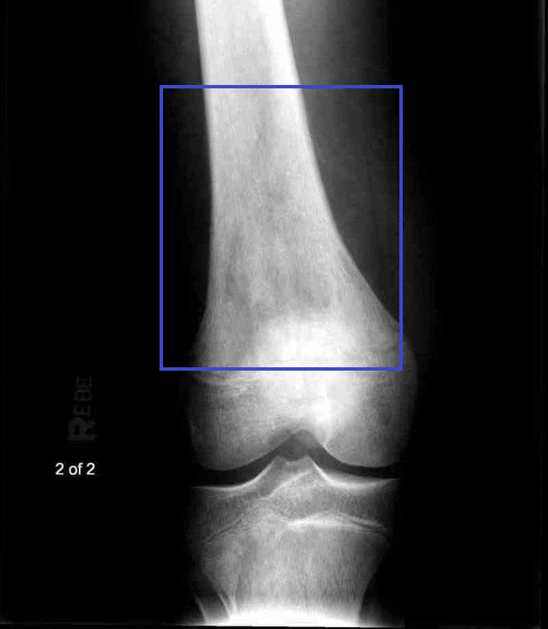
Location, age and the pattern of matrix and margin identify most of the common tumours, and the two tables set out the features that make each diagnosis. Osteosarcoma is the prototype of the aggressive lesion: a metaphyseal tumour in a young patient with osteoid matrix, a sunburst periosteal reaction and a Codman triangle.
- Location
- Cortex of long bones
- Age
- 10-25y
- Classic Features
- Less than 1.5cm nidus with reactive sclerosis, nocturnal pain relieved by NSAIDs
- Location
- Metaphysis, away from joint
- Age
- Less than 30y
- Classic Features
- Cortex and medulla continuous with parent bone, pedunculated or sessile
- Location
- Hands, metaphysis long bones
- Age
- 20-40y
- Classic Features
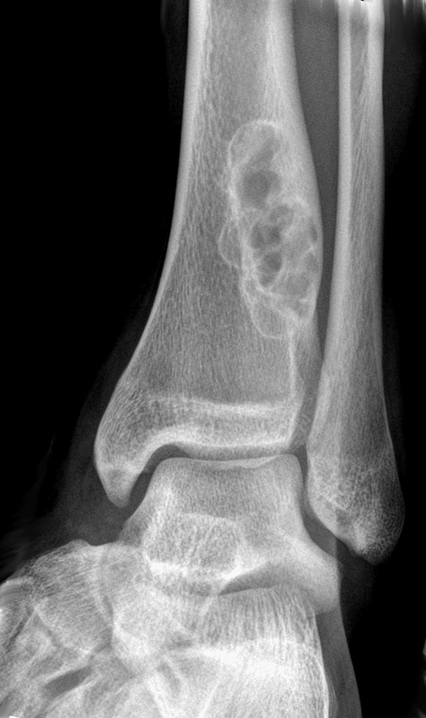
- Central, rings and arcs calcification, Lodwick IA
- Location
- Epiphysis, extends to subchondral
- Age
- 20-40y
- Classic Features
- Eccentric, subarticular, no sclerotic margin (IB), closed physis
- Location
- Proximal humerus, prox femur
- Age
- Less than 20y
- Classic Features
- Central, fallen fragment sign, slightly expansile
- Location
- Metaphysis, eccentric
- Age
- Less than 20y
- Classic Features
- Well-defined, sclerotic rim, asymptomatic, resolves
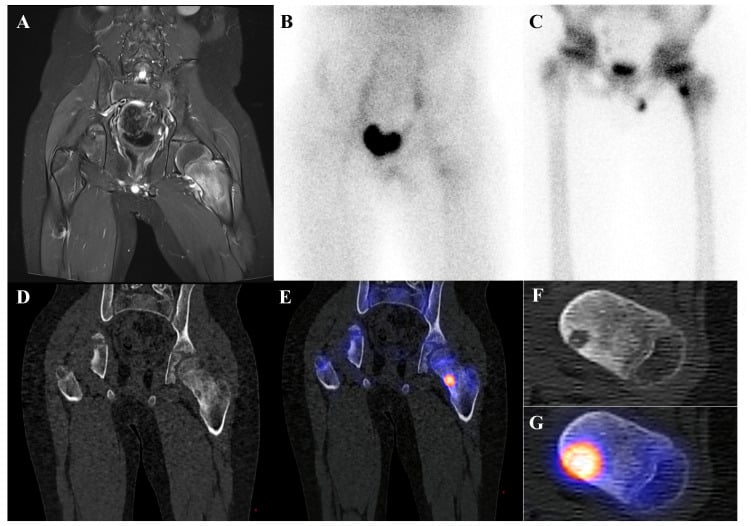
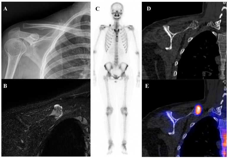
Soft Tissue Tumour Imaging
The red flags. Deep location, large size and rapid growth are the features that matter. Any deep soft-tissue mass greater than 5 cm warrants urgent specialist referral and staging MRI before biopsy. The table sets the benign and malignant indicators side by side.
- Benign Indicators
- Less than 5 cm
- Malignant Indicators
- Greater than 5 cm
- Benign Indicators
- Superficial to fascia
- Malignant Indicators
- Deep to fascia
- Benign Indicators
- Well-defined, smooth
- Malignant Indicators
- Irregular, infiltrative
- Benign Indicators
- Homogeneous
- Malignant Indicators
- Heterogeneous, necrosis
- Benign Indicators
- Homogeneous signal
- Malignant Indicators
- Mixed signal, haemorrhage, necrosis
- Benign Indicators
- Uniform or none
- Malignant Indicators
- Heterogeneous, peripheral
Imaging-Guided Biopsy
Before the needle. Staging is complete before biopsy: whole-bone MRI for skip lesions and CT chest for lung metastases. The track is discussed with the tumour surgeon and planned in line with the definitive resection, so that the line of approach runs through the eventual incision and the whole track can be removed en bloc with the tumour.
The principles of the biopsy itself:
- Shortest path through normal tissue
- Avoid contaminating uninvolved compartments
- Mark the skin entry site for excision
- Core biopsy preferred over open when possible
- Haemostasis is critical
Why it matters. A poorly planned biopsy can convert a limb-salvage case to an amputation through track contamination. In the MSTS survey in the evidence base the diagnostic-error rate from biopsy was 17.8%, in 19.3% a biopsy problem forced a different or more complex operation, and errors, complications and adverse changes in outcome were 2 to 12 times more common when the biopsy was performed at a referring institution rather than a treatment centre.
Surgical Margins
Why imaging defines the margin. Imaging does not just diagnose; it defines whether a tumour can be removed with an oncologically adequate margin, which is why MRI assessment of compartmental boundaries is mandatory. MRI-defined tumour extent, compartmental boundaries and neurovascular relations determine whether a wide margin (limb salvage) is achievable or a more radical procedure or amputation is needed. The planned biopsy track must lie within the tissue that will be excised, or it contaminates the margin and the resection must be widened.
Enneking described four margins by the plane of dissection relative to the tumour and its reactive zone.
- Plane of dissection
- Through the tumour itself (e.g. curettage, debulking)
- Oncological result
- Macroscopic tumour left behind
- Plane of dissection
- Through the reactive zone / pseudocapsule (shell-out)
- Oncological result
- Risks residual satellite or reactive-zone tumour
- Plane of dissection
- En bloc with a cuff of normal tissue, within the same compartment
- Oncological result
- Standard for malignant limb salvage
- Plane of dissection
- Removal of the entire involved compartment(s), or amputation above it
- Oncological result
- Whole compartment cleared
Differential Diagnosis: Aggressive vs Non-Aggressive
The decisive radiographic discriminator is the zone of transition combined with periosteal reaction and cortical status, integrated with patient age. The table collapses the most common pattern-based decisions a candidate must make at the viva.
- Most Likely Group
- Benign, slow (NOF, fibrous dysplasia, enchondroma)
- Key Discriminators
- Sharp pencil-traceable margin, no soft-tissue mass
- Common Mimics / Traps
- Low-grade chondrosarcoma can look enchondroma-like
- Most Likely Group
- Giant cell tumour
- Key Discriminators
- Closed physis, subchondral extension
- Common Mimics / Traps
- ABC (fluid-fluid levels), chondroblastoma (open physis)
- Most Likely Group
- Osteosarcoma
- Key Discriminators
- Codman triangle, soft-tissue ossification, age 10-25
- Common Mimics / Traps
- Osteomyelitis and stress fracture can mimic
- Most Likely Group
- Ewing sarcoma / round cell
- Key Discriminators
- Age 5-25, systemic symptoms, fever
- Common Mimics / Traps
- Osteomyelitis, eosinophilic granuloma, lymphoma
- Most Likely Group
- Metastases / myeloma
- Key Discriminators
- Known primary; vertebral body or pedicle involvement is not a reliable discriminator
- Common Mimics / Traps
- Brown tumours of hyperparathyroidism
- Most Likely Group
- Chondrosarcoma
- Key Discriminators
- Pain, cortical destruction, deep endosteal scalloping
- Common Mimics / Traps
- Enchondroma (asymptomatic, no deep scalloping)
WIDE = WorryAggressive vs Non-Aggressive at a Glance
Hook:Each WIDE feature independently suggests aggression. Any one in an at-risk age group means refer and stage before you touch the lesion.
Guidelines, Registries & Global Practice
Primary bone sarcomas are rare worldwide (osteosarcoma incidence approximately 3-4 per million per year, peaking in adolescence, with a second peak in older adults often related to Paget disease or prior radiation). Ewing sarcoma is rarer still and shows marked geographic and ancestral variation, being notably less common in populations of African ancestry. Soft-tissue sarcomas are commoner than bone sarcomas (roughly 4-5 per 100,000 per year) but still account for under 1% of adult malignancies. Bone metastases vastly outnumber primary bone tumours overall and dominate the differential in patients over 40.
- Region
- UK
- Key Position
- Urgent direct-access radiograph for suspected bone sarcoma; refer suspected sarcoma to a specialist centre BEFORE biopsy; MRI of whole compartment for local staging
- Region
- Europe
- Key Position
- Any deep or over-5cm soft-tissue mass imaged with MRI and referred to a reference centre before biopsy; biopsy planned by the treating sarcoma team
- Region
- US / international
- Key Position
- Cross-sectional local imaging plus chest CT for staging; image-guided core biopsy along a track that can be excised; multidisciplinary review
- Region
- US
- Key Position
- Radiograph first-line for a suspected primary bone lesion; MRI for local extent; bone scan/PET-CT for skeletal and systemic staging
Controversies and Areas of Uncertainty
Reliability of radiographic classification. Despite their teaching value, the Lodwick and Enneking radiographic classifications show only poor-to-moderate interobserver agreement (kappa 0.23-0.28) and fair-to-moderate intra-observer reproducibility (0.42-0.45), and training level made no measurable difference, so this is not a gap experience closes. The accuracy figure is the one to carry: across the three systems, lesions were correctly called malignant in only 73%, 59% and 62% of cases. A framework that is right about two-thirds of the time guides thinking; it cannot be the sole basis for a benign-versus-malignant decision, which must integrate clinical data and cross-sectional imaging.
Enchondroma versus low-grade chondrosarcoma. No single imaging feature is definitive. Deep endosteal scalloping (over two-thirds of the cortical thickness), cortical destruction, a soft-tissue mass, pain and an axial or proximal location favour chondrosarcoma, but the overlap is substantial, and biopsy of cartilage tumours is itself notoriously unreliable for grade.
Whole-body MRI versus PET-CT. Both detect skip and distant disease; the optimal staging algorithm, and the place of whole-body diffusion-weighted MRI, is still evolving and varies between centres. CT chest remains mandatory for pulmonary metastases regardless, as small lung nodules may be PET-negative.
Surveillance imaging after sarcoma resection. The value of routine post-treatment MRI for detecting local recurrence is debated, and the numbers are sharper than the debate suggests. Among 366 patients, 17% developed a local recurrence; 66% were found by clinical signs and symptoms and only 34% by surveillance imaging, and the tumours found by imaging were not smaller than those found clinically (4.5 cm versus 3.9 cm), which removes the main rationale for scanning, since earlier detection is only worth having if it means smaller disease. No patient subgroup has been reliably shown to benefit preferentially from imaging surveillance over examination. The case for continuing surveillance MRI within current guidelines rests on the third of recurrences that were imaging-only, not on earlier detection.
Clinical Imaging Modalities
The sequence. Radiograph first, always. Then MRI of the whole involved bone for local staging, CT chest for pulmonary metastases, and PET-CT or bone scan for whole-body skeletal staging. Imaging is completed before biopsy, so that the biopsy track can be planned and skip lesions identified.
- Primary Role
- First-line characterisation
- Strengths
- Matrix, periosteal reaction, zone of transition, aggressiveness
- Limitations
- Insensitive to marrow and soft-tissue extent
- Primary Role
- Cortical detail, matrix, staging chest
- Strengths
- Subtle cortical breach, mineralisation, lung metastases
- Limitations
- Ionising radiation, poor soft-tissue contrast
- Primary Role
- Local staging (marrow + soft tissue)
- Strengths
- Marrow extent, skip lesions, neurovascular relations
- Limitations
- Cost/access; metal artefact; less specific for matrix
- Primary Role
- Skeletal survey, polyostotic disease
- Strengths
- Whole-skeleton sensitivity
- Limitations
- Low specificity; myeloma often photopenic
- Primary Role
- Whole-body staging, response, recurrence
- Strengths
- Metabolic activity, distant disease, restaging
- Limitations
- False positives (infection, healing); resolution limits
Clinical Decision Scenarios
Practise clinical reasoning and management decisions out loud
“A 16-year-old presents with a 2-month history of knee pain. X-ray shows a mixed lytic and sclerotic lesion in the distal femoral metaphysis with periosteal reaction.”
“A 55-year-old woman presents with back pain. Spinal X-ray shows a lytic lesion in L3 vertebral body. She has a history of breast cancer 5 years ago.”
“A 30-year-old presents with knee pain. X-ray shows an expansile lytic lesion in the proximal tibial epiphysis extending to the subchondral bone. The lesion has no sclerotic margin.”
ABCDS Framework
- Age: Most important factor - metastases if greater than 40
- Border: Narrow = benign, Wide = aggressive
- Cortex: Intact/expanded vs destroyed
- Depth: Medullary/cortical/surface location
- Soft tissue: Mass indicates aggressive behaviour
Lodwick Classification
- IA: Geographic + sclerotic rim = very benign
- IB: Geographic, well-defined = slow growing
- IC: Geographic, ill-defined = moderate
- II: Moth-eaten = aggressive
- III: Permeative = very aggressive (Ewing, lymphoma)
Periosteal Reaction
- Solid/continuous = slow, benign
- Lamellated (onion-skin) = Ewing, infection
- Sunburst = osteosarcoma
- Codman triangle = aggressive
Pre-Biopsy Rules
- Whole bone MRI BEFORE biopsy (skip lesions)
- CT chest for lung metastases
- Discuss track with tumour surgeon
- Plan track in line with definitive resection
- Poor biopsy can convert salvage to amputation
Evidence Base
Surgical Staging System for Musculoskeletal Sarcoma (Enneking)
- Introduced the GTM staging system stratifying sarcomas by histological grade (G1 low / G2 high), anatomic compartment (T1 intracompartmental / T2 extracompartmental) and metastasis (M0/M1).
- Defined the three surgical stages (I low-grade, II high-grade, III metastatic) each subdivided by compartmental status, and linked them to surgical margins (intralesional, marginal, wide, radical).
- Provided the conceptual basis for relating imaging-defined compartmental anatomy to oncologically appropriate resection.
Lodwick and Enneking Classifications Have Poor Reliability
- Across 48 radiolucent lesion cases rated twice by 20 observers, interobserver agreement was poor for the original Lodwick (39%, kappa 0.23), modified Lodwick (39%, kappa 0.25) and Enneking benign classification (53%, kappa 0.28).
- Intra-observer reproducibility was only fair to moderate (kappa 0.42-0.45), and training level had no measurable effect.
- Lesions were correctly called malignant in only 73%, 59% and 62% of cases respectively across the three systems.