Sole Motor Nerve to Gluteus Maximus
- Sacral plexus roots L5, S1, S2; purely motor, with no cutaneous branch; the sole motor supply to gluteus maximus
- Exits the greater sciatic foramen below piriformis; the superior gluteal nerve exits above
- Gluteus maximus is the principal hip extensor, most important from flexion (rising from sitting, stairs, running), and a lateral rotator
- Injury gives weak hip extension, a gluteus maximus lurch and buttock atrophy, not a Trendelenburg gait
- Enters the deep surface of gluteus maximus about 5 cm from the greater trochanter tip, so the posterior-approach split is limited proximally
- Partial gluteus maximus denervation is most associated with the posterior approach, but clinical sequelae in primary THA are usually not significant
- “Below versus above piriformis, extension versus abduction, lurch versus Trendelenburg
- “Test gluteus maximus prone with the knee flexed to 90 degrees, which takes the hamstrings out of hip extension
- “Weak extension with sensory loss, a reduced ankle reflex or distal weakness: look beyond the nerve to an S1 root or a proximal sciatic/sacral-plexus lesion
Origin and Course
One nerve, one muscle. The inferior gluteal nerve arises from the sacral plexus from the roots L5, S1 and S2 and supplies exactly one muscle: it is the sole motor supply to gluteus maximus. That single fact organises everything else. It is purely motor, with no cutaneous branch, so an injury is silent on sensory testing and shows up only as weak hip extension.
The course. The nerve leaves the pelvis through the greater sciatic foramen below piriformis, as does the sciatic nerve, and immediately breaks up into branches that enter the deep (anterior) surface of gluteus maximus. The superior gluteal nerve leaves above piriformis, which makes piriformis the key landmark between the two.
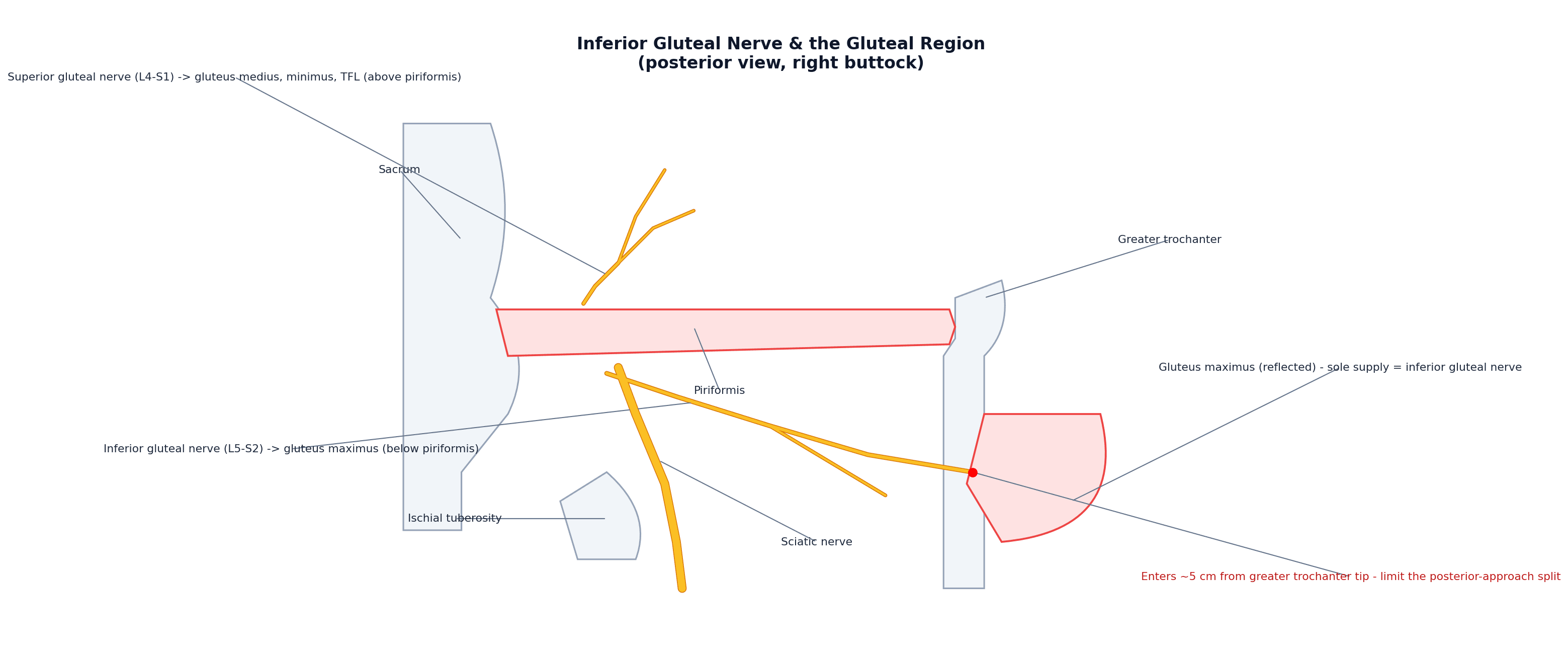
Its neighbour. Learn the nerve as a pair with the superior gluteal nerve: below versus above piriformis, extension versus abduction, lurch versus Trendelenburg.
- Inferior gluteal nerve
- L5, S1, S2
- Superior gluteal nerve
- L4, L5, S1
- Inferior gluteal nerve
- Below piriformis
- Superior gluteal nerve
- Above piriformis
- Inferior gluteal nerve
- Gluteus maximus (sole supply)
- Superior gluteal nerve
- Gluteus medius, gluteus minimus, tensor fasciae latae
- Inferior gluteal nerve
- Hip extension (from flexion), lateral rotation
- Superior gluteal nerve
- Hip abduction (pelvic stabiliser in stance)
- Inferior gluteal nerve
- Gluteus maximus lurch (backward trunk thrust)
- Superior gluteal nerve
- Trendelenburg gait/lurch
Gluteus Maximus: The Muscle the Nerve Powers
Because the nerve is the sole supply of gluteus maximus, its functional importance follows from the muscle's attachments. Gluteus maximus is the largest and most superficial gluteal muscle, and it arises broadly from:
- The posterior gluteal line of the ilium and the bone behind it
- The posterior aspect of the lower sacrum and coccyx
- The sacrotuberous ligament
- The thoracolumbar fascia
A dual insertion. The upper roughly three-quarters of the muscle inserts into the iliotibial tract, and the deep lower quarter into the gluteal tuberosity of the femur. Through the iliotibial tract it tenses the fascia lata and helps stabilise the extended knee and the lateral hip, so its weakness affects more than straight-line extension.
Its actions. It is the body's most powerful hip extensor and the only powerful extensor of the flexed hip, which is why it matters most for rising from sitting, climbing stairs and running or jumping, exactly the activities lost when the nerve is injured. The whole muscle is a lateral rotator. The upper fibres, above the axis of the hip joint, assist abduction through the iliotibial band, and the lower fibres assist adduction.
Injury, Examination and the Differential
The deficit. Injury to the nerve weakens hip extension: the patient has difficulty rising from a low chair, climbing stairs and running, and the buttock atrophies and flattens.
The lurch. The gait sign is the gluteus maximus lurch, a backward thrust of the trunk at heel strike. It keeps the hip extended over the stance limb by bringing the line of gravity, and the ground reaction force, behind the hip, so the weak extensor is not needed.
The gluteus maximus lurch (trunk thrown backwards at heel-strike) signals inferior gluteal nerve / maximus weakness, whereas the trunk thrown sideways over the stance leg (Trendelenburg) signals superior gluteal nerve / abductor weakness.
Testing the muscle. Examine the patient prone with the knee flexed to 90 degrees, which slackens the hamstrings so they cannot substitute as hip extensors, and ask them to lift the thigh off the couch against resistance. What extension power remains is the maximus. Inspect and palpate the buttock for flattening and atrophy, comparing sides.
The differential of weak hip extension. If weak extension comes with sensory loss, a reduced ankle reflex or distal weakness, look beyond the nerve to an S1 root or a proximal sciatic or sacral-plexus lesion.
- Isolated inferior gluteal nerve palsy - pure motor maximus weakness, no sensory loss, normal hamstrings and ankle
- S1 radiculopathy - adds sensory loss (posterolateral leg and sole), a reduced ankle jerk and plantarflexion weakness, with back or leg pain (developed in the lumbar radiculopathy topic)
- Proximal sciatic nerve or sacral plexus lesion - a wider deficit, with the hamstrings and the sciatic distribution (foot drop, sensory loss) involved alongside the maximus
- Hamstring injury - knee-flexion weakness, with hip extension largely preserved by the maximus (developed in the hamstring injury topics)
Surgical Relevance and the Safe Split
The 5 cm rule. The nerve enters the deep surface of gluteus maximus about 5 cm from the tip of the greater trochanter. In the posterior approach to the hip the muscle split should not extend more than about 5 cm proximal to the trochanter, because beyond that the nerve can be damaged before it is seen. Hence the recommendation of a modified, "safe-split" posterior approach that limits the proximal extent of the split.
Where the danger lies. The skin incision is not the hazard. The danger is how far proximally the muscle is split and how hard it is retracted apart, since the nerve lies on the undersurface and is injured before anyone sees it. Keep the split short and distal, and part the fibres rather than levering them.
How often it matters. Partial denervation of gluteus maximus is most associated with the posterior approach: EMG finds it in about seven of ten posterior-approach patients, whereas the anterolateral approach mainly threatens the superior gluteal nerve branch to tensor fasciae latae. Abductor power is not measurably reduced, and in primary THA the clinical sequelae of these partial lesions are usually not significant. Respect the anatomy because a complete lesion is disabling, but do not tell a patient that a posterior approach will weaken their hip.
Beyond the hip. The nerve is also at risk in sacral and pelvic surgery and in deep gluteal procedures around the greater sciatic foramen.
Mnemonics & Memory Aids
IGN
Hook:IGN: Inferior to piriformis / Innervates gluteus maximus, Gluteus maximus extends the hip (lurch if injured), Near the GT (~5 cm safe split).
Clinical Decision Scenarios
Practise clinical reasoning and management decisions out loud
“Describe the inferior gluteal nerve and the effect of injuring it.”
Origin & course
- Sacral plexus (L5, S1, S2); purely motor (no cutaneous branch)
- Exits greater sciatic foramen BELOW piriformis
- Enters the deep surface of gluteus maximus (its sole motor supply)
Function
- Gluteus maximus = principal hip extensor (from flexion: rising/stairs/running)
- Lateral rotator; upper fibres assist abduction (via IT band)
- Powers powerful hip extension
Injury
- Weak hip extension (rising from chair, stairs, running)
- Gluteus maximus lurch (backward trunk thrust) + buttock atrophy
- NOT Trendelenburg (that is superior gluteal nerve/abductors)
Surgical relevance
- Enters gluteus maximus ~5 cm from the greater trochanter tip
- Posterior hip approach: limit the split to <~5 cm from the GT (safe split)
- Posterior approach most associated with gluteus maximus partial denervation
Evidence & Key Studies
The course of the inferior gluteal nerve in the posterior approach to the hip
- In cadaveric dissection, the inferior gluteal nerve enters the deep surface of gluteus maximus approximately 5 cm from the tip of the greater trochanter.
- Its close relationship to the deep surface of gluteus maximus explains its susceptibility to injury during the posterior approach to the hip - it is easily damaged before it is seen if the muscle is split and parted more than 5 cm from the greater trochanter tip.
- A modified posterior approach limiting the proximal split is recommended to avoid damaging the nerve.
Lesion of gluteal nerves and muscles in THA through three surgical approaches (EMG study)
- Partial denervation of gluteus maximus was most associated with the posterior approach (about 71% of patients), whereas the transgluteal approach mostly affected gluteus medius and the anterolateral approach the superior gluteal nerve branch to tensor fasciae latae.
- The power of the hip abductors was not significantly reduced in partially denervated muscles, and clinical sequelae were not significant in primary THA.
- The approach-specific pattern of gluteal nerve/muscle lesions reflects the underlying nerve anatomy.
The inferior gluteal nerve entering the deep surface of gluteus maximus about 5 cm from the greater trochanter tip and its consequent vulnerability in the posterior approach (with the recommendation to limit the split) come from the cited Ling & Kumar cadaveric study; the approach-specific pattern of gluteal nerve/muscle denervation (posterior approach most associated with gluteus maximus denervation) and the usually insignificant clinical sequelae in primary THA from the cited Chomiak EMG study. The roots (L5-S2), the sole-motor-supply-to-gluteus-maximus, the exit below piriformis, the hip-extension function, and the gluteus maximus lurch (versus Trendelenburg) are standard, well-established anatomical teaching. (See also our Superior Gluteal Nerve and Posterior Hip Approach topics.)