The Posterior Approach to the Hip: Planes, Short External Rotators, Sciatic Nerve and Viva Questions
The posterior approach to the hip, layer by layer: the gluteus maximus split, the short external rotators, sciatic nerve protection, repair and viva answers.
By OrthoVellum Editorial TeamPublished 13 min read
Educational content for clinicians, not medical advice. Editorial policy
Key points
- There is no true internervous plane. Gluteus maximus is split along its fibres within the territory of the inferior gluteal nerve, which enters the muscle's deep surface about 5 cm from the tip of the greater trochanter.
- The short external rotators run piriformis, superior gemellus, obturator internus, inferior gemellus, then quadratus femoris. Tag and release only what the exposure needs, near the trochanter, and keep quadratus femoris where you can.
- The sciatic nerve is the main structure at risk, and its relationship to piriformis varies. Protect it with retractors on bone under vision, limited traction and a check that no suture or cable has caught it, not with a memorised distance.
- In a meta-analysis of five comparative studies, dislocation after posterior THA was 0.49% with soft-tissue repair and 4.46% without (relative risk 8.21). Repair the capsule and rotators when the tissue is viable.
On this page11 sections
The posterior approach to the hip is the exposure most trainees learn first and the one examiners ask about most, because almost everything that can go wrong with it is anatomical. The question in theatre and in the viva is the same: what do you divide, what do you protect, and what do you put back? The short answer is that you split gluteus maximus, release the short external rotators and posterior capsule close to the trochanter, keep the sciatic nerve out of harm's way, and repair what you released. Dislocation is the price of the approach, covered in full on the THA dislocation topic page.
Where does the posterior approach fit?
It gives broad access to the femoral head and neck, the acetabulum, the posterior capsule, the posterior wall and column, and the proximal femur, and a standard exposure leaves the abductor insertion alone. That makes it a common choice for primary THA, hemiarthroplasty and revision, and a close relative of the older Kocher-Langenbeck exposure used for posterior acetabular fractures.
Its weaknesses follow from the same anatomy. The sciatic nerve lies close to the field, and posterior stability depends on structures the approach divides. Lateral positioning can also hide pelvic roll that changes the meaning of a cup guide.
Name the structures, not the eponym. "Moore", "Southern", "posterolateral" and "posterior" are used inconsistently. An operation note should say which of piriformis, obturator internus, the gemelli, obturator externus, quadratus femoris and the capsule were preserved, divided and repaired.
Positioning and incision
The patient lies in lateral decubitus, operative side up, with the pelvis held square to the table by supports that resist roll without compressing soft tissue. Pad the axilla, the dependent fibular head, knees and ankles. Record the starting relationship of the anterior superior iliac spines, sacrum and table before draping, and recheck it after retractors go in and after the femur is delivered. Drape the leg free so the hip can be flexed and internally rotated, without traction on the sciatic nerve.
The incision is centred on the greater trochanter. Its distal limb follows the femoral shaft and its proximal limb curves towards the posterior superior iliac spine. Length follows habitus, pathology and the exposure needed, not a fixed number.
Is there an internervous plane in the posterior approach?
No true internervous plane. The first deep step is a blunt split of gluteus maximus in the line of its fibres, after the fascia lata has been opened over the trochanter. Both halves of the split are supplied by the inferior gluteal nerve (L5-S2), so the manoeuvre is intramuscular. The deep exposure then comes from the rotators and capsule, not from a plane between two nerve territories.
Why the split is safe, and where it stops. The inferior gluteal nerve leaves the pelvis below piriformis and enters the deep surface of gluteus maximus. In a cadaveric study of 12 specimens it entered the muscle about 5 cm from the tip of the greater trochanter, and the authors warned that it is easily damaged before it is seen if the split goes further proximally. The danger is a long proximal split and forceful parting of the muscle, not the skin incision.
How much it matters. In an EMG study, partial denervation of gluteus maximus was found in 71.4% of the posterior-approach group (15 patients) after THA, yet abductor power was not significantly reduced and the authors judged the clinical sequelae not significant in primary THA. Respect the anatomy, but do not tell a patient the approach will weaken the hip.
A trap in the textbooks. Some operative texts describe the posterior or Kocher-Langenbeck exposure as working between gluteus maximus (inferior gluteal nerve) and gluteus medius (superior gluteal nerve). The superior gluteal nerve is indeed furthest from the field in a posterior approach. But the step you actually perform, the maximus split, sits inside one nerve's territory, and that is the answer to give.
The layers, from skin to joint
| Layer | What you do | Structure at risk |
|---|---|---|
| Skin and fat | Incise over the trochanter, curving towards the PSIS | Skin flaps if undermined |
| Fascia lata and gluteal fascia | Incise in line with the incision | None specific |
| Gluteus maximus | Split bluntly along its fibres; self-retaining retractor | Inferior gluteal nerve and vessels if the split runs too far proximally |
| Trochanteric bursa | Excise or reflect only what you need | None specific |
| Short external rotators | Identify, tag and release near the trochanter (or preserve selected tendons) | Sciatic nerve; medial femoral circumflex artery near quadratus femoris |
| Posterior capsule | Longitudinal, L- or T-shaped capsulotomy with stay sutures | Sciatic nerve medially, especially in revision scar |
| Joint | Dislocate in flexion, adduction and internal rotation, or cut the neck in situ | Sciatic nerve on traction; greater trochanter |
The short external rotators: order, supply and release
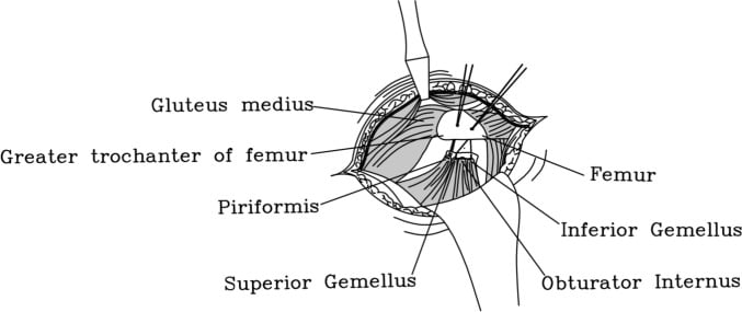
From proximal to distal the rotators are piriformis, superior gemellus, obturator internus, inferior gemellus and quadratus femoris, with obturator externus deep and inferior. The gemelli and obturator internus form the conjoint tendon. Piriformis is the key landmark: the superior gluteal nerve and vessels leave the pelvis above it, and the sciatic and inferior gluteal nerves usually leave below it. Piriformis, the gemelli, obturator internus and quadratus femoris are supplied by small branches of the sacral plexus (the nerve to piriformis, the nerve to obturator internus and the nerve to quadratus femoris); obturator externus is supplied by the obturator nerve.
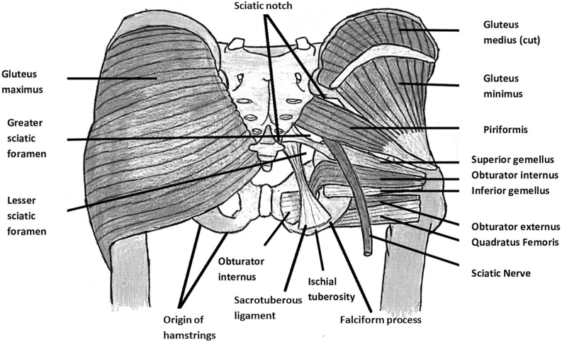
Release. Internal rotation of the hip puts the rotators on tension so they are easier to see and divide. Tag piriformis and the conjoint tendon separately, then divide them close to their femoral insertion while keeping enough length to repair. Reflected medially, the tagged tendons lie between the retractor and the sciatic nerve as a protective layer.
Quadratus femoris and the blood supply. The deep branch of the medial femoral circumflex artery runs at the proximal border of quadratus femoris, passes posterior to obturator externus and anterior to the conjoint tendon, and is the main supply to the femoral head (see hip blood supply). Taking quadratus femoris down too proximally can damage it. That matters little when the head is being replaced, but greatly when it is being kept, as in acetabular fracture surgery; there, the Kocher-Langenbeck guide divides the rotators at least 1.5 cm from the trochanter. Preserve quadratus femoris unless extensile femoral exposure demands otherwise.
Tissue-sparing variants. Piriformis can be preserved when the anatomy and the view allow, releasing only what is needed to reach the capsule. It is an option, not a requirement. Convert to a standard release early if visualisation, femoral delivery, cup position or nerve safety starts to suffer.
How do you protect the sciatic nerve?
The anatomy. The sciatic nerve (L4-S3) leaves through the greater sciatic foramen, descends deep to gluteus maximus with piriformis as its roof, and rests on the gemelli, obturator internus and quadratus femoris, with the greater trochanter lateral. Its common peroneal division is lateral, has fewer and larger fascicles with less connective tissue, and is tethered at the sciatic notch and the fibular neck. It fails first under stretch, so a "sciatic" injury often presents as foot drop.
It is not always below piriformis. In the Beaton and Anson scheme the undivided nerve passes below piriformis in roughly 85%. A meta-analysis of 18 studies and 6,062 cadavers found a piriformis or sciatic nerve anomaly in 16.9%. Expect variation rather than assume the textbook picture.
The practical rules.
- Document sciatic and common peroneal function before surgery.
- Place every deep retractor on bone under direct vision; never lever a posterior retractor towards the sciatic notch.
- Limit sustained traction and extremes of position; knee flexion with the hip extended slackens the nerve across both joints.
- Do not rely on a memorised nerve-to-rim distance. Published figures differ, and the reviewed guidance is to protect the nerve by technique, not by measurement.
- Expose the nerve formally when scar, deformity, revision or an uncertain plane makes indirect protection unreliable.
- Before closing, check that no repair suture, cable or implant threatens it.
How often it goes wrong. Nerve palsy complicates roughly 0.2-1% of primary THAs, and the sciatic nerve accounts for about 80% of these, its peroneal division more often than the tibial (THA neurovascular injury). In a Mayo Clinic series of 27,004 primary THAs, 47 patients (0.17%) had a motor palsy; developmental dysplasia, post-traumatic arthritis, the posterior approach, lengthening and cementless femoral fixation were associated with it, and only 10 of 28 complete palsies (36%) recovered fully. Approach as a risk factor is contested: another cited series found no difference between approaches.
Lengthening. No amount of lengthening is universally safe. Risk rises with the size and acuteness of the change, dysplasia, scar and pre-existing neuropathy. The 4 cm figure often quoted is a planning trigger, not a validated threshold.
Capsulotomy and dislocation
Separate the released tendons from the capsule without shredding either, unless you are raising them as one sleeve. Design a longitudinal, L-shaped or T-shaped capsulotomy that leaves viable flaps, and place stay sutures before the tissue retracts. In revision surgery, tell capsule and scar from nerve before any medial dissection.
Dislocate a mobile primary hip with controlled flexion, adduction and internal rotation once the release is complete. If it needs force, stop and find the residual block. An in-situ neck cut is safer when fracture, stiffness, protrusio, deformity or nerve tension makes dislocation hazardous.
For the acetabulum, keep the anterior retractor on bone deep to iliopsoas, because the femoral nerve and vessels lie anteriorly, and do not lever an inferior retractor through the obturator membrane.
Posterior repair, and why dislocation rates fell
Posterior dislocation, in flexion, adduction and internal rotation, makes up 75-80% of THA dislocations, and the posterior capsule and short rotators are the restraint against it. Repairing them is the main approach-specific answer to that weakness, alongside component position and the other stability factors below.
The evidence. Two surgeons who adopted an identical enhanced repair reported dislocation falling from 4% to 0% (395 patients each) and from 6.2% (160 hips) to 0.8% (124 hips). A meta-analysis of five comparative studies found 0.49% with repair against 4.46% without, a relative risk of 8.21 (95% CI 4.05-16.67) for no repair. In a separate review of 11 studies, rates with repair were 1.01% for the posterior approach against 0.70% anterolateral and 0.43% direct lateral. These are older observational series with different heads, implants and rehabilitation, so treat the numbers as direction and size of effect, not a modern rate for every patient.
The technique. Repair the capsule and the released tendons to bone through transosseous tunnels or anchors, or to each other, matching the repair to what was divided. An intact piriformis does not need detaching to make a conventional repair. Friable tissue will not hold sutures, and stability must then come from component position, offset, tension, removal of impingement and articulation choice, including dual mobility where indicated.
Repair is one part of stability. In a series of 206 dislocated primary THAs, 58% had a cup inside the Lewinnek zone of 40° ± 10° inclination and 15° ± 10° anteversion. Combined version, impingement, soft-tissue tension and spinopelvic behaviour matter as much (see hip-spine syndrome and instability). And routine precautions add little: a review of 6,900 posterior-approach patients found dislocation in 2.2% with precautions and 2.0% without.
| Key number | Source and population |
|---|---|
| ~5 cm from greater trochanter tip | Inferior gluteal nerve entry into gluteus maximus; 12 cadavers |
| 71.4% | Partial gluteus maximus denervation on EMG after posterior THA; posterior group of 15 patients |
| 16.9% | Piriformis or sciatic nerve anomaly; 6,062 cadavers, 18 studies |
| 0.49% vs 4.46% | Dislocation with vs without posterior repair; meta-analysis of 5 studies |
| 0.17% | Motor nerve palsy after primary THA; 27,004 hips, one institution |
| 58% | Dislocated THAs with the cup inside the Lewinnek zone; 206 hips |
| 2.2% vs 2.0% | Dislocation with vs without hip precautions; 6,900 patients |
Extensile options
- Proximally, the posterior ilium and posterior column, protecting the gluteal neurovascular structures at the notch.
- Distally, along the posterior or posterolateral femur, controlling the perforating branches of profunda femoris.
- Trochanteric slide or extended trochanteric osteotomy for femoral revision, keeping a reconstructable abductor-trochanteric unit.
- Full Kocher-Langenbeck for posterior wall and column fractures. It is a trauma-specific exposure with its own nerve planning, not the arthroplasty approach under another name.
- Trochanteric flip (digastric) osteotomy to extend a Kocher-Langenbeck for superior dome fragments or femoral head lesions. Obturator externus is left intact to shield the deep branch of the medial femoral circumflex artery, at the cost of trochanteric non-union and heterotopic ossification risk.
Viva questions with answers
"What is the internervous plane?" There is none. Gluteus maximus is split along its fibres within the inferior gluteal nerve territory, and the deep exposure comes from releasing the short rotators and capsule.
"How far can you split gluteus maximus?" About 5 cm proximal to the tip of the greater trochanter, where the inferior gluteal nerve reaches the muscle's deep surface. Beyond that it can be torn before it is seen.
"Name the short external rotators in order." Piriformis, superior gemellus, obturator internus, inferior gemellus, quadratus femoris, with obturator externus deep and inferior. Piriformis separates the superior gluteal bundle above from the sciatic and inferior gluteal nerves below.
"Why keep quadratus femoris?" The deep branch of the medial femoral circumflex artery runs at its proximal border and is the main supply to the femoral head. A proximal release also brings bleeding and puts the nerve nearer the field.
"Where is the sciatic nerve and how do you protect it?" Deep to gluteus maximus, on the gemelli, obturator internus and quadratus femoris, usually below piriformis, though about one cadaver in six has an anomaly. Retractors on bone under vision, limited traction, rotators reflected over the nerve, and a check before closure.
"A patient has foot drop in recovery. What do you do?" Examine and compare with the baseline, map peroneal and tibial function, remove tight dressings, look for haematoma, and X-ray for fracture, cement or implant problems. Explore urgently for a compressive or clearly correctable cause. Otherwise give an ankle-foot orthosis and involve nerve specialists. The THA neurovascular injury page covers when to revise for lengthening.
"Why does this approach dislocate more, and what do you do about it?" It divides the posterior restraints. Repair them: in pooled data, dislocation fell from 4.46% to 0.49% with repair. Then check cup and stem together, offset, length and impingement, and choose the articulation for the patient's risk.
In the exam
The full operative sequence, with failure plans and aftercare, is in the posterior approach operative guide, and the trauma version in the Kocher-Langenbeck guide. To see the layers and structures at risk in three dimensions, use the 3D posterior hip approach walkthrough.
Frequently asked questions
What is the internervous plane of the posterior approach to the hip?
There is no true internervous plane. The key superficial step is a split of gluteus maximus in the line of its fibres, and both sides of the split are supplied by the inferior gluteal nerve. The deep exposure comes from releasing or preserving the short external rotators and opening the posterior capsule. Some operative texts describe a plane between gluteus maximus and gluteus medius, but the split itself is intramuscular.
Which nerve is most at risk in the posterior approach to the hip?
The sciatic nerve. It leaves the pelvis through the greater sciatic foramen, usually below piriformis, and runs down behind the hip on the short external rotators. Its common peroneal division is lateral and more vulnerable to stretch, so a sciatic injury often shows as foot drop. The inferior gluteal nerve is also at risk if gluteus maximus is split too far proximally.
Why does the posterior approach have a higher dislocation rate?
The posterior capsule and short external rotators restrain the hip against posterior dislocation, which happens in flexion, adduction and internal rotation. The approach divides exactly those structures. Repairing them closes most of the gap: a meta-analysis found posterior dislocation rates of 0.49% with soft-tissue repair against 4.46% without, and rates with repair close to those of the anterolateral and direct lateral approaches.
Do patients need hip precautions after a posterior approach hip replacement?
Not routinely. A systematic review of 6,900 patients after primary posterior-approach THA found dislocation in 2.2% of those given hip precautions and 2.0% of those without, a difference that was not statistically significant. Restrictions are best kept for selected patients, such as those with poor tissue, unstable intraoperative testing, cognitive impairment or neuromuscular disease.
References
- Kwon MS, Kuskowski M, Mulhall KJ, Macaulay W, Brown TE, Saleh KJ. Does surgical approach affect total hip arthroplasty dislocation rates? Clin Orthop Relat Res 2006;447:34-38. DOI (opens in a new tab)
- Pellicci PM, Bostrom M, Poss R. Posterior approach to total hip replacement using enhanced posterior soft tissue repair. Clin Orthop Relat Res 1998;(355):224-228. DOI (opens in a new tab)
- Ling ZX, Kumar VP. The course of the inferior gluteal nerve in the posterior approach to the hip. J Bone Joint Surg Br 2006;88(12):1580-1583. DOI (opens in a new tab)
- Chomiak J, Hurácek J, Dvořák J, et al. Lesion of gluteal nerves and muscles in total hip arthroplasty through 3 surgical approaches. An electromyographically controlled study. Hip Int 2015;25(2):176-183. DOI (opens in a new tab)
- Smoll NR. Variations of the piriformis and sciatic nerve with clinical consequence: a review. Clin Anat 2010;23(1):8-17. DOI (opens in a new tab)
- Farrell CM, Springer BD, Haidukewych GJ, Morrey BF. Motor nerve palsy following primary total hip arthroplasty. J Bone Joint Surg Am 2005;87(12):2619-2625. DOI (opens in a new tab)
- Lewinnek GE, Lewis JL, Tarr R, Compere CL, Zimmerman JR. Dislocations after total hip-replacement arthroplasties. J Bone Joint Surg Am 1978;60(2):217-220. PubMed (opens in a new tab)
- Abdel MP, von Roth P, Jennings MT, Hanssen AD, Pagnano MW. What safe zone? The vast majority of dislocated THAs are within the Lewinnek safe zone for acetabular component position. Clin Orthop Relat Res 2016;474(2):386-391. DOI (opens in a new tab)
- Crompton J, Osagie-Clouard L, Patel A. Do hip precautions after posterior-approach total hip arthroplasty affect dislocation rates? A systematic review of 7 studies with 6,900 patients. Acta Orthop 2020;91(6):687-692. DOI (opens in a new tab)
- Díaz-Ponte S, Aristizabal-Jaramillo C, Palencia-Escorihuela M, Bonilla G, Ortiz-Martinez JG. Use of soft tissue repair as a hip dislocation preventive strategy following a total hip arthroplasty by posterior and posterolateral approach in patients with osteoarthritis: a systematic scoping review. J Orthop Surg Res 2025;20(1):648. DOI (opens in a new tab)
Study the full topic
Included in Pro
Go deeper with Pro
- Operative guide: posterior approach to the hip (Moore/Southern)
- Operative guide: Kocher-Langenbeck approach to the posterior acetabulum
- 3D anatomy: posterior hip approach walkthrough
Share this article
Cite this article
OrthoVellum Editorial Team. The Posterior Approach to the Hip: Planes, Short External Rotators, Sciatic Nerve and Viva Questions [Internet]. OrthoVellum; 2026 Oct 19 [cited 2026 Oct 19]. Available from: https://www.orthovellum.com/blog/posterior-approach-hip
About this content
Prepared by the OrthoVellum Editorial Team from cited sources, under our editorial policy.
For education and exam preparation; not medical advice or a substitute for clinical judgement and local guidance.
Spotted an error? Tell us; every report is read.
Get new articles by email
New clinical explainers, landmark-trial summaries and exam guidance from the OrthoVellum blog. No account needed; unsubscribe from any email.