Hallux IPJ and Lesser Toe IPJ | Post-Traumatic and Degenerative
- Post-traumatic arthritis is the most common cause in younger patients - previous fracture, dislocation, or turf toe injury
- Hallux IPJ arthritis more symptomatic than lesser toes - essential for push-off in gait
- Arthrodesis (fusion) is gold standard - eliminates pain, stable construct, minimal functional loss
- Fusion position critical: hallux IPJ fused in 10-15° plantar flexion to clear ground during gait
- Inflammatory arthritis (RA, psoriatic) can affect multiple IPJs simultaneously
- “Hallux IPJ fusion position: 10-15° plantar flexion, slight valgus (matches contralateral)
- “Lesser toe PIPJ: arthroplasty (resection) acceptable as motion less critical
- “K-wire fixation sufficient for lesser toes, plate/screw preferred for hallux IPJ
- “Nonunion is NOT rare at the hallux IPJ - the largest series (n=227) reports ~25% radiographic nonunion and ~21% reoperation; an unquantified proportion are painless fibrous unions that behave acceptably
Overview and Epidemiology
Interphalangeal joint arthritis of the foot is less common than first MTP arthritis, and no source cited on this page quantifies its share of forefoot arthritis. When it is symptomatic, especially at the hallux, it significantly affects gait and footwear.
Where. The hallux IPJ accounts for over 80% of cases and the lesser toe PIPJ for under 20%. The hallux IPJ matters more because it contributes push-off power in gait; the lesser toe IPJs have a minimal functional role, so their arthritis can often be managed conservatively.
Who. Post-traumatic arthritis presents at 20-50 years and affects men and women equally; it is the most common cause under 50. Degenerative arthritis presents over 50, with a female predominance, and predominates in older adults. Bilateral disease is uncommon unless the cause is inflammatory.
Why fusion works here. Finger IPJs need their motion; toe IPJs have little normal motion to lose. That is why arthrodesis is an excellent treatment for the symptomatic toe IPJ, reliably relieving pain with minimal functional deficit.
Anatomy and Pathophysiology
The joints. The interphalangeal joints of the foot are hinge joints with minimal normal motion compared with the fingers. The collateral ligaments provide mediolateral stability. The plantar plate belongs to the MTP joint and does not extend to IPJ level.
- Normal Motion
- 40-60° dorsiflexion, 0-10° plantar flexion
- Functional Role
- Push-off power in gait, significant
- Arthritis Impact
- High impact - pain with walking, shoe pressure
- Normal Motion
- 30-50° flexion
- Functional Role
- Minimal functional role
- Arthritis Impact
- Low impact - mostly cosmetic and shoe fitting
- Normal Motion
- 20-30° flexion
- Functional Role
- Negligible functional role
- Arthritis Impact
- Rarely symptomatic
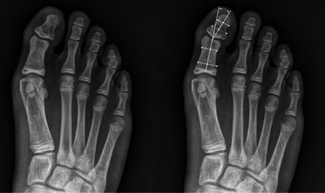
From injury to arthritis. The initiating event in post-traumatic disease is an intra-articular fracture, a dislocation or a severe hyperextension (turf toe) injury that damages the articular cartilage; even if the joint is anatomically reduced, the cartilage is permanently injured. Over months to years the damaged cartilage degenerates progressively: its proteoglycan and collagen structure is lost, chondrocytes undergo apoptosis, and subchondral bone becomes exposed in places.
Established disease. Once the cartilage is lost completely, exposed subchondral bone produces mechanical pain with motion and weight-bearing, osteophytes form at the joint margins, and the joint space narrows on X-ray. At end stage the joint may collapse, chronic pain produces an antalgic gait, dorsal osteophytes cause shoe pressure, and surgery is required for symptom relief.
Post-traumatic arthritis. The history is of a specific injury, with unilateral, progressive pain and stiffness. Symptoms develop 6 months to 5 years after the injury, earlier after a more severe one, and X-ray changes may precede symptoms. The causes:
- Intra-articular fracture, even if well reduced, and the missed undisplaced intra-articular fracture
- IPJ dislocation, which reduces but leaves a cartilage shear injury
- Severe turf toe, a hyperextension injury that contuses the hallux IPJ cartilage
- Repeated microtrauma in athletes (soccer, ballet)
- Chronic mallet or hammer toe deformity, which leads to secondary arthritis
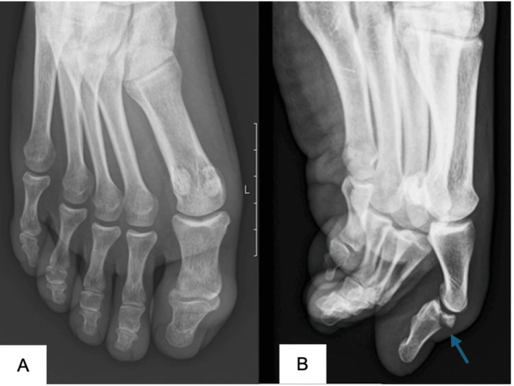
Degenerative arthritis. Age-related cartilage wear, chronic mechanical overload and genetic predisposition produce an insidious onset over years, typically after 60, with slow progression and no specific injury. These patients may have arthritis elsewhere (hands, knees), and the condition is associated with hallux rigidus (MTP arthritis). Risk factors:
- Age over 60 years
- Obesity, through increased forefoot load
- High-impact activities (running, jumping)
Inflammatory arthritis. Rheumatoid arthritis is the most common inflammatory cause; psoriatic arthritis is another, and gout is rare at the IPJ. The signature is multiple IPJ involvement, bilateral and symmetric, with morning stiffness, swelling and synovitis, and systemic disease markers (RF, anti-CCP). Always screen for inflammatory arthritis if multiple IPJs are affected. Medical management (DMARDs, biologics) comes first, fusion follows if it fails, and these patients often require multiple joint fusions.
Clinical Presentation and Examination
History. Pain localises to a specific IPJ, hallux or lesser toe, and is aggravated by walking, push-off (at the hallux) and shoe pressure. Onset is months to years after an injury in post-traumatic disease and insidious in degenerative disease. Morning stiffness suggests an inflammatory cause, end-of-day stiffness a degenerative one. Patients have often already tried NSAIDs or shoe modifications, and the functional impact falls on walking distance and athletic activities.
Examination. Look for swelling, a dorsal prominence from osteophytes and malalignment: hyperextension or flexion contracture at the hallux IPJ, hammer or mallet toe in the lesser toes. The affected IPJ is tender, and warm if inflamed; motion is reduced and painful at end range, with crepitus. The gait is antalgic with reduced push-off in hallux IPJ arthritis. Check shoe wear pattern and pressure areas.
- ROM - compare with the contralateral side (normally 40-60° dorsiflexion)
- Grind test - axially load and rotate the IPJ - crepitus and pain suggest arthritis
- Dorsal osteophyte - palpate the dorsal joint - a prominence indicates advanced disease
- Push-off test - single-leg toe raise - weakness or avoidance if painful
These findings confirm symptomatic arthritis requiring treatment.
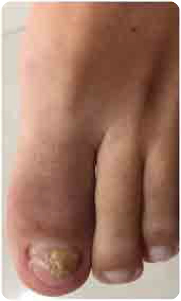
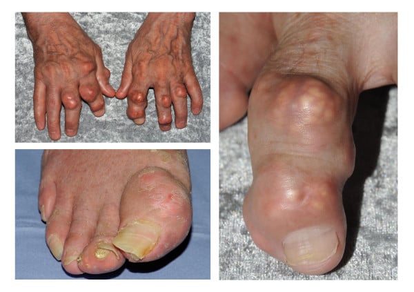
Differential diagnosis. Nail dystrophy with oedema and erythema centred on the hallux IPJ should raise suspicion of psoriatic onycho-pachydermo periostitis rather than isolated mechanical osteoarthritis. Chronic tophaceous gout produces asymmetric nodular swelling and yellow-white deposits around the great toe and other interphalangeal joints.
- Key Distinguishing Features
- Acute onset, severe pain, erythema, asymmetric
- Diagnostic Test
- Serum uric acid, joint aspiration (urate crystals)
- Key Distinguishing Features
- Acute, fever, systemic illness, recent trauma/surgery
- Diagnostic Test
- Joint aspiration (cell count, culture)
- Key Distinguishing Features
- Acute hyperextension injury, plantar plate tear
- Diagnostic Test
- MRI (plantar plate injury, no chronic arthritis)
- Key Distinguishing Features
- Pain at nail, paronychia, ingrown nail
- Diagnostic Test
- Inspection of nail and nail bed
Assessing Union: Painless Fibrous Union vs Symptomatic Nonunion
Because radiographic nonunion is common and only some of those toes are symptomatic, the decisive clinical skill after hallux IPJ arthrodesis is telling a benign fibrous union, left alone, from a symptomatic mobile nonunion, revised. The radiograph still counts: in Braswell's cohort it was the patients labelled radiographic nonunion who carried the higher reoperation risk, but reoperation was driven by symptoms and mobility, not the radiograph alone. The judgement is made on the patient in front of you.
- Radiographic features
- At least two cortices bridging the fusion site; no hardware failure; no lytic gapping around the implant
- Clinical features
- Painless, stable, no motion at IPJ
- Action
- None - solid fusion achieved
- Radiographic features
- Meets the radiographic nonunion definition (fewer than two bridging cortices) but stable-appearing hardware
- Clinical features
- PAINLESS toe with no detectable IPJ motion on examination - behaves like a fusion
- Action
- Accept - a successful clinical endpoint; do NOT revise
- Radiographic features
- Fewer than two bridging cortices, a lytic gap, or hardware loosening/breakage
- Clinical features
- Pain on weightbearing/push-off WITH detectable micromotion or instability at the fusion site
- Action
- Consider revision (re-preparation, compression, rigid re-fixation, bone graft)
How to assess it in clinic. Take weightbearing radiographs (CT only if union is genuinely equivocal or revision is being planned), stress the toe manually at the IPJ to detect micromotion, and correlate strictly with the patient's pain.
Confirm the pain generator. A diagnostic local-anaesthetic infiltration of the IPJ that abolishes the pain confirms the joint as the source before committing to revision. This matters because adjacent first-MTP arthritis (hallux rigidus) or a neighbouring nail or soft-tissue problem can mimic IPJ pain, and radiographic nonunion at a nearby fused MTP can coexist.
Radiographic nonunion is not an operative indication by itself. Revise a hallux IPJ nonunion only when it is BOTH symptomatic (pain on push-off/weightbearing) AND mobile (detectable motion or hardware failure at the fusion site). A painless, stable fibrous union is an accepted successful outcome and should be left alone - operating on it exposes the patient to the risks of surgery with little to gain.
Setting and Checking the Fusion Position Intra-operatively
The target. At the hallux IPJ the target is roughly 10-15° plantar flexion, slight physiological valgus to match the contralateral great toe, neutral rotation, and alignment with the first metatarsal axis. It must be set and confirmed on the table, not eyeballed.
Why it matters. Malposition, rather than nonunion, is the most avoidable cause of a disappointed patient.
- Excessive dorsiflexion - the nail catches the ground at toe-off, painfully
- Excessive plantar flexion - the toe tip and dorsal joint are driven into the shoe, with dorsal toe pressure and the nail driven dorsally
- Varus or valgus - medial or lateral shoe pressure and an unstable gait
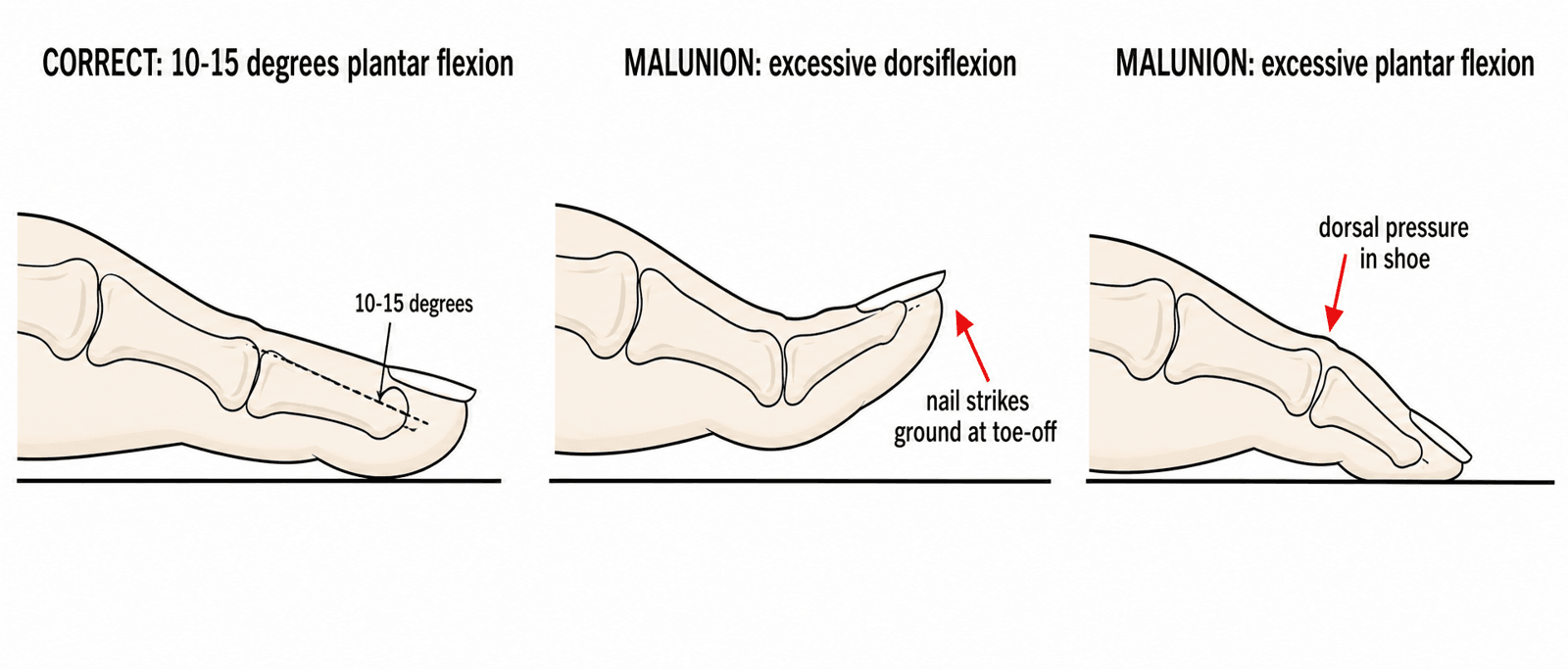
Setting and checking it. This is the model answer to the viva follow-up "how would you verify correct position intra-operatively?", and doing it on the table is what prevents the excessive-dorsiflexion malunion that produces nail ground-strike.
- Reference the contralateral hallux - keep the opposite foot in the field, or note its cascade pre-operatively, and reproduce its resting angle and valgus
- Simulate stance - press a flat rigid plate (an instrument tray lid, or the surgeon's flat hand) against the whole plantar forefoot as though the foot were flat on the floor. The pulp should just contact the plate with the nail facing forwards, not skywards; too much plantar flexion and the pulp digs in hard, too much dorsiflexion and the toe lifts off the plate and will catch the ground in gait
- Check rotation - the nail plate should lie in the same plane as the lesser-toe nails, with no pronation or supination
- Provisional K-wire, then mini-fluoroscopy (AP and lateral) to confirm alignment, surface coaptation and hardware placement before definitive fixation
- Re-assess clearance once definitive fixation is applied, before closure
Combined first-ray fusions. Set the first MTP position first, then tailor the IPJ to it and re-check whole-ray pulp-to-floor clearance. Immobilising the MTP shifts load and geometry onto the IPJ.
PLANTHallux IPJ Fusion Position
Hook:PLANT the hallux IPJ in slight plantar flexion!
Investigations
Radiographs. X-rays are sufficient for diagnosis in most cases. The standard foot series:
- AP - IPJ alignment and joint space
- Lateral - osteophytes and the dorsal prominence
- Oblique - additional detail of the IPJs
A subtle IPJ dislocation can be missed without a true lateral radiograph, so obtain a dedicated lateral when alignment is uncertain.
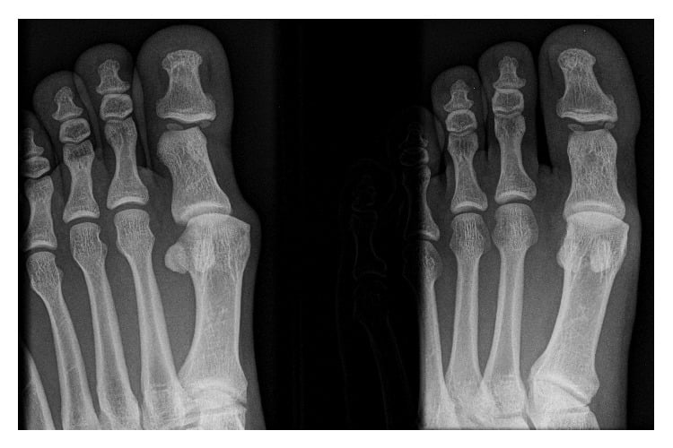
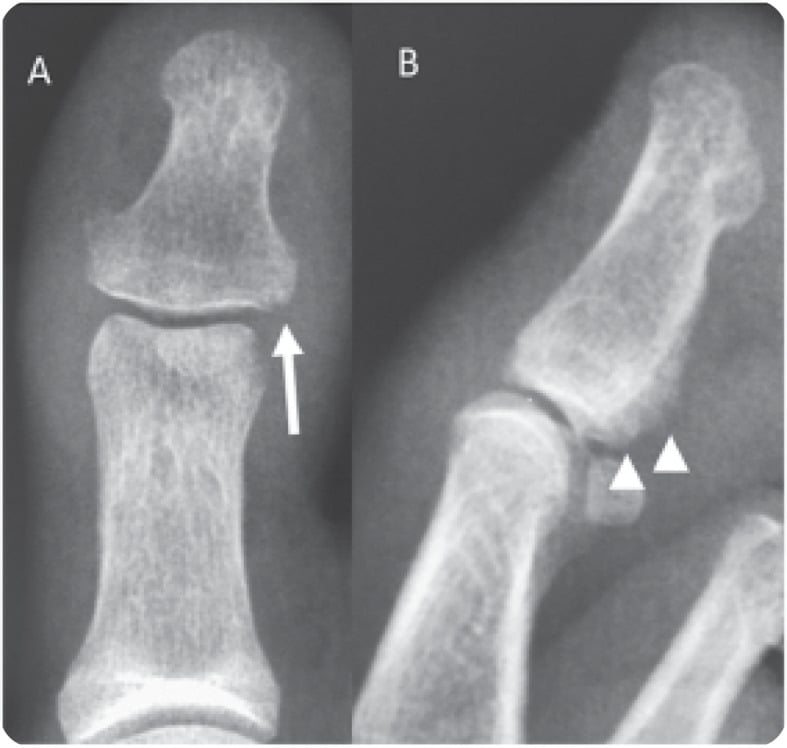
The findings are joint space narrowing (bone-on-bone in severe cases), osteophytes (especially dorsal), subchondral sclerosis, subchondral cysts, and malalignment into flexion or extension. Severity is graded on the same features:
- Mild - joint space narrowing, small osteophytes
- Moderate - significant narrowing, larger osteophytes, sclerosis
- Severe - bone-on-bone contact, collapse, deformity
MRI is rarely needed; clinical examination and X-ray are usually sufficient. It shows cartilage thinning and full-thickness defects, subchondral bone marrow oedema, synovitis (fluid, thickened synovium), osteophytes and loose bodies. Order it when:
- The diagnosis is uncertain and other pathology must be ruled out
- Cartilage status needs assessing for pre-operative planning
- An osteochondral defect is suspected
- Inflammatory arthritis is suspected and synovitis needs assessing
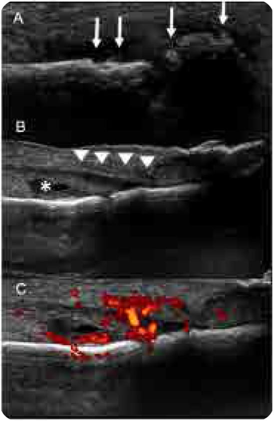
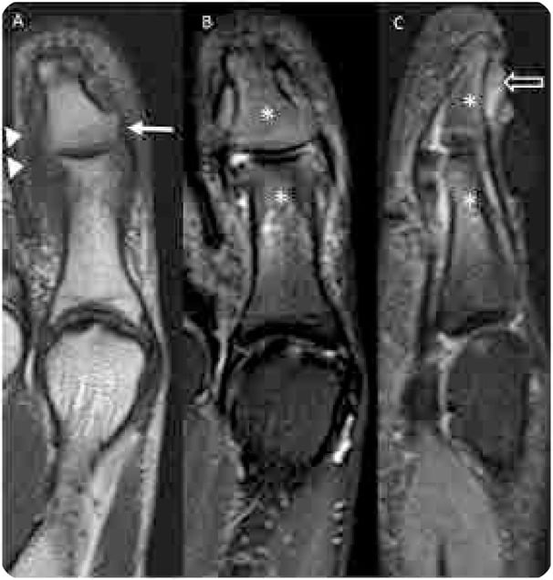
Laboratory tests help differentiate inflammatory from degenerative arthritis. They are indicated when inflammatory arthritis (multiple IPJs involved), septic arthritis (acute, systemically ill) or gout is suspected:
- ESR and CRP - elevated in RA and infection
- Rheumatoid factor and anti-CCP - RA screening
- Serum uric acid - gout screening
- Joint aspiration - cell count, culture and crystal analysis if sepsis or gout is suspected
Management Algorithm
The goal is pain relief and restoration of function. Conservative treatment is first-line for mild symptoms. For symptomatic hallux IPJ arthritis that has failed it, arthrodesis is the gold standard. Lesser toe IPJ arthritis can often be managed conservatively, or with resection arthroplasty if surgery is needed.
Non-operative management suits mild to moderate symptoms, the patient medically unfit for surgery and the patient who refuses it. It is palliative rather than curative: 40-50% achieve acceptable symptom control, and most eventually progress to surgery.
- Footwear - a stiff sole reduces IPJ motion; a rocker-bottom sole offloads the forefoot and reduces push-off stress; an extra-depth toe box accommodates dorsal osteophytes
- Orthotics - a carbon fibre plate insole stiffens the forefoot; a Morton extension limits hallux motion
- Medication - NSAIDs; intra-articular corticosteroid injection gives temporary relief for 3-6 months
- Activity - avoid high-impact activity; low-impact exercise such as swimming or cycling
Hallux IPJ arthrodesis. The indications are symptomatic hallux IPJ arthritis that has failed conservative treatment, pain limiting daily activities, and arthritis confirmed on X-ray. The technique:
- Exposure - dorsal longitudinal incision over the IPJ, protecting the neurovascular bundles (medial and lateral digital nerves)
- Joint preparation - excise all articular cartilage from the proximal and distal phalanx and curette the opposing surfaces to bleeding subchondral bone; a power burr may be used for precise preparation
- Position - set and confirmed on the table (see the fusion position section below)
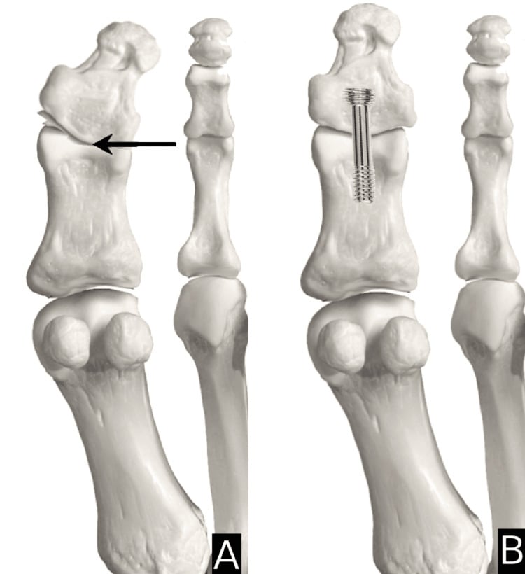
- Fixation - plate and screw, two or three crossed K-wires (less expensive), or a single lag screw for compression
- Closure - layered, with a subcuticular skin suture
Choosing the fixation. Plate and screw fixation is preferred at the hallux for its rigidity, and K-wires suffice for the lesser toes. The comparative data, however, do not show any single construct (single screw, multiple screws, screw plus plate, non-screw) reliably eliminating nonunion at this joint, and in the largest series implant type made no significant difference. Meticulous joint preparation to bleeding bone matters more than the specific implant.
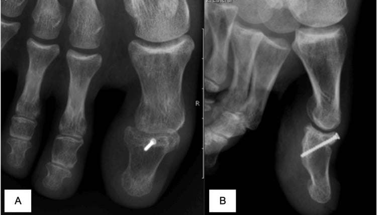
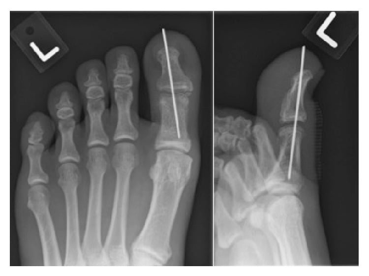
Lesser toe surgery has good outcomes with simple techniques.
- PIPJ arthrodesis - for severe arthritis or rigid deformity; technique as for the hallux but with simpler fixation, K-wires usually sufficing; fused in slight flexion (neutral or 10° flexion)
- PIPJ resection arthroplasty - excision of the proximal phalanx head creates a pseudoarthrosis that preserves some motion; less stable than fusion but acceptable in a lesser toe; used for a flexible hammer toe with PIPJ arthritis
- DIPJ arthrodesis - rarely needed as DIPJ arthritis is uncommon; K-wire fixation, fused in neutral
Rehabilitation. Union is expected at 8-12 weeks with rigid fixation.
- Weeks 0-2 - post-operative shoe with a rigid sole, weight-bearing as tolerated through the heel only, elevation, ice and dressing changes
- Weeks 2-6 - continue the post-operative shoe and progress to full forefoot weight-bearing; remove K-wires at 3-4 weeks if used; X-ray at 6 weeks to assess healing
- Weeks 6-12 - stiff-soled athletic shoe, full weight-bearing, low-impact activities
- Months 3-6 - regular shoes (most patients by 3-4 months) and most activities; high-impact sport at 4-6 months
Complications
- Incidence
- ~25% (largest HIPJ series); up to 40% if combined with MTP fusion
- Risk Factors
- Diabetes (significant risk factor), inadequate fixation, poor bone quality
- Prevention/Management
- Joint preparation to bleeding bone, compression; revise ONLY if symptomatic mobile nonunion (painless fibrous union is acceptable)
- Incidence
- 5-10%
- Risk Factors
- Incorrect positioning during surgery
- Prevention/Management
- Careful positioning (10-15° plantar flexion hallux), check alignment intra-op
- Incidence
- 5-10%
- Risk Factors
- K-wire fixation
- Prevention/Management
- Pin care, early removal at 3-4 weeks, antibiotics if infected
- Incidence
- Rare
- Risk Factors
- Altered gait mechanics post-fusion
- Prevention/Management
- Proper fusion position, orthotics if symptomatic
- Incidence
- 5%
- Risk Factors
- Excessive plantar or dorsiflexion fusion
- Prevention/Management
- Correct positioning, may need nail removal
- Incidence
- Under 2%
- Risk Factors
- Iatrogenic during exposure
- Prevention/Management
- Careful dissection, protect digital nerves
IPJ stiffness after fusion is expected, since ankylosis is the goal of the operation. Malunion and its consequences are covered with the fusion position below.
Outcomes and Prognosis
Hallux IPJ fusion. Pain relief and patient satisfaction are generally high despite imperfect radiographic union, and the functional deficit is minimal because the IPJ has limited normal motion. Older texts quote over 90% union. The largest contemporary series (Braswell 2023, 227 fusions) reports ~25% radiographic nonunion and ~21% reoperation, and in that series radiographic nonunion strongly predicted reoperation (p less than 0.0001). When the IPJ is fused alongside a first MTP fusion, nonunion reaches up to 40% (Brodsky 2021).
How far fibrous union reassures. A stable, painless fibrous union (fibrous ankylosis) is an accepted clinical endpoint that behaves like a solid fusion. The evidence that it is tolerable comes from Brodsky's 20 feet, where IP nonunion did not predict AOFAS score or satisfaction. Braswell defines clinical fibrous union but does not report how many of its 58 nonunions were painless. So a fibrous union can be an acceptable outcome, and a quarter of these fusions will not unite radiographically, but "most nonunions are painless" is not something either paper establishes.
Prognostic factors. Favourable factors are rigid fixation (plate preferred over K-wires), good bone quality, non-smoking, normal body weight, compliance with the post-operative protocol and correct fusion position. Unfavourable ones are smoking (it impairs bone healing), poor bone quality (osteoporosis, RA), inadequate fixation, infection, non-compliance with weight-bearing restrictions and inflammatory arthritis (higher nonunion risk). The largest series tested several of these against nonunion: smoking status, inflammatory arthritis and implant type made no significant difference to nonunion, whereas diabetes was a significant risk factor.
Lesser toe surgery. Fusion and resection arthroplasty are both used; resection is less predictable but acceptable because motion is non-critical. Correcting a hammer or mallet toe improves appearance and footwear.
Guidelines, Registries & Global Practice
Global epidemiology. IPJ arthritis of the foot is uncommon relative to first MTP (hallux rigidus) and lesser-toe MTP disease. The hallux IPJ accounts for the large majority of symptomatic cases; isolated lesser-toe IPJ arthritis is usually incidental. Post-traumatic disease (turf toe / hyperextension injury, intra-articular fracture, dislocation) predominates in younger and athletic populations worldwide, while primary degenerative and inflammatory (rheumatoid, psoriatic) disease predominate with age.
Society guidance (side by side). No orthopaedic society publishes an IPJ-arthritis-specific guideline; recommendations are extrapolated from first-ray degenerative-disease and inflammatory-arthritis pathways. Practice converges on a non-operative-first approach with arthrodesis for refractory disease.
- Relevant position
- Stepwise non-operative care for forefoot OA before surgery; arthrodesis for advanced first-ray degeneration
- Practical implication
- Document failed conservative trial before fusion
- Relevant position
- Foot & ankle arthrodesis for end-stage forefoot arthritis; honest consent on nonunion
- Practical implication
- Quote realistic, series-based nonunion figures at consent
- Relevant position
- Optimise DMARD/biologic therapy before elective forefoot surgery in inflammatory arthritis
- Practical implication
- Co-manage with rheumatology; time surgery to disease control
- Relevant position
- Stable internal fixation with adequate joint-surface preparation and compression
- Practical implication
- Preparation to bleeding bone emphasised over specific implant
Registry note. Toe IPJ arthrodesis is not separately tracked by national joint registries (NJR, AOANJRR, AJRR, SHAR), which capture replacement arthroplasty rather than small-joint fusion - so the best evidence remains institutional case series rather than registry data. This is itself an exam-worthy point about the limits of the evidence base.
High- vs limited-resource practice variation. In well-resourced settings, weightbearing radiographs, occasional MRI/ultrasound, plate or screw fixation, and formal gait/footwear rehabilitation are standard. In limited-resource settings, plain radiographs and K-wire fixation (inexpensive, effective) predominate, and accommodative footwear plus simple analgesia carry much of the non-operative burden. Outcomes hinge on joint preparation and patient selection rather than implant cost.
Controversies and Areas of Uncertainty
The dominant controversy. Dedicated series report ~25% radiographic nonunion (Braswell 2023) and up to 40% when combined with MTP fusion (Brodsky 2021). A painless fibrous union is widely accepted as a successful clinical endpoint, so routine revision for an asymptomatic radiographic nonunion is not justified. But hold the evidence precisely: the finding that nonunion did not predict AOFAS or satisfaction comes from Brodsky's 20 feet, which is far too small to exclude an effect, while in Braswell's 227 cases radiographic nonunion strongly predicted reoperation (p less than 0.0001). Neither paper reports what proportion of radiographic nonunions were painless. So the honest position is that a fibrous union can be an acceptable endpoint and the radiograph alone should not trigger revision - not that radiographic nonunion is benign.
No construct has been shown to reliably reduce IPJ nonunion (single screw vs multiple screws vs screw+plate vs non-screw all comparable). At the first ray more broadly, implant survival is similar across two-screw, plate, and plate+lag-screw constructs; plates carry a higher hardware-removal rate. Joint preparation likely matters more than implant choice.
Arthrodesis is gold standard for the hallux IPJ; resection arthroplasty is reserved for lesser toes where motion is non-critical. There is no robust evidence base for IPJ implant arthroplasty or cheilectomy at the toe IPJ, unlike the better-studied first MTP.
When both joints need fusing, IP nonunion was lowest when the IPJ was fused FIRST (17%) versus MTP-first or simultaneous (both 50%) - a small-series signal, not a guideline, but a plausible mechanical argument (immobilising the MTP loads the IPJ).
MCQ Practice Points
Q: What is the most common cause of hallux IPJ arthritis in patients under 50 years? A: Post-traumatic arthritis - previous intra-articular fracture, IPJ dislocation, or severe turf toe injury causing articular cartilage damage.
Q: What is the gold standard surgical treatment for symptomatic hallux IPJ arthritis? A: Arthrodesis (fusion) - reliably relieves pain with minimal functional deficit (IPJ has limited normal motion). Note: radiographic union is NOT guaranteed (~25% nonunion in the largest series), but a painless fibrous union behaves clinically like a solid fusion.
Q: What is the correct position for hallux IPJ arthrodesis? A: 10-15° plantar flexion, slight valgus to match contralateral great toe, neutral rotation. Excessive dorsiflexion causes nail ground strike; excessive plantar flexion causes shoe pressure.
Q: What fixation method is preferred for hallux IPJ arthrodesis? A: Plate and screw or lag screw - provides rigid fixation with highest union rate. K-wires acceptable for lesser toe IPJs but plate preferred for hallux.
Exam Viva Scenarios
Practise clinical reasoning and management decisions out loud
“A 35-year-old footballer presents with 2 years of progressive hallux IPJ pain. He sustained a severe turf toe injury 3 years ago. Examination shows reduced hallux IPJ ROM (20° vs normal 60°), crepitus with motion, and tenderness. X-rays demonstrate joint space narrowing, osteophytes, and subchondral sclerosis. What is your diagnosis and management?”
“A 48-year-old female with known rheumatoid arthritis presents with bilateral painful hallux and second toe IPJ swelling and stiffness. She has tried DMARDs and biologics with partial control. X-rays show erosive changes at multiple IPJs bilaterally. She requests surgical treatment for pain relief. How do you approach this?”
“A 52-year-old male presents 6 months after hallux IPJ fusion performed elsewhere. The fusion has healed (solid union on X-ray) but he complains that his great toe nail catches on the ground when walking and is painful. X-rays show the IPJ fused in 30° dorsiflexion. What is the problem and how do you manage it?”
DEFINITION
- IPJ arthritis: hallux IPJ, lesser toe PIPJ/DIPJ
- Hallux IPJ most symptomatic (essential for push-off)
- Post-traumatic (under 50y) vs degenerative (over 60y)
- Inflammatory arthritis: bilateral, multiple IPJs
- Limited normal IPJ motion = fusion causes minimal deficit
CAUSES
- Post-traumatic: fracture, dislocation, turf toe
- Degenerative: age-related (over 60y)
- Inflammatory: RA, psoriatic (bilateral)
- Timeline: symptoms 6mo-5y post-injury
CLINICAL FEATURES
- Pain at specific IPJ with walking
- Reduced ROM, crepitus with motion
- Dorsal osteophytes (palpable prominence)
- X-ray: joint space narrowing, osteophytes, sclerosis
CONSERVATIVE TREATMENT
- Stiff-soled shoes or rocker-bottom
- Carbon fiber plate insole
- NSAIDs, intra-articular steroid
- 40-50% achieve symptom control
HALLUX IPJ FUSION
- Gold standard for failed conservative
- Position: 10-15° plantar flexion, slight valgus
- Fixation: plate/screw or lag screw (no construct eliminates nonunion)
- Radiographic nonunion ~25% (largest series); painless fibrous union is acceptable
- Time to union: typically 8-12 weeks
KEY POINTS
- Hallux IPJ more significant than lesser toes
- Fusion eliminates pain, minimal functional loss
- Correct position critical (10-15° plantar flexion)
- Nonunion ~25% radiographically; diabetes is the key risk factor
- Malunion causes nail or shoe pressure problems
Evidence Base and Key Studies
The Braswell (2023) series below is the landmark contemporary dataset and directly overturns the "over 90% union" figure repeated in older textbooks. Know the ~25% radiographic nonunion / ~21% reoperation numbers AND the reconciling concept (painless fibrous union behaves like a solid fusion) for the viva.
Outcomes After Hallux Interphalangeal Joint Arthrodesis (largest series to date)
- 227 primary hallux IPJ arthrodeses - largest single-centre series published
- Radiographic nonunion rate 25.5% (58/227); reoperation rate 21.1% (48/227)
- Diabetes was a significant risk factor for nonunion (p=0.014)
- No significant difference by smoking status, inflammatory arthritis, or implant type (single screw vs multiple screws vs screw+other vs non-screw)
- Prior first MTP arthrodesis did NOT significantly raise IPJ nonunion rate in this cohort
Ipsilateral Hallux MTP + IP Joint Arthrodesis
- 20 feet (14 rheumatoid arthritis, 5 failed hallux valgus surgery, 1 hallux rigidus)
- All MTP arthrodeses healed, but 8/20 (40%) failed to heal at the IP joint
- IP nonunion lowest when IP fused first (17%) vs MTP first (50%) vs simultaneous (50%)
- IP nonunion did NOT predict AOFAS score or satisfaction; 18/20 patients satisfied
- Median hallux AOFAS rose from 25 to 68; fibrous ankylosis was an acceptable clinical outcome
Fixation Technique and Implant Survival in First MTP/IP Arthrodesis
- 83 patients / 89 fusions comparing two crossed screws vs dorsal plate vs dorsal plate + lag screw
- Overall implant survival 96.5% at 1 year and 94.0% at 10 years - no significant difference between constructs
- Highest union rate with dorsal plate plus lag screw (93.1%)
- Hardware removal higher with plate constructs (10.3%) than two-screw (3.1%)
- Mean AOFAS 83 with no significant difference between fixation groups
Immediate Weightbearing After First-Ray Fusion (HIPJ arthralgia signal)
- 25 first MTP arthrodeses with interfragmentary screw plus dorsal locking plate, immediate full weightbearing
- Overall union 96%; clinical healing ~5.9 weeks, radiographic fusion ~6.8 weeks
- Mean VAS pain fell from 6.6 to 0.6
- 2 patients developed symptomatic hallux IPJ arthralgia after MTP fusion (adjacent-joint load transfer)
First MTP Joint Pain and Turf Toe in Athletes (etiology review)
- Reviews turf toe, sand toe, sesamoiditis, FHL/EHL tendinopathy, gout and hallux rigidus as causes of first-ray pain in athletes
- Turf toe is a hyperextension plantar-plate injury of the first MTP - a recognised precursor to post-traumatic forefoot arthritis
- Weightbearing radiographs and point-of-care ultrasound aid diagnosis
- First-line management is non-surgical: footwear/activity modification, physical therapy, selective injection
Arthrodesis for Hallux Rigidus (contemporary review)
- First MTP arthrodesis remains the cornerstone for advanced first-ray degeneration with favourable long-term outcomes and low revision when fusion succeeds
- Interphalangeal arthritis is an explicitly recognised complication of MTP fusion
- Nonunion and malunion are the principal failure modes requiring revision
- Careful patient selection is emphasised