ATFL and CFL Insufficiency
- ATFL: Weakest lateral ligament. Resists inversion in plantarflexion. Most commonly injured.
- CFL: Injured in severe sprains. Resists inversion in dorsiflexion. Crosses both ankle and subtalar joints.
- Anterior Drawer: Tests ATFL integrity. Compare to contralateral side.
- Talar Tilt: Tests CFL integrity. Performed in neutral dorsiflexion.
- Anatomic repair: Direct ATFL ± CFL repair, with retinacular augmentation when appropriate; not every case requires a Gould modification
- “ATFL injury is common, but mechanism and injury sequence are not absolute in every sprain
- “Anterior drawer emphasises ATFL; talar tilt loads CFL plus capsular/subtalar structures
- “Rehabilitation is first-line and judged by function, giving-way episodes and objective progress rather than a mandatory duration
- “Choose repair when tissue is repairable; reconstruction when tissue, prior surgery, deformity or instability pattern makes repair unreliable
- “High athletic demand alone does not automatically require reconstruction
Overview and Epidemiology
Lateral ankle instability is the most common sports injury worldwide, affecting athletes across all sports and age groups. It is chronic mechanical and/or functional instability of the lateral ankle complex after an ankle sprain. This page covers established instability. The acute injury that precedes it - grading, Ottawa rules and functional rehabilitation - is covered in lateral ankle sprains.
How common. There are about 23,000 ankle sprains a day in the United States, 1 per 10,000 person-days in the general population, and they make up up to 30% of all sporting injuries. They are most common in basketball, soccer and volleyball. Incidence peaks at 15-35 years, in the active population; the sex ratio is equal in the general population, with a higher female incidence in specific sports.
How many become unstable. Persistent symptoms and recurrent giving way are common, but reported rates vary widely with the population, the definition used and the length of follow-up. Use validated patient-reported instability measures and functional testing rather than any one population percentage.
Who is at risk.
- Previous ankle sprain, the strongest predictor
- Incomplete or inadequate rehabilitation
- Generalised ligamentous laxity: a Beighton score of 4 or more of 9 is the screening threshold used in the instability literature; the age-adjusted diagnostic cut-off for generalised joint hypermobility is 6/5/4 by age (see generalised ligamentous laxity)
- High-demand sports (basketball, soccer, netball)
- Hindfoot varus malalignment
- Cavovarus foot deformity
- Peroneal weakness or dysfunction
Anatomy and Mechanism of Injury
The ligaments. Three ligaments arise from the lateral malleolus and stabilise the ankle against inversion and anterior translation of the talus. They are injured in order of strength, weakest first: ATFL, then CFL, then PTFL, although the sequence is not absolute in every sprain. The PTFL, the strongest, fails only in severe trauma or ankle dislocation.
- Origin
- Anterior border of lateral malleolus
- Insertion
- Talar neck (anterolateral)
- Function
- Resists anterior translation + inversion in plantarflexion
- Injury Order
- 1st (weakest)
- Origin
- Tip of lateral malleolus
- Insertion
- Lateral calcaneus
- Function
- Resists inversion in dorsiflexion; crosses ankle + subtalar
- Injury Order
- 2nd
- Origin
- Medial surface of lateral malleolus
- Insertion
- Posterior talus
- Function
- Resists posterior translation; strongest ligament
- Injury Order
- 3rd (rarely injured)
The ATFL is the one that matters most. It is the weakest of the three, failing at 58-90N, and the most commonly injured. It measures 20-25mm long and 10-12mm wide, has one to three bands (two most commonly), and lies horizontal in plantarflexion, becoming vertical in dorsiflexion.
The CFL is also 20-25mm long and runs 10-45° posterior to the long axis of the fibula, deep to the peroneal tendons. Its unique feature is that it crosses two joints, the ankle and the subtalar joint, so a CFL injury affects subtalar stability as well. That fact returns in concurrent subtalar instability, below.
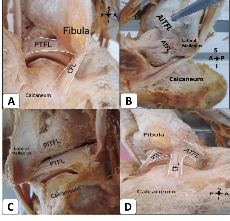
Mechanism. The typical injury is inversion in plantarflexion, and combined injuries are common. In plantarflexion the narrower posterior talus sits in the mortise, so there is less bony stability, and the ATFL becomes the primary restraint and the first to fail. With greater force the CFL is injured next, by inversion in dorsiflexion.
ACPLateral Ligaments - ACP
Hook:ACP = Anterior to Posterior = Weakest to Strongest. Like the alphabet, A comes first (injured first).
Dynamic stabilisers. Peroneus longus and brevis are the primary dynamic stabilisers, with extensor digitorum longus as secondary support. Proprioceptive afferents are critical for neuromuscular control, and peroneal reaction time is delayed in chronic instability.
Classification Systems
- Pathology
- Microscopic tear, stretch injury
- Clinical Findings
- Mild swelling, tenderness
- Stability
- Stable
- Treatment
- RICE, early mobilisation
- Pathology
- Partial macroscopic tear
- Clinical Findings
- Moderate swelling, ecchymosis
- Stability
- Mild laxity
- Treatment
- Brace 4-6 weeks, physio
- Pathology
- Complete rupture ATFL ± CFL
- Clinical Findings
- Severe swelling, ecchymosis
- Stability
- Frank instability
- Treatment
- Brace, physio ± surgery
Grade I and II injuries typically heal with conservative treatment. Grade III injuries may require surgery if chronic instability develops.
Clinical Assessment
History. Start with the index injury: its mechanism, and how it was treated and rehabilitated. Then establish the current picture:
- Symptoms: giving-way episodes, pain, swelling
- Functional limitation: sport, uneven ground, stairs
- Prior treatment: duration of physiotherapy, bracing
Red flags. Locking suggests a loose body, medial pain the deltoid, and persistent swelling an osteochondral lesion.
Look for swelling at the lateral ankle and in the anterolateral gutter, and for ecchymosis, which may indicate a recent episode. Hindfoot varus predisposes to lateral sprains and a cavus foot is associated with instability. Peroneal wasting suggests chronic injury.
Investigations
Radiographs. Standard weight-bearing views are the essential first-line imaging:
- Weight-bearing AP: mortise alignment, osteochondral lesion
- Mortise view: lateral clear space
- Lateral: talar dome, anterior impingement
Stress radiographs are for selected cases, when objective documentation will change treatment or clarify the examination. Anterior translation and talar tilt measurements are technique- and position-dependent; side-to-side comparison and image quality matter more than a universal cut-off. Normal stress values do not exclude functional instability.
MRI. Clinical examination remains the primary diagnostic tool, even though MRI has high sensitivity. MRI is indicated for:
- Diagnostic uncertainty
- Suspected osteochondral lesion
- Preoperative planning
- Associated pathology (peroneal, syndesmosis)
The ATFL shows discontinuity, thickening or signal change, and the CFL is often better seen on axial images. MRI also shows talar dome osteochondral lesions, a common association, and peroneal tendon tears or subluxation.
CT and ultrasound are reserved for specific clinical scenarios. CT characterises an osteochondral lesion, assesses bony morphology and plans malalignment correction. Ultrasound is operator dependent but assesses the ligaments and peroneal subluxation dynamically, and is a cost-effective screening test.
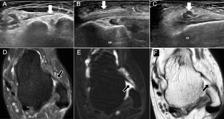
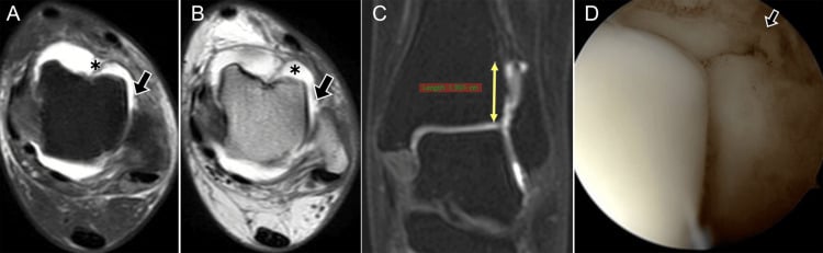
Associated pathology. These are the conditions to exclude:
- Frequency
- 20-25%
- Investigation
- MRI, CT
- Clinical Significance
- Address at surgery
- Frequency
- 15-20%
- Investigation
- MRI, ultrasound
- Clinical Significance
- May need concurrent repair
- Frequency
- 10-15%
- Investigation
- Lateral X-ray, MRI
- Clinical Significance
- Debride at arthroscopy
- Frequency
- 5-10%
- Investigation
- X-ray, MRI
- Clinical Significance
- Different treatment
- Frequency
- Variable
- Investigation
- Stress views
- Clinical Significance
- Consider reconstruction
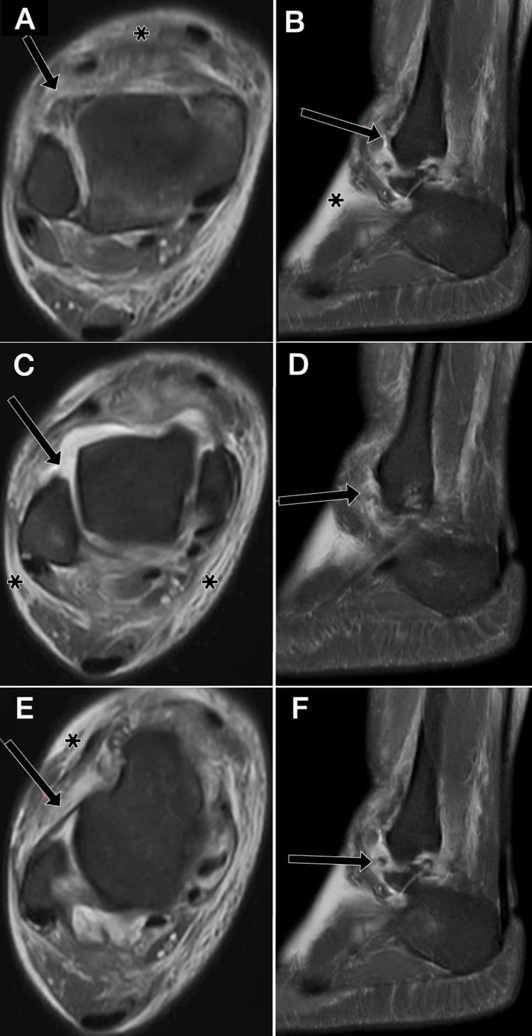
- Key Distinguishing Feature
- Recurrent giving-way; positive anterior drawer/talar tilt
- Best Test
- Clinical stress tests; stress radiographs
- Why It Matters
- Index diagnosis - guides rehab vs surgery
- Key Distinguishing Feature
- Retromalleolar pain/clicking; pain on resisted eversion, not true giving-way
- Best Test
- Dynamic ultrasound; MRI
- Why It Matters
- Needs tendon repair/groove deepening, not ligament repair alone
- Key Distinguishing Feature
- Deep ankle pain, catching/locking, effusion rather than instability
- Best Test
- MRI / CT
- Why It Matters
- Often coexists; must address arthroscopically at surgery
- Key Distinguishing Feature
- Pain proximal to joint line; positive external rotation/squeeze test
- Best Test
- ER stress test; weight-bearing CT
- Why It Matters
- Different treatment - stabilisation, not lateral repair
- Key Distinguishing Feature
- Anterolateral pain on dorsiflexion, no objective laxity
- Best Test
- MRI; diagnostic arthroscopy
- Why It Matters
- Treated by debridement, not ligament repair
- Key Distinguishing Feature
- Hindfoot 'rolling' on uneven ground; CFL involvement
- Best Test
- Broden/stress views; clinical exam
- Why It Matters
- May need reconstruction crossing the subtalar joint
Management
The decision runs in four steps:
- Confirm the phenotype: functional, mechanical or combined, and any associated pathology.
- Rehabilitate progressively, judging the response by function rather than a fixed number of months.
- Repair when tissue is repairable; reconstruct when the pattern or the tissue makes repair unreliable.
- Correct deforming forces and concomitant pathology that would otherwise cause failure.
Rehabilitation is first-line and remains the foundation for mechanical, functional and combined instability. It is progressive and has five components:
- Proprioceptive and perturbation training
- Peroneal and global lower-limb strength
- Range restoration and calf capacity
- Bracing or taping for selected activities
- Running, cutting, landing and sport-specific progression
When has it failed? Duration is response- and goal-dependent, and a calendar alone does not define failure. Document giving-way frequency, patient-reported instability, balance, hop and cutting tasks, strength and confidence. The 3-6 month threshold is pragmatic rather than evidence-defined.
Surgical indications. Consider surgery when:
- Recurrent giving way or instability limits desired function despite a well-delivered rehabilitation programme
- Mechanical laxity or repairable pathology correlates with symptoms
- Alignment and associated osteochondral, peroneal, syndesmotic, impingement or subtalar disease have been assessed
- Patient goals and postoperative rehabilitation capacity support the plan
Athletic level, laxity and patient preference modify treatment but are not stand-alone indications.
Choosing the operation. The options, and when each applies:
- Anatomic repair - direct ATFL ± CFL repair when local tissue quality and excursion permit. Inferior extensor retinaculum augmentation is selective, not mandatory, and open and arthroscopic approaches both have supporting evidence.
- Anatomic reconstruction - consider for poor tissue, failed prior repair, major deformity or laxity, or a multiligament pattern that cannot reliably be repaired. The graft and tunnel strategy should recreate the required ATFL and CFL functions while protecting subtalar motion.
- Alignment and associated pathology - correct clinically important cavovarus or another deforming force when it would overload the repair, and treat osteochondral lesions, impingement, syndesmosis and peroneal pathology in the same plan when indicated.
Historical non-anatomic tenodeses restrict motion and are now salvage rather than routine choices.
Surgical Technique
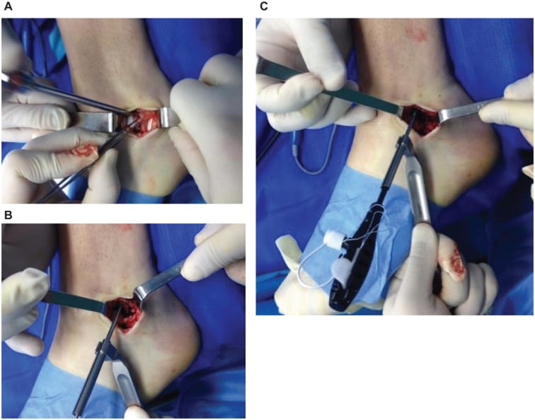
The Broström-Gould repair is the gold standard: an anatomic repair of the ATFL, and of the CFL when needed, reinforced by advancing the inferior extensor retinaculum (IER).
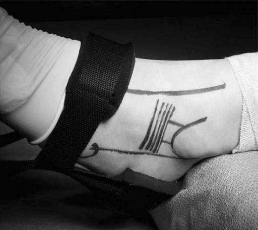
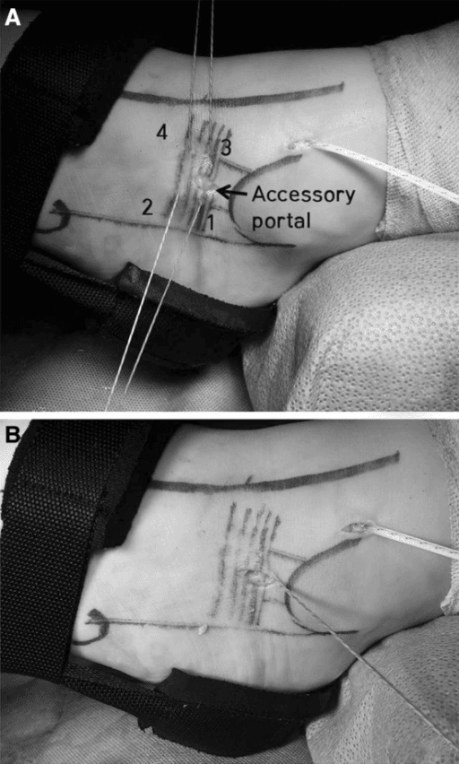
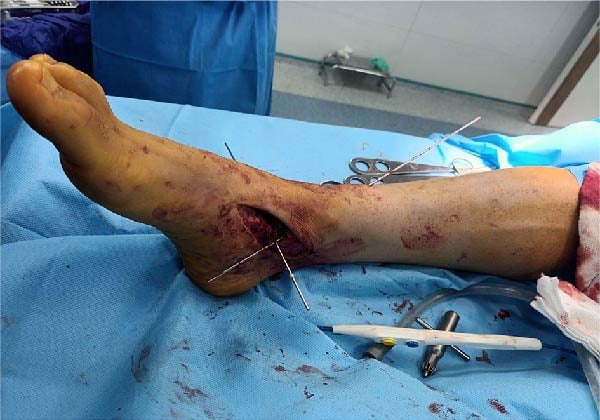
Set-up. Supine, with a bump under the ipsilateral hip and the leg in slight internal rotation for lateral access, under a high-thigh tourniquet at 250-300mmHg. Use a standard foot and ankle set, 2.4-3.0mm suture anchors and non-absorbable braided suture; fluoroscopy is optional.
Approach. A curvilinear incision of 4-6cm over the lateral malleolus. Map the superficial peroneal nerve before the incision, and identify and protect its branches.
The superficial peroneal nerve is at risk with lateral ankle approaches. It typically crosses the surgical field 5-10cm proximal to the tip of the lateral malleolus. Always identify and protect this nerve to avoid sensory deficit.
Steps.
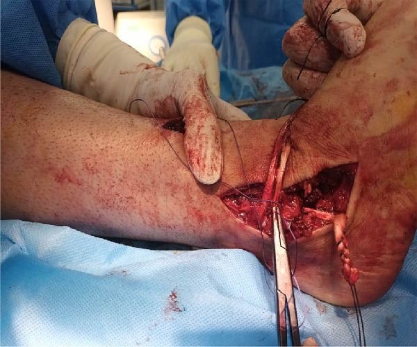
- Exposure. Incise through the subcutaneous tissue, protecting the superficial peroneal nerve; identify and preserve the IER, then open the ankle capsule.
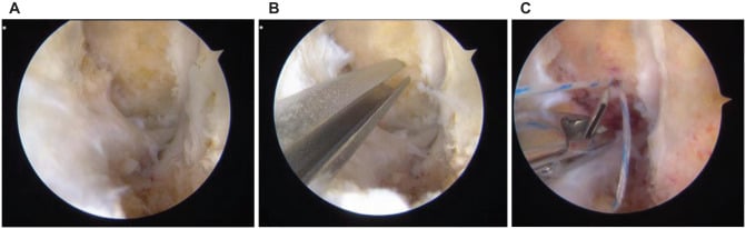
- Find the ATFL. It lies within the anterolateral capsule and is often attenuated and scarred; identify its talar and fibular insertions.
- Repair the ATFL. Imbricate the remnant pants-over-vest, with a suture anchor at the fibular origin (the anatomic footprint). Tension the repair with the foot in eversion and dorsiflexion, enough to eliminate the anterior drawer.
- Gould modification. Advance the IER over the ATFL repair and anchor it to the fibula with sutures, for additional restraint and proprioceptive input.
- Repair the CFL if needed. Find it deep to the peroneal tendons and repair it the same way, with an anchor at its fibular footprint.
- Close. Irrigate, close in layers to prevent wound dehiscence, and apply a below-knee backslab in neutral.
The Gould step. Retinacular augmentation is standard practice with most open repairs, but it is selective rather than mandatory: not every case requires it. When it is done, the IER advancement must be adequate.
Pitfalls. Over-tightening causes stiffness and subtalar restriction. Assess both ligaments so that a CFL injury is not missed, and place the anchors at the anatomic footprints.
Complications
Intraoperative. Nerve identification is the key to preventing intraoperative complications; the superficial peroneal nerve is the nerve most commonly injured.
- Incidence
- 2-5%
- Prevention
- Careful dissection, identify before incision
- Management
- Neurolysis, reassurance
- Incidence
- 1-2%
- Prevention
- Posterior incision extension awareness
- Management
- Reassurance (usually neuropraxia)
- Incidence
- 5-10%
- Prevention
- Pre-op MRI assessment
- Management
- Convert to reconstruction
- Incidence
- Rare
- Prevention
- Proper technique, bone quality assessment
- Management
- Re-anchor or transosseous sutures
Early (less than 6 weeks). Wound dehiscence occurs in 2-5%, superficial infection in 1-3%, deep infection in less than 1% and haematoma in 1-2%. Careful surgical technique minimises them, and prevention is layered closure, appropriate immobilisation, early mobilisation within the protocol, and DVT prophylaxis. Treat with wound care and antibiotics as needed, and return to theatre for a deep infection.
Late (greater than 6 weeks). Late complications often require revision surgery. Recurrence risk varies with tissue, alignment, laxity, sport and rehabilitation.
- Incidence
- 5-10%
- Cause and risk
- Usually dorsiflexion restriction; prevent with early mobilisation and by avoiding over-tightening
- Treatment
- Physiotherapy, manipulation under anaesthesia
- Incidence
- 5-10%
- Cause and risk
- Failure of repair or stretch-out; non-compliance and generalised laxity are risk factors
- Treatment
- Revision with reconstruction
- Incidence
- 5-15%
- Cause and risk
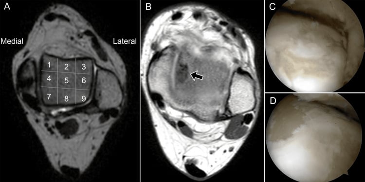
- May indicate missed pathology (osteochondral lesion, impingement); investigate with MRI and diagnostic arthroscopy
- Treatment
- Address the underlying cause
- Incidence
- -
- Cause and risk
- More common with non-anatomic reconstructions; Broström repair preserves subtalar motion
- Treatment
- -
Postoperative Care
Rehabilitation protocol. Early immobilisation protects the healing repair, and protected mobilisation from two weeks allows healing while preventing stiffness. Proprioceptive training is critical for functional recovery, and a gradual return to sport prevents re-injury.
- Goals
- Wound healing, pain and swelling control, DVT prevention
- Protection and loading
- Below-knee backslab or CAM boot; non-weight-bearing or touch weight-bearing; elevation, ice
- Exercise
- Ankle pumps (within the splint if able)
- Milestones
- Wound check at 10-14 days, sutures removed, transition to CAM boot
- Goals
- Early range of motion avoiding inversion, progressive weight-bearing, maintained muscle function
- Protection and loading
- CAM boot for walking, weight-bearing as tolerated in the boot; boot off for exercises and sleeping by week 4; no inversion stress, no running or jumping
- Exercise
- Active dorsiflexion and plantarflexion, avoiding inversion; isometric peroneal exercises
- Milestones
- -
- Goals
- Full range, progressive strengthening, start of proprioceptive training
- Protection and loading
- Wean from the boot; supportive ankle brace for activities
- Exercise
- Full range including inversion; progressive resistance; proprioception (wobble board, single-leg stance); stationary cycling, swimming
- Milestones
- Full pain-free range by week 8; single-leg stance for 30 seconds by week 10
- Goals
- Full activity, sport-specific training, long-term stability
- Protection and loading
- Ankle brace for sport for the first 6-12 months
- Exercise
- Jogging from week 12, agility drills weeks 14-16, sport-specific training weeks 16-20; ongoing proprioceptive maintenance
- Milestones
- Full return to sport at 4-6 months
Return to sport requires:
- Full pain-free range of motion
- Strength equal to the contralateral side
- Completed agility testing
- Sport-specific drills without symptoms
Outcomes
Rehabilitation. Without surgery, 70-80% stabilise. Results are best with supervised physiotherapy; proprioceptive training is key to success and peroneal strengthening essential. Bracing for sport may be permanent.
Repair and reconstruction. The Broström-Gould repair has a lower revision rate than non-anatomic procedures. Anatomic reconstruction has a higher revision rate than a primary Broström and a longer rehabilitation.
- Brostrom-Gould
- 85-90%
- Anatomic Reconstruction
- 80-90%
- Brostrom-Gould
- 85-95%
- Anatomic Reconstruction
- 80-90%
- Brostrom-Gould
- 5-10%
- Anatomic Reconstruction
- 5-15%
- Brostrom-Gould
- 4-6 months
- Anatomic Reconstruction
- 6-9 months
Prognostic factors.
- Unfavourable
- Combined ATFL + CFL injury
- Unfavourable
- Generalised ligamentous laxity
- Unfavourable
- Hindfoot varus or cavovarus
- Unfavourable
- Poor compliance
- Unfavourable
- Associated osteochondral lesion (OCD)
Guidelines, Registries & Global Practice
Global Epidemiology
- Ankle sprain is among the most common musculoskeletal injuries worldwide; the lateral ankle sprain is the dominant subtype (Doherty meta-analysis of 181 prospective studies).
- Incidence is higher in females than males (13.6 vs 6.94 per 1000 exposures) and in indoor/court sports (basketball, volleyball, netball, handball).
- Approximately 20-40% of acute sprains progress to chronic instability; the Cochrane review cites ~10-20% developing chronic lateral instability.
- A prior sprain remains the single strongest predictor of recurrence regardless of region or healthcare setting.
Side-by-Side Guidelines & Consensus
- Diagnostic Position
- History of ≥1 significant sprain plus recurrent giving-way, recurrent sprains, or feelings of instability
- Management Emphasis
- Distinguish mechanical vs functional instability; structured outcome measures
- Diagnostic Position
- Clinical diagnosis; stress imaging for objective laxity
- Management Emphasis
- Functional treatment + neuromuscular/proprioceptive rehabilitation first-line for acute and chronic injury
- Diagnostic Position
- Ottawa rules for acute imaging; clinical exam primary
- Management Emphasis
- Early functional rehabilitation; surgery for refractory instability
- Diagnostic Position
- Stress views and MRI for surgical planning
- Management Emphasis
- Anatomic repair (Broström-Gould) as gold standard; reconstruction for failures/laxity
- Diagnostic Position
- Exclude associated osteochondral and syndesmotic injury
- Management Emphasis
- Anatomic restoration of ATFL/CFL footprints; address concomitant pathology
Registry & Outcome Notes
- Unlike arthroplasty, lateral ankle stabilisation is not tracked in national joint registries (NJR, AOANJRR, SHAR); evidence derives from cohort series, meta-analyses, and consensus statements rather than registry survivorship.
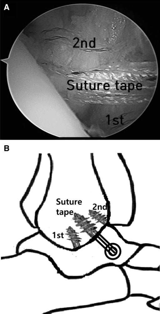
- Reported recurrence after anatomic repair is broadly 5-15%, higher with generalised laxity - the rationale for suture-tape augmentation or reconstruction in selected patients.
High- vs Limited-Resource Practice Variation
- High-resource settings: Ready access to MRI, stress imaging, arthroscopy, and suture-tape/InternalBrace augmentation; arthroscopic Broström increasingly favoured for lower wound morbidity.
- Limited-resource settings: Diagnosis is largely clinical (Ottawa rules, stress testing); supervised neuromuscular rehabilitation and bracing carry the management burden, with open anatomic repair the mainstay where surgery is indicated.
- The conservative-first, rehabilitation-led pathway is universal and equally valid across all settings - the major global driver of good outcomes.
The Varus Hindfoot: Why the Ligament Repair Alone Fails, and When to Add a Calcaneal Osteotomy
The biomechanical problem. In a varus or cavovarus hindfoot the ground-reaction force passes medial to the subtalar joint axis, generating a constant inversion (supination) moment across the ankle and hindfoot. The lateral ligaments are chronically loaded, which predisposes to the original recurrent sprains.
Why the repair fails. After surgery the same moment places a persistent stretching force on a Broström-Gould repair, so an isolated soft-tissue repair on an uncorrected varus hindfoot is prone to gradual stretch-out and recurrent instability. Failing to recognise and correct hindfoot varus is one of the leading causes of an otherwise technically sound repair failing.
Sorting out the deformity. The assessment itself belongs to the cavovarus foot work-up: standing hindfoot alignment, a standing hindfoot-alignment (long-axial) radiograph, and the Coleman block test. The block decides whether the varus is forefoot-driven, a plantarflexed first ray that corrects on the block and is flexible, or a fixed hindfoot varus that does not correct. This topic's job is deciding which bony procedure, if any, must accompany the ligament surgery.
The surgical corollary. The ligament repair or reconstruction is done on a balanced, plantigrade foot:
- Fixed hindfoot varus - add a lateralising, valgus-producing calcaneal osteotomy (Dwyer lateral closing-wedge, or a lateral-translation/Z-type osteotomy) to move the weight-bearing axis lateral to the subtalar joint and unload the lateral repair
- Forefoot-driven varus (Coleman block positive, correcting on the block) - a dorsiflexion (dorsal closing-wedge) osteotomy of the first metatarsal addresses the plantarflexed first ray
- Peroneal balance - consider a peroneus longus-to-brevis transfer, strengthening eversion, where longus overdrive plantarflexing the first ray is driving the varus
Shown a patient with recurrent lateral instability and a varus or cavovarus hindfoot, the trap is to offer a Broström in isolation. Realign first, then stabilise the ligaments.
Concurrent Subtalar Instability: Recognise It and Recreate the CFL
Symptoms. The patient describes the hindfoot "rolling" or giving way on uneven ground, sometimes with a medial-hindfoot ache, persisting despite an apparently competent ATFL. The instability may feel deeper or more posterior than a pure ankle giving-way.
Examination is difficult because ankle and subtalar motion couple. Look for a medial subtalar glide or tilt and reproduction of the "rolling" sensation; CFL insufficiency (a positive talar tilt) should raise suspicion.
Imaging. Broden views and subtalar stress radiographs are described to demonstrate talocalcaneal tilt, but normative values are poorly standardised and the tilt is difficult to separate from ankle (talar) tilt, so the diagnosis remains substantially clinical. CT and MRI mainly exclude other hindfoot pathology and coalition.
Treatment. Rehabilitation comes first, exactly as for the lateral ankle, with proprioceptive and peroneal work. At surgery, because the CFL is the shared restraint, an isolated ATFL-only Broström is inadequate: the reconstruction must extend distally to the calcaneus and recreate the CFL, and an anatomic reconstruction reproducing both the ATFL and CFL footprints is preferred.
Why not a tenodesis. The older non-anatomic tenodeses (Chrisman-Snook, Evans) did restrain subtalar motion, but at the cost of subtalar stiffness and later arthritis, a key reason anatomic techniques that preserve subtalar motion are now favoured.
Still giving way on uneven ground after a competent ATFL repair: think concurrent subtalar instability, and recreate the CFL rather than re-tension the ATFL.
Controversies and Areas of Uncertainty
Open versus arthroscopic repair. Meta-analyses show comparable stability with lower wound morbidity arthroscopically. Longer-term durability and learning-curve data are still maturing, and no high-level RCT establishes clear superiority of either approach.
Suture-tape (InternalBrace) augmentation. It is biomechanically stronger and allows earlier loading, but its clinical benefit over standard repair is small and short-term. Concern about over-constraint and cost-effectiveness remains unresolved.
Repair versus reconstruction in laxity. Direct repair has a higher failure rate in generalised laxity (Beighton ≥4), but the optimal threshold for choosing graft reconstruction is not standardised. Augmented repair may bridge the gap but lacks long-term comparative data.
Early surgery in elite athletes. Whether acute repair of complete grade III ruptures in high-level athletes shortens return to sport compared with functional treatment remains debated; most evidence still favours rehabilitation first.
Rehabilitation dose. The ideal duration, supervision intensity and balance or neuromuscular protocol are not standardised across guidelines.
Imaging. The role of weight-bearing CT and dynamic imaging in routine assessment is evolving and not yet consensus practice.
MCQ Practice Points
Q: Which lateral ankle ligament is injured first during an inversion injury?
A: The ATFL (anterior talofibular ligament) is the weakest and most commonly injured. It is injured with inversion in plantarflexion - the most common ankle sprain mechanism. Remember ACP order: Anterior → Calcaneofibular → Posterior (weakest to strongest).
Q: A patient has a positive talar tilt test but negative anterior drawer. Which ligament is injured?
A: The CFL (calcaneofibular ligament) is injured. The talar tilt test assesses CFL integrity (inversion stress in dorsiflexion), while the anterior drawer tests ATFL. This pattern is unusual as ATFL is typically injured first, so consider other pathology.
Q: What does the Gould modification add to the original Brostrom repair?
A: The Gould modification adds augmentation with the inferior extensor retinaculum (IER). The IER is advanced over the ATFL repair and anchored to the fibula, providing additional restraint and improved proprioceptive input. This is now standard with all Brostrom repairs.
Q: When is anatomic reconstruction preferred over Brostrom repair for lateral ankle instability?
A: Reconstruction is indicated for: (1) Failed Brostrom repair, (2) Generalized ligamentous laxity (Beighton score greater than 4), (3) Heavy/high-demand athletes, and (4) Poor quality native tissue. These patients have higher failure rates with direct repair.
Q: What is unique about the calcaneofibular ligament compared to other lateral ankle ligaments?
A: The CFL crosses two joints - both the ankle joint AND the subtalar joint. This means CFL injury affects subtalar stability as well as ankle stability. The ATFL and PTFL only cross the ankle joint.
Self-Assessment Quiz
Viva Scenarios
Practise clinical reasoning and management decisions out loud
“A 25-year-old basketball player presents with recurrent left ankle 'giving way' episodes despite 6 months of physiotherapy. He reports 3-4 episodes per month during games. Examination shows positive anterior drawer test compared to the contralateral side. What is your assessment and management plan?”
“A 30-year-old female netball player presents 18 months after a Brostrom repair with recurrent instability. She has hypermobile joints (Beighton score 6/9). Stress testing shows persistent ATFL laxity. How do you approach this case?”
“A 22-year-old soccer player sustains an inversion injury during a match. He has significant lateral swelling, ecchymosis, and tenderness over the ATFL. He cannot weight bear. Anterior drawer appears positive but is difficult to assess due to guarding. How do you manage this acute injury?”
ANATOMY - ACP
- ATFL: Weakest, injured first, resists inversion in plantarflexion
- CFL: Crosses ankle AND subtalar, injured second
- PTFL: Strongest, only injured in dislocations
- ACP order = Anterior to Posterior = Weakest to Strongest
EXAMINATION
- Anterior drawer = ATFL (anterior translation)
- Talar tilt = CFL (inversion in dorsiflexion)
- External rotation = Syndesmosis (NOT lateral ligaments)
- Always compare to contralateral side
TREATMENT ALGORITHM
- FIRST: Rehabilitation 3-6 months (majority stabilize)
- SECOND: Brostrom-Gould repair (85-90% success)
- THIRD: Reconstruction if failed Brostrom/laxity/heavy athlete
- Gould modification = IER augmentation (always do this)
KEY NUMBERS
- 20-40%: Acute sprains → chronic instability
- 70-80%: Stabilize with rehabilitation alone
- 85-90%: Good results with Brostrom-Gould
- 4-6 months: Return to sport after Brostrom
Evidence Base
Broström L - Original Anatomic Repair
- Original description of direct delayed anatomic repair of ruptured lateral ligaments
- Established that scarred ligament ends could be re-apposed years after injury with good results
- Foundation for all modern anatomic lateral ankle stabilization
Gould N, Seligson D, Gassman J - Gould Modification
- Added advancement of the inferior extensor retinaculum (IER) over the repaired ATFL
- Reinforces the repair and limits inversion, adding a proprioceptive contribution
- Converted the Broström into the 'Broström-Gould' construct
Doherty C et al - Incidence and Prevalence of Ankle Sprain (Meta-analysis)
- Meta-analysis of 181 prospective studies; ankle sprain incidence higher in females than males (13.6 vs 6.94 per 1000 exposures)
- Highest incidence in indoor/court sports; children at higher risk than adolescents or adults
- Lateral ankle sprain was the most common subtype overall
Gribble PA et al - International Ankle Consortium Position Statement
- Consensus selection criteria defining chronic ankle instability (CAI) for research and practice
- Requires a history of at least one significant sprain plus recurrent 'giving way', recurrent sprains, or feelings of instability
- Standardised CAI definition to harmonise the literature
de Vries JS et al - Interventions for Chronic Ankle Instability (Cochrane Review)
- Seven randomised trials; insufficient high-quality evidence to favour any single surgical or conservative technique
- After surgical reconstruction, early functional rehabilitation gave earlier return to work and sport than 6 weeks of immobilisation
- Its background - not its results - is the source of the widely quoted 10-20% of acute sprains developing chronic instability
Moorthy V et al - Open vs Arthroscopic Broström (Meta-analysis)
- Six comparative studies; arthroscopic repair gave slightly higher AOFAS and Karlsson scores and lower VAS pain
- No difference in postoperative anterior drawer, talar tilt, or overall complications
- Wound-related complications were significantly lower with arthroscopic repair (OR 0.25)
Cho BK et al - Suture-Tape Augmented Modified Broström in Generalised Laxity
- 28 patients with generalised ligamentous laxity (Beighton ≥4); 2-year follow-up
- FAOS improved 63.2 to 90.6 and talar tilt 16.2° to 3.6°; anterior translation 12.1 mm to 4.2 mm
- Only 1 patient (3.6%) had recurrent subjective and mechanical instability
Wang AH et al - Arthroscopic vs Open Modified Broström in Joint Laxity
- 64 CLAI patients with generalised joint laxity (Beighton ≥4); 24-month follow-up
- No difference in functional scores or stress radiographs between arthroscopic and open repair
- Arthroscopic group returned to work earlier; failure rates 23.3% vs 16.1% (not significant)