The hypermobile child — benign variant, syndromic marker, or aortic time bomb
- Beighton score of 6 or more out of 9 in a prepubertal child, 5 or more after puberty, and 4 or more beyond age 50 defines generalised joint hypermobility - the threshold falls because laxity declines with age.
- Beighton measures laxity only — it does not diagnose a syndrome. The diagnosis is made by what accompanies the laxity.
- Ectopia lentis is superotemporal in Marfan and inferonasal in homocystinuria; the latter also carries thrombosis risk under anaesthesia.
- Translucent skin with visible veins, easy bruising and thin lips in a hypermobile child is vascular EDS until disproven — avoid arteriography and elective joint surgery.
- Blue sclerae plus laxity plus fractures with wormian bones equals osteogenesis imperfecta, not EDS.
- Atlantoaxial instability in Down syndrome is screened by symptoms and examination; routine flexion-extension films no longer mandated for sport participation in most guidance.
- Laxity is a major driver of recurrent patellar dislocation, atraumatic multidirectional shoulder instability and pes planovalgus.
- “Ask three questions out loud: is it generalised, is there a second tissue involved, is there a family history of sudden death?
- “Marfan is the tall hypermobile child with lens dislocation; Loeys-Dietz adds hypertelorism, bifid uvula and tortuous arteries with earlier dissection.
- “Down syndrome laxity is global and low tone; the hip and the C1-C2 articulation are the joints that get you into trouble.
- “Larsen syndrome: multiple congenital large-joint dislocations with a flat face and cervical kyphosis.
- “Never operate on a shoulder for atraumatic instability before a rehabilitation programme has been completed.
A normal-looking 8-year-old with a Beighton of 8 may still be Marfan. Height percentile, arm span to height ratio, palate, sclerae, skin and a cardiac murmur must be documented before the word benign is used.
Marfanoid habitus with ectopia lentis is not automatically Marfan. Homocystinuria adds learning difficulty, osteoporosis and, critically, perioperative thromboembolism. Urine amino acids are cheap.
Tissue will not hold suture, arterial access can be fatal and wound dehiscence is the rule. Skin translucency and acrogeria change the plan before the knife.
Hypermobility spectrum disorder causes diffuse pain, but a single hot joint, night pain or a limp still demands a radiograph and inflammatory markers. Laxity does not protect against sepsis or malignancy.
Recognising the Pattern
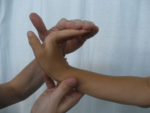
Definition. Generalised ligamentous laxity is joint range of motion beyond the accepted normal in multiple joints across multiple limbs, in the absence of an acute injury or neurological disorder. It is quantified by the Beighton score out of 9.
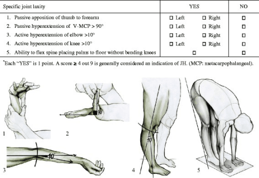
The Beighton nine points, scored formally:
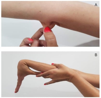
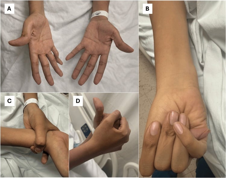
- Passive dorsiflexion of the fifth metacarpophalangeal joint beyond 90 degrees — one point per hand.
- Passive apposition of the thumb to the flexor forearm — one point per hand.
- Hyperextension of the elbow beyond 10 degrees — one point per arm.
- Hyperextension of the knee beyond 10 degrees — one point per leg.
- Forward flexion of the trunk with knees straight, palms flat on the floor — one point.
Thresholds. 6 or more out of 9 in a prepubertal child, 5 or more in a pubertal or adult subject up to age 50, and 4 or more over age 50. Laxity is greatest in infancy, declines through childhood, and is greater in girls and in individuals of African and Asian descent.
Why you will also see a flat "Beighton 4 or more", and when that is legitimate. The sports and instability literature - shoulder multidirectional instability, recurrent lateral ankle instability - commonly screens with a single cut-point of 4 or more out of 9, and lists it as a risk factor favouring reconstruction over repair. That is a screening threshold for a risk factor, deliberately set low to be sensitive, and it is not the same instrument as the age-adjusted diagnostic threshold above, which exists to classify generalised joint hypermobility. Both are defensible for their own purpose; quoting one in the other's setting is the error. If you are asked to define generalised joint hypermobility, give 6/5/4 by age. If you are asked what makes an instability recurrence more likely, 4 or more is the figure that literature uses.
Confirming it is genuinely generalised. Test at least one upper limb, one lower limb, the spine and one axial or peripheral joint not in the Beighton set — shoulder external rotation beyond 85 degrees, hip abduction, ankle dorsiflexion, or a positive Gorlin sign (tongue to nose tip). Isolated shoulder or patellar laxity is not generalised laxity.
How to say it in a viva. "This child has generalised joint hypermobility with a Beighton score of 8 out of 9. I would now determine whether this is isolated hypermobility or part of a heritable connective tissue disorder, by examining skin extensibility and scarring, sclerae, palate and dentition, cardiovascular system including a search for aortic regurgitation, and by taking a three-generation family history focusing on sudden death, aortic surgery and lens dislocation."
Mimics and false positives.
- Post-injury laxity in a single joint — unilateral, with a mechanism; Beighton elsewhere is normal.
- Hypotonia of neuromuscular origin — cerebral palsy of the hypotonic type, spinal muscular atrophy or a congenital myopathy give an apparently lax child with reduced power and reflex change. Test power and tone before scoring laxity.
- Trained flexibility — gymnasts and dancers acquire range at trained joints; the untrained fifth MCP and elbow discriminate.
- Juvenile idiopathic arthritis in remission or rickets — apparent malalignment mistaken for laxity; radiographs of the physis settle rickets.
- Chronic recurrent effusion — capsular distension gives pseudolaxity in a single joint.
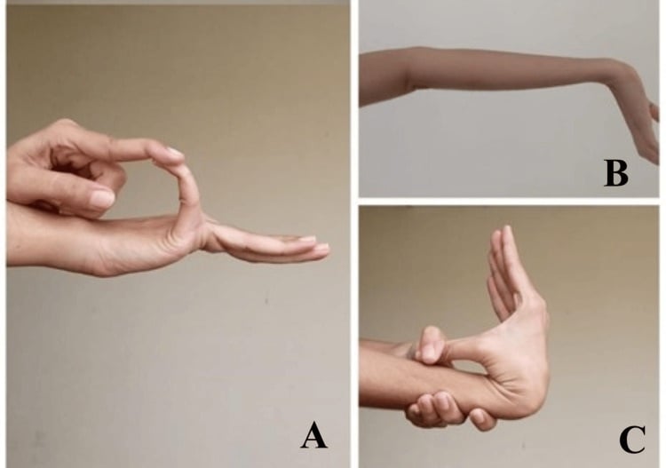
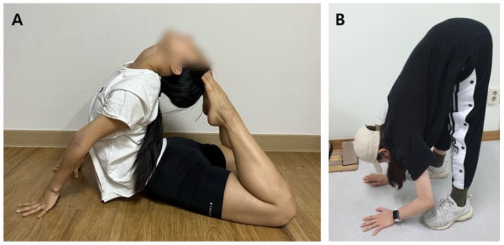
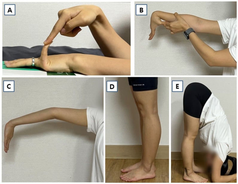
Next Investigation
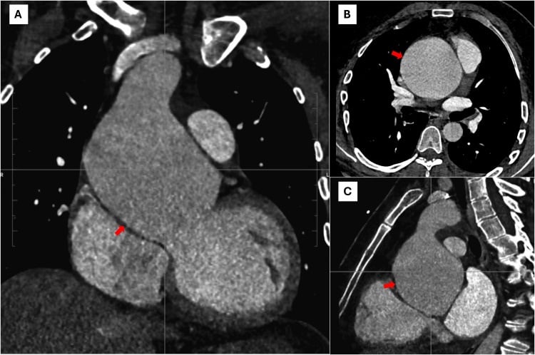
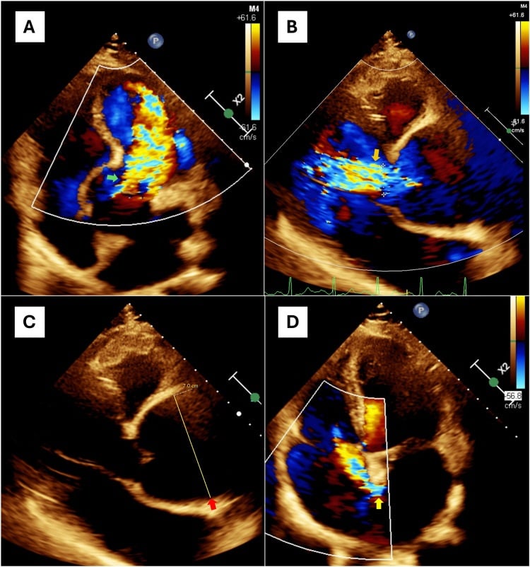
Aortic root measurement must be indexed to body surface area and age as a Z-score, not compared with an adult absolute diameter. A 12-year-old with a 38 mm root is grossly abnormal even though 38 mm is near-normal in a large adult.
5-4-3-2-1 becomes 'Pinkies, Thumbs, Elbows, Knees, Spine'Beighton — the nine points
Hook:Two points each for fifth MCP hyperextension beyond 90 degrees, thumb to forearm, elbow hyperextension beyond 10 degrees and knee hyperextension beyond 10 degrees, plus one point for palms flat on the floor with knees straight. Total 9. Threshold 6 or more prepubertal, 5 or more in adults up to 50.
The Differential
- Typical age / setting
- Adolescence; tall, thin, arm span exceeding height
- Discriminating feature
- Superotemporal ectopia lentis with aortic root dilatation at the sinuses of Valsalva
- What confirms it
- Ghent criteria; echocardiogram Z-score; FBN1 mutation
- Typical age / setting
- Teens to third decade; may present with bowel or arterial rupture
- Discriminating feature
- Thin translucent skin with visible subcutaneous veins, acrogeria, thin lips — joint laxity often limited to digits
- What confirms it
- COL3A1 mutation; avoid arteriography
- Typical age / setting
- Childhood; marfanoid but distinct
- Discriminating feature
- Hypertelorism, bifid or broad uvula, cleft palate, arterial tortuosity with dissection at smaller aortic diameters
- What confirms it
- CT or MR angiography head to pelvis; TGFBR1/2, SMAD3 testing
- Typical age / setting
- Early childhood; marfanoid habitus
- Discriminating feature
- Inferonasal lens dislocation, learning difficulty, osteoporosis, thromboembolism risk under anaesthesia
- What confirms it
- Urine and plasma homocysteine; cystathionine beta-synthase assay
- Typical age / setting
- Infancy to adolescence
- Discriminating feature
- Laxity plus low-energy fractures, blue or grey sclerae, dentinogenesis imperfecta, wormian bones at the lambdoid suture
- What confirms it
- Radiographic survey; COL1A1/COL1A2 sequencing
- Typical age / setting
- School age to adolescence, often familial, girls more than boys
- Discriminating feature
- Beighton positive with soft velvety skin but no lens, aortic or skin-fragility features; chronic diffuse pain and fatigue
- What confirms it
- Clinical 2017 criteria after excluding other heritable disorders; no confirmatory gene test
- Typical age / setting
- Age 3 to 10; asymptomatic, picked up incidentally
- Discriminating feature
- Laxity only — normal habitus, skin, eyes, heart, and an equally lax parent
- What confirms it
- Diagnosis of exclusion; reassurance and no imaging
- Typical age / setting
- Infancy onward
- Discriminating feature
- Global laxity with hypotonia, brachycephaly, single palmar crease; hip subluxation and atlantoaxial instability
- What confirms it
- Karyotype; lateral cervical spine flexion-extension if myelopathic signs
- Typical age / setting
- Childhood
- Discriminating feature
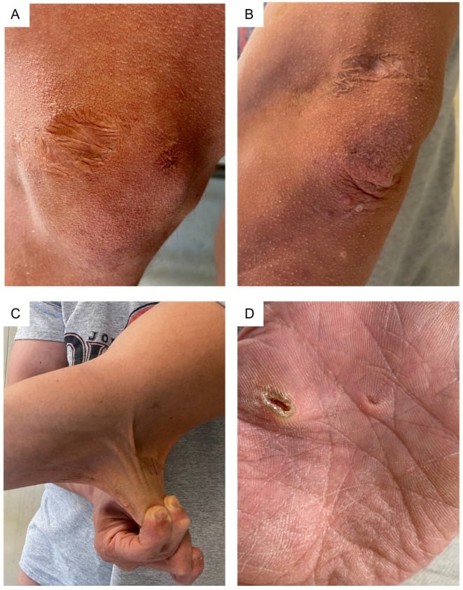
- Marked skin hyperextensibility with atrophic cigarette-paper scars over knees and forehead; molluscoid pseudotumours
- What confirms it
- COL5A1/COL5A2 mutation
- Typical age / setting
- Neonate to toddler; breech, female, firstborn
- Discriminating feature
- Laxity localises the risk — Ortolani and Barlow positive, limited abduction, Galeazzi sign
- What confirms it
- Ultrasound before 4-6 months; AP pelvis thereafter
- Typical age / setting
- Neonatal
- Discriminating feature
- Multiple congenital large-joint dislocations (knees, hips, elbows), flat facies, cervical kyphosis with instability
- What confirms it
- FLNB mutation; cervical spine radiographs mandatory before intubation
- Typical age / setting
- Infancy to early childhood
- Discriminating feature
- Distinctive facies with cardiac lesion — supravalvular aortic stenosis in Williams; long palpebral fissures in Kabuki
- What confirms it
- Microarray or targeted panel; cardiology referral
- Typical age / setting
- Infancy
- Discriminating feature
- Congenital hypotonia with progressive early scoliosis and scleral fragility
- What confirms it
- Urinary lysyl pyridinoline to hydroxylysyl pyridinoline ratio; PLOD1
- Typical age / setting
- Neonatal
- Discriminating feature
- Bilateral congenital hip dislocation with extreme laxity; redundant, doughy, sagging skin in dermatosparaxis
- What confirms it
- COL1A1/COL1A2 exon 6 skipping; ADAMTS2
Narrowing It Down
- 1Step 1 - Is it truly generalised?
Score Beighton formally and test at least one joint outside the set.
A score of 6 or more in a prepubertal child, 5 or more after puberty, or 4 or more beyond 50 qualifies as generalised. If laxity is confined to one joint, stop the syndromic search and treat the joint: recurrent patellar dislocation, atraumatic shoulder instability or a lax ankle.
- 2Step 2 - Is tone and power normal?
Examine tone, power, reflexes, tongue and motor milestones before attributing everything to ligament.
Reduced power, absent reflexes, tongue fasciculation or delayed milestones redirect to a neuromuscular or metabolic cause, and to creatine kinase and neurology review, not to a connective tissue panel. Laxity and hypotonia look alike across a room and are different problems.
- 3Step 3 - Is a second tissue involved?
Examine skin (extensibility, atrophic scars, translucency, bruising), eyes (lens position, high myopia, blue sclerae), teeth, palate and hearing.
Laxity plus skin fragility is classical or vascular EDS. Laxity plus blue sclerae plus fractures is osteogenesis imperfecta. Laxity alone with soft skin is hypermobility spectrum disorder. This single question is what separates a benign variant from a syndrome.
- 4Step 4 - What is the habitus and the face?
Measure height, arm span and the upper-to-lower segment ratio, and look at the palate, chest and face.
Tall with arm span exceeding height, arachnodactyly (positive wrist and thumb signs), high-arched palate and pectus points to Marfan. Add hypertelorism and a bifid uvula and it is Loeys-Dietz. Short stature with brachycephaly and hypotonia is Down. Flat facies with congenital dislocations is Larsen.
- 5Step 5 - Is the cardiovascular system involved?
Auscultate for aortic regurgitation and mitral prolapse, palpate all peripheral pulses, and take a family history of sudden death or aortic surgery.
Any marfanoid or vascular feature mandates an echocardiogram with an aortic root Z-score. A family history of sudden death escalates urgency REGARDLESS of examination findings - a normal praecordium does not exclude an enlarging root.
- 6Step 6 - Is the cervical spine at risk?
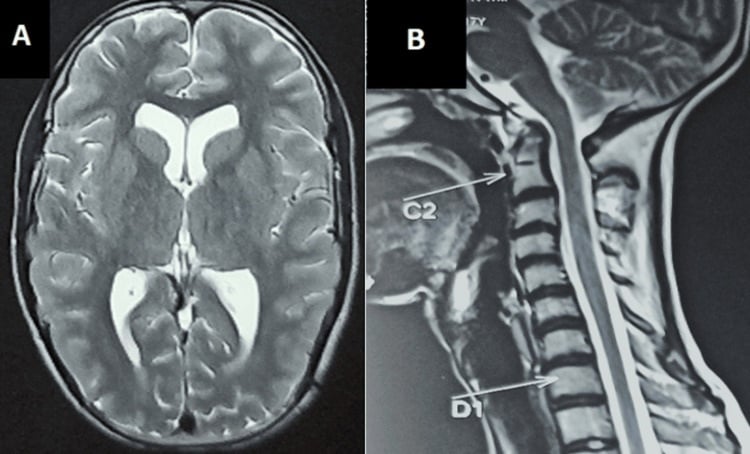
Ask specifically about gait deterioration, new clumsiness, torticollis and sphincter change, and examine for long-tract signs.
Down syndrome, Larsen, kyphoscoliotic EDS and the skeletal dysplasias all carry upper cervical instability. SYMPTOMS AND SIGNS, not routine screening films, drive flexion-extension radiographs and MRI.
- 7Step 7 - Does the answer change management?
Decide what the label buys the patient before pursuing it.
Benign laxity needs education, proprioceptive and closed-chain strengthening, and orthotics for symptomatic planovalgus. Syndromic laxity needs genetics, cardiology, an anaesthetic pre-warning and a raised threshold for elective soft-tissue surgery, which fails more often in lax tissue.
MCQ Practice Points
Q: What Beighton score defines generalised hypermobility in a 9-year-old?
A: 6 or more out of 9. The threshold falls to 5 or more after puberty and to 4 or more beyond age 50, because ligamentous laxity declines with age. Note that these are consensus cut-points on an instrument whose supporting studies were all rated only "fair" or "poor" on COSMIN - the number is a convention, not a validated boundary.
Q: A marfanoid teenager has inferonasal lens dislocation and mild learning difficulty. Diagnosis and anaesthetic implication?
A: Homocystinuria. Perioperative thromboembolism risk demands hydration, thromboprophylaxis and avoidance of nitrous oxide. Marfan's ectopia lentis is superotemporal.
Q: Which laxity syndrome most requires cervical spine imaging before intubation?
A: Larsen syndrome, because of cervical kyphosis and instability at C4-C5. Down syndrome requires imaging when there are symptoms or long-tract signs, rather than routinely.
Q: Which finding separates osteogenesis imperfecta from Ehlers-Danlos in a lax child?
A: Low-energy fractures with blue or grey sclerae, dentinogenesis imperfecta and wormian bones in the lambdoid suture. EDS gives skin and vessel fragility without the fracture-and-wormian-bone pattern.
Q: Which feature separates Loeys-Dietz from Marfan?
A: Hypertelorism with a bifid or broad uvula, and generalised arterial tortuosity. Loeys-Dietz dissects at smaller aortic diameters, so surgical thresholds are lower; ectopia lentis is characteristically absent.
Exam Viva Scenarios
Practise clinical reasoning and management decisions out loud
“You are shown clinical photographs of a 14-year-old boy, 190 cm tall, hyperextending both elbows and both knees, with the thumb apposed to the forearm. He has had three lateral patellar dislocations in a year. Skyline radiographs show a shallow trochlea.”
“You are shown radiographs of a newborn with both knees in fixed hyperextension and anterior tibial translation, and both hips dislocated. The face is flat with a depressed nasal bridge.”
“A 17-year-old girl is referred with a painful, atraumatically subluxing shoulder. You are shown a photograph of her forearm: the skin is thin and translucent with clearly visible subcutaneous veins, and there is extensive bruising. Her digits are hypermobile but her Beighton is only 4.”
Beighton (out of 9)
- Fifth MCP beyond 90 degrees, both hands — 2
- Thumb to flexor forearm, both — 2
- Elbow beyond 10 degrees, both — 2
- Knee beyond 10 degrees, both — 2
- Palms flat on floor, knees straight — 1
- Threshold: 6 prepubertal, 5 adult, 4 over 50
Must-not-miss discriminators
- Superotemporal lens dislocation — Marfan
- Inferonasal lens dislocation — homocystinuria
- Translucent skin, visible veins, thin lips — vascular EDS
- Hypertelorism, bifid uvula, tortuous arteries — Loeys-Dietz
- Blue sclerae, fractures, wormian bones — osteogenesis imperfecta
- Congenital knee and hip dislocation, flat face — Larsen
Orthopaedic consequences of laxity
- Developmental dysplasia of the hip in the neonate
- Recurrent patellar dislocation with trochlear dysplasia
- Atraumatic multidirectional shoulder instability
- Flexible pes planovalgus and hallux valgus
- Scoliosis, protrusio and pectus in Marfan
- Higher failure rate of soft-tissue stabilisation procedures
Investigations
- Echocardiogram with aortic root Z-score — any marfanoid feature
- Dilated slit-lamp examination — lens position
- Plasma and urinary homocysteine — marfanoid plus learning difficulty
- Hip ultrasound before 4-6 months, AP pelvis thereafter
- Cervical flexion-extension plus MRI — symptomatic instability only
- Targeted gene panel when phenotype is specific; none for hypermobile EDS
Management principles
- Education, pacing and closed-chain proprioceptive strengthening first
- Orthoses for symptomatic planovalgus only
- Rehabilitation before any stabilisation surgery
- Genetics, cardiology and anaesthetic alert for syndromic laxity
- Avoid arteriography and elective surgery in vascular EDS
Evidence Base
Articular Mobility in an African Population
- The paper that gives the score its name - an epidemiological survey of articular mobility carried out in a South African population
- PubMed indexes no abstract for it; what the record does confirm through its MeSH indexing is that AGE FACTORS and SEX FACTORS were examined alongside the fingers, elbows, knees and spine
- That age and sex dependence is the reason the modern cut-points differ by age at all, rather than being a single number
- The nine-point scoring set in universal use today - fifth metacarpophalangeal joints, thumbs, elbows, knees and forward flexion - derives from this survey
Measurement Properties of Clinical Assessment Methods for Classifying Generalized Joint Hypermobility - a Systematic Review
- Systematic review of four test methods (Beighton score, Carter and Wilkinson, Hospital del Mar, Rotes-Querol) and two questionnaires (the five-part questionnaire and self-reported Beighton), appraised against the COSMIN checklist
- EVERY INCLUDED STUDY WAS RATED 'FAIR' OR 'POOR' - not one study of any method reached good methodological quality
- For the Beighton score itself, reliability showed 'limited positive to conflicting evidence', with acknowledged shortcomings in the validity studies; the other three test methods lack satisfactory information on both
- The recommendation for clinical use is nonetheless the Beighton score: a cut-point of 5 of 9 in ADULTS including historical information, and at least 6 of 9 in CHILDREN
- The authors state plainly that more studies are needed before evidence-based recommendations can be made about the best method
The 2017 International Classification of the Ehlers-Danlos Syndromes
- The International EDS Consortium replaced the 1998 Villefranche nosology of six subtypes with a revised classification recognising THIRTEEN subtypes, each with a set of suggestive clinical criteria
- THE CRITICAL PRACTICAL POINT: definite diagnosis of every EDS subtype EXCEPT THE HYPERMOBILE TYPE relies on molecular confirmation of a causative genetic variant
- The clinical criteria for hypermobile EDS were deliberately revised and tightened to allow better distinction from other joint hypermobility disorders
- A pathogenetic scheme regroups subtypes whose causative proteins act in the same pathway, for research purposes
Hypermobile Ehlers-Danlos Syndrome - Clinical Description and Natural History
- hEDS is described as likely the MOST COMMON hereditary disorder of connective tissue, presenting largely with musculoskeletal complaints - hypermobility, subluxation and dislocation, with skin and soft tissue manifestations
- Many patients report activity-related pain and a proportion progress to DAILY pain, which is the feature that brings them to an orthopaedic clinic
- The 2017 nosology merged two previously separate labels - joint hypermobility syndrome and hEDS - into a single entity
- Both were, and the merged entity remains, CLINICAL diagnoses made in the absence of other causation