Intra-Articular | Nonunion Risk | ORIF Often Required
WEISS/JAKOB CLASSIFICATION
Critical Must-Knows
- INTRA-ARTICULAR fracture (elbow and wrist joint)
- Greater than 2mm displacement = surgical indication
- High risk of nonunion due to synovial fluid bathing fracture
- Late complications: nonunion, malunion, cubitus valgus, tardy ulnar nerve palsy
- Lateral approach - do NOT dissect posterior (blood supply)
Clinical Pearls
- "Milch Type I = Salter-Harris IV (lateral to trochlear ridge)
- "Milch Type II = Salter-Harris II (through trochlear ridge, unstable)
- "Nonunion causes lateral spur and cubitus valgus
- "Tardy ulnar nerve palsy occurs years later due to valgus
Clinical Imaging
Imaging Gallery


Critical Exam Concepts
Intra-Articular Fracture
Crosses the articular surface. Unlike supracondylar, this is an intra-articular fracture. Anatomic reduction required to prevent degenerative changes and angular deformity.
The 2mm Rule
Greater than 2mm displacement = ORIF. Less than 2mm can be treated non-operatively but needs close follow-up (can displace in cast). Some advocate surgery for greater than 2mm on any view.
High Nonunion Risk
Synovial fluid prevents healing. Fracture bathed in synovial fluid from elbow joint. Combined with pull of extensor origin, high risk of nonunion. This is why surgery is often needed.
Tardy Ulnar Nerve Palsy
Delayed complication of cubitus valgus. Nonunion causes lateral spur and progressive valgus. Ulnar nerve is stretched over time. May present years later with ulnar neuropathy.
Quick Decision Guide
| Type | Displacement | Articular Step | Treatment |
|---|---|---|---|
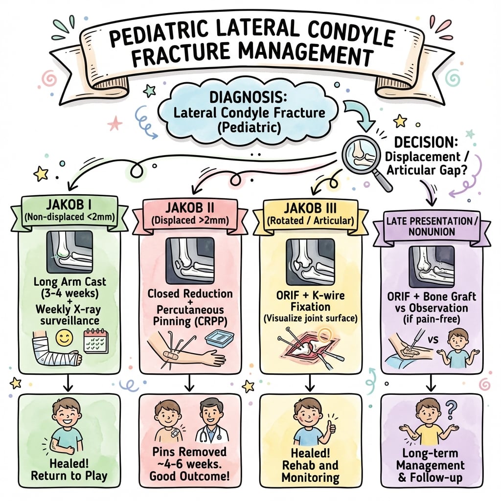
| Weiss Type I | Less than 2mm | Intact cartilage hinge | Cast, weekly XR for 3 weeks |
| Weiss Type II | 2-4mm | Gap but some contact | ORIF with K-wires |
| Weiss Type III | Greater than 4mm | Complete articular disruption | ORIF with K-wires |
1 OUT, 2 THROUGHMilch Classification
| 1 | Type I = Lateral to trochlear ridge Elbow is stable (Salter-Harris IV) |
| 2 | Type II = Through trochlear ridge Elbow potentially unstable (Salter-Harris II) |
| 1 | Type I = Lateral to trochlear ridge Elbow is stable (Salter-Harris IV) |
| 2 | Type II = Through trochlear ridge Elbow potentially unstable (Salter-Harris II) |
Hook:Type 1 stays OUT (lateral), Type 2 goes THROUGH (trochlear ridge)
NUTSLateral Condyle Complications
| N | Nonunion Most common serious complication due to synovial fluid |
| U | Ulnar nerve palsy (tardy) Delayed onset due to progressive cubitus valgus |
| T | Tilted (cubitus valgus) Angular deformity from lateral physeal arrest |
| S | Stiffness From prolonged immobilization or malunion |
| N | Nonunion Most common serious complication due to synovial fluid | T | Tilted (cubitus valgus) Angular deformity from lateral physeal arrest |
| U | Ulnar nerve palsy (tardy) Delayed onset due to progressive cubitus valgus | S | Stiffness From prolonged immobilization or malunion |
Hook:Don't go NUTS - treat lateral condyle fractures properly!
STAY ANTERIORSurgical Approach
| S | Stay anterior and lateral Lateral Kocher approach |
| A | Avoid posterior dissection Posterior blood supply to fragment |
| S | Stay anterior and lateral Lateral Kocher approach |
| A | Avoid posterior dissection Posterior blood supply to fragment |
Hook:STAY ANTERIOR - blood supply from posterior, don't strip it!
Overview and Epidemiology
Why This Fracture Matters
The lateral condyle fracture is the second most common pediatric elbow fracture (after supracondylar) but has a higher complication rate. It is intra-articular, prone to nonunion, and can lead to progressive cubitus valgus and tardy ulnar nerve palsy.
Epidemiology
- 15% of pediatric elbow fractures
- Second only to supracondylar
- Peak age 5-7 years
- Slight male predominance
- Usually fall onto outstretched hand
Anatomy Involved
- Lateral condyle (capitellum + part of lateral trochlea)
- Extends through articular surface
- Physis involved (Salter-Harris pattern)
- Lateral collateral ligament attaches to fragment
Anatomy and Biomechanics
Blood Supply - Do Not Strip Posterior
The blood supply to the lateral condyle enters posteriorly. During ORIF, do NOT dissect or strip the soft tissues from the posterior aspect of the fragment. Use an anterior/lateral approach and visualize the articular surface from the front.
Key Anatomical Points
The lateral condyle includes:
- Capitellum (articulates with radial head)
- Lateral portion of trochlea (variable extent)
- Lateral epicondyle (common extensor origin)
Blood supply: Enters posteriorly. The fragment has NO anterior blood supply once fractured. Protect posterior soft tissues during surgery.
Deforming forces: Extensor muscles (attached to lateral epicondyle) pull the fragment distally and rotate it, causing displacement.
Classification Systems
Weiss/Jakob Classification (Most Clinically Useful)
| Type | Displacement | Description | Treatment |
|---|---|---|---|
| Type I | Less than 2mm | Minimally displaced, cartilage hinge intact | Long arm cast 90°, weekly XR x3 |
| Type II | 2-4mm | Partial articular disruption, some contact | ORIF with K-wires |
| Type III | Greater than 4mm | Complete displacement, no articular contact | ORIF with K-wires |
This classification is more practical for guiding treatment than Milch.
Clinical Assessment
History
- Fall mechanism (FOOSH with varus stress)
- Time since injury
- Any swelling or deformity
- Neurovascular symptoms
- Previous elbow injury
Examination
- Lateral elbow swelling and tenderness
- Ecchymosis (may be extensive)
- Full neurovascular exam
- Check elbow stability (compare to opposite)
- ROM likely limited by pain
Beware the Occult Fracture
In young children, the lateral condyle ossific nucleus may not yet be visible (capitellum ossifies around age 1-2). An effusion (positive fat pad sign) with lateral tenderness but no obvious fracture line suggests an occult lateral condyle fracture. Obtain an internal oblique view, consider ultrasound/MRI or arthrogram, or treat as a fracture and follow closely with repeat films.
Differential Diagnosis of the Painful, Swollen Paediatric Elbow
Distinguishing Lateral Condyle Fracture from Mimics
| Condition | Mechanism / Age | Key Distinguishing Feature | Pitfall to Avoid |
|---|---|---|---|
| Lateral condyle fracture | FOOSH, varus stress; peak 5-7y | Lateral metaphyseal fragment, intra-articular, displaces in cast | Underestimating displacement on AP/lateral only |
| Supracondylar fracture | FOOSH, hyperextension; peak 5-7y | Transverse metaphyseal line, anterior humeral line abnormal, extra-articular | Missing concurrent NV injury (AIN/median) |
| Medial epicondyle avulsion | Valgus/throwing; older child 9-14y | Medial fragment, may be incarcerated in joint | Forgetting to count ossification centres (CRITOE) |
| Transphyseal distal humeral separation | Birth/NAI in infants under 3y | Whole epiphysis displaced medially, radiocapitellar line maintained to capitellum | Mistaking for elbow dislocation; consider non-accidental injury |
| Radial neck/head fracture | FOOSH valgus | Radial head angulation, point tenderness over radial neck | Attributing all lateral pain to the condyle |
| Pulled elbow (radial head subluxation) | Axial pull; toddler 1-4y | No swelling, refusal to use arm, normal radiographs | Over-imaging; reduces with supination-flexion |
CRITOE and the Lateral Condyle
Use the CRITOE ossification sequence (Capitellum, Radial head, Internal/medial epicondyle, Trochlea, Olecranon, External/lateral epicondyle) to interpret the immature elbow. Comparison views of the contralateral elbow are invaluable - a displaced ossific fragment lateral to the metaphysis confirms the diagnosis when the cartilaginous fracture itself is radiolucent.
Investigations
Radiological Investigations
| View | What to Check | Key Finding |
|---|---|---|
| AP elbow | Fragment size and displacement | Measure gap on AP |
| Lateral elbow | Rotation of fragment | Fragment often rotates posteriorly |
| Internal oblique | Better visualization of fracture | 45° internal rotation |
| Comparison views | Opposite elbow | Helps in young children |
Internal oblique view is particularly helpful as it places the fracture line in profile.
Management
Greater than 2mm = ORIF
The threshold for surgery is greater than 2mm displacement. Some authors are more aggressive and operate on any fracture greater than 2mm on ANY view. Non-operative fractures need weekly X-rays for 3 weeks as they can displace in cast.
Non-Operative Management (Weiss Type I)
Indications: Displacement less than 2mm, intact cartilage hinge.
Treatment: Long arm cast in 90 degrees elbow flexion.
Follow-up: Weekly X-rays for first 3 weeks. If any displacement occurs, convert to ORIF.
Duration: Cast for 4-6 weeks until union confirmed.
Key point: Close follow-up is mandatory. Up to 20% of initially non-displaced fractures may displace.
Screws vs K-wires
Traditional fixation is with smooth K-wires to avoid physeal injury. Some surgeons use a cannulated screw in older children (near skeletal maturity) for more stable fixation. Screws should NOT cross the physis in young children.
Surgical Technique Considerations
ORIF Technique for Lateral Condyle
Position: Supine with arm on hand table.
Approach: Lateral/Kocher interval between anconeus and ECU.
Critical step: Visualize the articular surface from ANTERIOR. Do NOT strip soft tissues from posterior fragment.
Reduction: Reduce articular surface anatomically. The metaphyseal reduction usually follows.
Fixation: Two smooth K-wires, usually 1.6mm. Divergent configuration. Cross fracture site but avoid olecranon fossa.
Complications
Complications of Pediatric Lateral Condyle Fractures
| Complication | Incidence | Cause | Management |
|---|---|---|---|
| Nonunion | Most serious | Synovial fluid, inadequate fixation | ORIF if early, osteotomy if late |
| Cubitus valgus | Common with nonunion | Lateral physeal arrest | Osteotomy if symptomatic |
| Tardy ulnar nerve palsy | Delayed (years) | Progressive valgus stretches nerve | Ulnar nerve transposition |
| Stiffness | Common | Prolonged immobilization | Physiotherapy, rarely need release |
| AVN | Rare | Posterior soft tissue stripping | Prevention - protect blood supply |
| Malunion | With late reduction | Missed or delayed diagnosis | Osteotomy if functional limitation |
Tardy Ulnar Nerve Palsy
Tardy ulnar nerve palsy is a delayed complication occurring years after the original injury. It develops because nonunion leads to cubitus valgus, which progressively stretches the ulnar nerve around the medial epicondyle. Treatment is ulnar nerve transposition (often anterior subcutaneous) and may require corrective osteotomy.
Postoperative Care
Post-Operative Protocol
Long arm backslab in 90 degrees elbow flexion. Neurovascular checks. Elevate and ice.
Check wound and pin sites. X-ray to confirm maintained reduction. Convert to long arm cast.
X-ray to assess healing. If good callus, may remove pins (in clinic). Continue cast.
X-ray confirms union. Remove cast. Begin active ROM exercises. Avoid passive stretching.
Full return to activities. Final check of motion, alignment, carrying angle. Follow long-term if any concern.
Outcomes and Prognosis
Prognosis Depends on Prompt Diagnosis and Treatment
Good outcomes when:
- Fracture recognized early
- Displacement greater than 2mm treated surgically
- Anatomic articular reduction achieved
- Union confirmed before discharge from follow-up
Poor outcomes when:
- Diagnosis delayed
- Fracture displaces in cast and missed
- Nonunion develops
Special Considerations
Management of Nonunion
Early nonunion (less than 3-6 months): ORIF with bone grafting still possible. Better outcomes than late reconstruction.
Established nonunion (greater than 6 months): In situ fixation with bone grafting or corrective osteotomy. The nonunion may be too sclerotic for direct healing without osteotomy.
Late nonunion with cubitus valgus: May require supracondylar osteotomy for angular correction plus ulnar nerve transposition.
Controversies and Areas of Uncertainty
Closed vs Open Reduction
Traditional teaching mandated open reduction for displacement over 2-4mm to confirm articular congruity. Song's prospective series and later cohorts show closed reduction with percutaneous pinning can succeed even in fully displaced/rotated fractures, with intraoperative arthrogram to confirm joint reduction. The trade-off is the inability to directly inspect the articular surface.
The 2mm vs 4mm Threshold
The operative threshold is debated. Many use over 2mm on the maximally displaced view; others reserve open surgery for at least 4mm (articular disruption), treating 2-4mm fractures with closed pinning after arthrogram. Weiss data link the 4mm point to articular incongruity and higher complications.
Implant Choice
Smooth K-wires remain standard to spare the open physis, but cannulated/headless screws are increasingly used near maturity for compression and earlier motion. Comparative data show no clear outcome superiority of one implant over another.
Buried vs Exposed Pins
Leaving pins percutaneous (exposed) allows clinic removal but carries pin-site infection risk; buried pins reduce infection but need a second anaesthetic for removal. Practice varies; pin-site infection is usually superficial and resolves with oral antibiotics and pin removal.
Late-Presenting Nonunion: Repair or Observe?
Whether to repair an established, minimally symptomatic nonunion is contested. Historic concern about osteonecrosis and stiffness favoured observation, but contemporary series (e.g. Eamsobhana 2015) report high union rates and good Mayo scores even with mild symptoms, supporting earlier osteosynthesis - ideally before the nonunion is neglected beyond ~28 months. Aggressive dissection of a fibrosed, displaced fragment still risks avascular necrosis, so meticulous posterior soft-tissue preservation is essential.
Evidence Base and Key Studies
Weiss et al. - Displacement-Based Classification Predicts Complications
- 158 operatively treated fractures - largest operative series at the time
- Type I less than 2mm; Type II at least 2mm with intact cartilage (arthrogram); Type III at least 2mm with disrupted articular surface
- Complication rate 11% (Type II) versus 34% (Type III) - more than 3-fold higher
- Every Type II fracture had under 4mm displacement; every Type III had at least 4mm on radiographs
Song et al. - Five-Stage Classification and Treatment Algorithm
- Prospective study of 63 unstable fractures graded on four radiographic views
- Closed reduction and internal fixation succeeded in 13 of 17 stage-3 and 30 of 40 stage-4 fractures
- Open reduction reserved for residual displacement over 2mm after closed attempt
- No osteonecrosis, nonunion, malunion, or physeal arrest
Song et al. - Internal Oblique Radiograph for Assessment
- Prospective study of 54 fractures with AP, lateral, and oblique views
- 70% showed different displacement on AP versus internal oblique view
- Internal oblique view demonstrated more displacement in 30 cases and more instability in 20
- Classification should use the greatest displacement across at least three views
Song et al. - CRIF for Completely Displaced/Rotated Fractures
- Prospective, three Level I centres, 24 completely displaced and rotated (Jakob 3) fractures
- 18 of 24 (75%) reduced to within 2mm by closed technique with percutaneous fixation
- Closed reduction failed in 3 - converted to open reduction
- No osteonecrosis, nonunion, malunion, or physeal arrest
Exam Viva Scenarios
Use these scenarios to practise clinical reasoning and management decisions
Scenario 1: Displaced Lateral Condyle Fracture
"A 6-year-old child presents after a fall with lateral elbow pain and swelling. X-rays show a lateral condyle fracture with 3mm displacement. How would you manage this?"
Scenario 2: Late Presentation Nonunion
"A 10-year-old presents 4 months after a fall. He was treated at another hospital with casting. He now has an established nonunion of the lateral condyle with 15 degrees of cubitus valgus. There is no ulnar nerve dysfunction. How would you manage this?"
Scenario 3: Minimally Displaced Fracture - Non-Op
"A 5-year-old child presents with a lateral condyle fracture with 1.5mm displacement on the AP view and no displacement on the lateral view. How would you manage this?"
MCQ Practice Points
Displacement Threshold Question
Q: What is the displacement threshold for ORIF of pediatric lateral condyle fractures? A: 2mm. Greater than 2mm displacement, or any rotation of the fragment, requires ORIF. Less than 2mm can be treated non-operatively with close follow-up.
Classification Question
Q: In the Milch classification, what distinguishes Type I from Type II lateral condyle fractures? A: Exit point relative to trochlear ridge. Type I exits LATERAL to the trochlear ridge (elbow stable). Type II exits THROUGH the trochlear ridge (elbow potentially unstable). Type II is more common.
Blood Supply Question
Q: Why should posterior soft tissue stripping be avoided during ORIF of lateral condyle fractures? A: Blood supply enters posteriorly. The blood supply to the lateral condyle fragment comes from posterior. Stripping posterior soft tissues risks avascular necrosis.
Nonunion Cause Question
Q: Why is lateral condyle fracture at high risk of nonunion? A: Synovial fluid bathes the fracture. The intra-articular location means synovial fluid prevents hematoma formation and bone healing. This is compounded by the deforming force of the extensor muscles.
Tardy Ulnar Nerve Question
Q: What is tardy ulnar nerve palsy and how does it relate to lateral condyle fractures? A: Delayed ulnar neuropathy due to progressive cubitus valgus. Nonunion leads to lateral physeal arrest and valgus deformity, which stretches the ulnar nerve over years. Treatment is transposition.
Fixation Question
Q: What is the standard fixation for pediatric lateral condyle fractures? A: 2 divergent smooth K-wires. This avoids physeal damage. Screws may be considered in older children near skeletal maturity.
Guidelines, Registries & Global Practice
Global Epidemiology
- 15-20% of paediatric elbow fractures; second only to supracondylar
- Peak age 5-7 years; slight male predominance
- Mechanism: fall on outstretched hand with varus (pull-off) or push-off load
- Roughly 40-60% are displaced enough to warrant surgery across published series
Why Registry Data Are Limited
- National arthroplasty registries (NJR, AJRR, AOANJRR, SHAR) do not capture paediatric trauma
- Evidence base is observational cohorts and society position statements, not registry survival data
- No randomized trial defines the exact displacement threshold
Side-by-Side Society Guidance
Consensus Across Major Bodies (Principles Converge)
| Body / Region | Operative Threshold | Fixation Emphasis | Distinct Point |
|---|---|---|---|
| AAOS / POSNA (US) | Over 2mm or articular incongruity | Smooth K-wires; arthrogram to confirm reduction | CRPP acceptable for many displaced fractures |
| BOA / BSCOS (UK) | Over 2mm; close cast surveillance under 2mm | K-wires standard; structured follow-up | Emphasis on early review to detect late displacement |
| AO Foundation | Displacement / rotation or articular step | Anatomic articular reduction, lag screw near maturity | Protect posterior blood supply, anterolateral exposure |
| EFORT / European consensus | Over 2mm on maximal-displacement view | K-wires; selective screws | Internal oblique view to grade displacement |
High-Resource Settings
- Routine internal oblique views, fluoroscopy and intraoperative arthrogram
- Increasing use of closed reduction and percutaneous pinning
- MRI/CT available for occult or complex fractures
- Day-case surgery with structured pin-site care and early clinic review
Limited-Resource Settings
- Higher proportion of late presentation, nonunion and cubitus valgus
- Reliance on plain radiographs and comparison views; arthrogram less available
- Open reduction more common where image intensifiers are scarce
- Reconstruction (osteosynthesis +/- osteotomy +/- ulnar transposition) forms a larger share of workload
Globally Consistent Principles
Across every major society the core principles are identical: this is an intra-articular, nonunion-prone fracture; over 2mm displacement or articular incongruity is operative; reduction must be anatomic; the posterior blood supply must be preserved; and close radiographic follow-up is mandatory because non-displaced fractures can displace in cast. The main variation is the closed-versus-open debate and the precise 2mm-versus-4mm threshold.
LATERAL CONDYLE FRACTURES - PEDIATRIC
Clinical summary
Key Facts
- •15% of pediatric elbow fractures (2nd most common)
- •INTRA-ARTICULAR fracture
- •High nonunion risk (synovial fluid)
- •Blood supply from posterior - do NOT strip
Management
- •Less than 2mm: cast with weekly XR
- •Greater than 2mm: ORIF
- •Fix with 2 divergent K-wires
- •Lateral approach, stay ANTERIOR
Classification
- •Weiss I: less than 2mm - non-op
- •Weiss II: 2-4mm - ORIF
- •Weiss III: greater than 4mm - ORIF
- •Milch I: lateral to trochlear ridge
Complications - NUTS
- •Nonunion (most common serious)
- •Ulnar nerve palsy (tardy - years later)
- •Tilted (cubitus valgus)
- •Stiffness
Key Surgical Points
- •Lateral approach, stay ANTERIOR
- •Visualize articular surface
- •2 divergent smooth K-wires
- •Do NOT strip posterior soft tissues