ECRB Origin Pathology | 80-90% Respond to Conservative | 6-12 Months Before Surgery
- ECRB origin is the primary pathology - angiofibroblastic degeneration, not inflammation
- 80-90% respond to conservative management - physiotherapy, eccentric exercises, bracing
- Surgery only after 6-12 months of failed conservative treatment
- Open vs arthroscopic debridement - both effective, similar outcomes
- PRP superior to corticosteroid (durable to 2 years) but benefit over an active needling control is modest and delayed
- “Lateral epicondylitis is tendinosis (degeneration), not tendinitis (inflammation)
- “Cozen's test and Mill's test are provocative tests for diagnosis
- “ECRB origin at lateral epicondyle is most commonly affected structure
- “Natural history is self-limiting - most resolve within 12-18 months
Overview and Epidemiology
Lateral epicondylitis, tennis elbow, is a degenerative condition of the common extensor origin at the lateral epicondyle of the humerus. The name misleads twice over: only 5-10% of patients play tennis, and the "-itis" promises an inflammation that histology does not support. It is a tendinosis, not a tendinitis.
Who. The annual incidence is 1-3% of the general population, peaking at 35-50 years, the years of heaviest occupational demand. The dominant arm is affected in 75% of cases, and the typical patient is a manual worker, a computer user or a racquet-sports player.
Risk factors.
- Repetitive wrist extension and supination
- Manual labour: mechanics, carpenters, painters
- Computer work with poor ergonomics
- Racquet sports, especially with poor technique
- Smoking, which impairs tendon healing
- Diabetes, through its association with tendinopathy
Natural history and impact. The condition is self-limiting in most cases and resolves within 12-18 months, but it becomes chronic in 10-20%. While it lasts it hurts with lifting and gripping, keeps manual workers in particular off work, and carries an economic burden of lost working days and treatment costs.
Anatomy and Pathophysiology
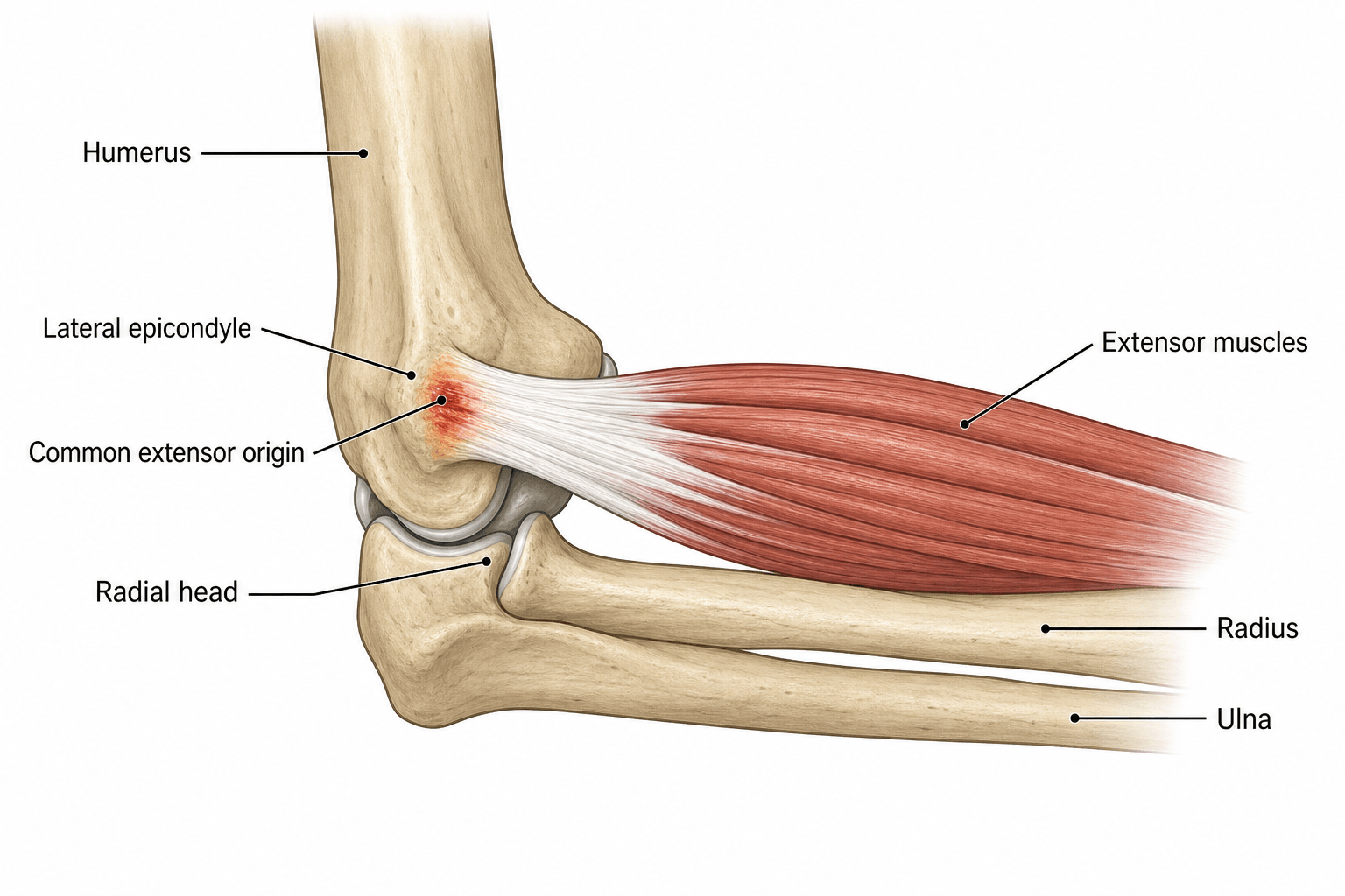
The common extensor origin. The lateral epicondyle is the attachment for four muscles, and their involvement is far from equal.
- Function
- Wrist extension, radial deviation
- Involvement in LE
- 90% - primary pathology
- Clinical Significance
- Deep surface at origin most affected
- Function
- Wrist extension, radial deviation
- Involvement in LE
- Sometimes involved
- Clinical Significance
- Superficial to ECRB, may need reflection
- Function
- Finger extension
- Involvement in LE
- Rarely involved
- Clinical Significance
- Posterior aspect of origin
- Function
- Wrist extension, ulnar deviation
- Involvement in LE
- Rarely involved
- Clinical Significance
- Ulnar aspect of origin
The lesion. It sits on the undersurface of the ECRB at its origin, Nirschl's original observation. That surface takes the maximum tensile stress and has a relatively poor blood supply, and the tissue found there is grey, friable and without normal tendon architecture: angiofibroblastic hyperplasia, the "Nirschl lesion".
How the load arrives. The ECRB origin is stressed most during wrist extension with the elbow extended, and pronation increases its tension. Eccentric loading in the deceleration phase of a tennis backhand or a hammer swing, repeated, causes cumulative microtrauma, and the healing response fails because the tendon-bone interface is poorly vascularised.
The degenerative cascade. The tendon passes through recognisable stages.
Tendinosis Progression
Organised type I collagen fibres, minimal vascularity, tenocytes aligned with the direction of load.
Tenocyte activation, increased proteoglycan and water content, reversible thickening. This is not lateral epicondylitis yet.
Angiofibroblastic degeneration: disorganised type III collagen, increased cellularity, neovascularisation with nerve ingrowth. This is lateral epicondylitis.
Collagen fibre disruption, partial tears, fibrosis, calcification. May progress to complete tendon failure in severe cases.
Histology. Fibroblasts are increased, without significant inflammatory cells. The collagen is disorganised type III where type I should be, there is neovascularisation with accompanying nerve ingrowth, and the pain mediators substance P and glutamate are increased. Prostaglandins are absent, which explains the poor response to NSAIDs.
Angio = new blood vessels (neovascularisation), fibroblastic = increased fibroblast activity, degeneration = breakdown of normal tendon structure. The term explains why anti-inflammatory treatments (cortisone, NSAIDs) provide only temporary relief: there is no inflammation to suppress.
The nerve. The posterior interosseous nerve lies deep to the common extensor origin and, about 4-5cm distal to the lateral epicondyle, passes deep to supinator at the level of the radial neck. That position matters twice: compression of the nerve can coexist with lateral epicondylitis and mimic it, and the nerve is at risk during surgical release. Telling the two conditions apart is critical, and the clinical distinction is set out with the assessment below.
Classification Systems
Nirschl's staging is the most widely used. Nirschl was the first to recognise the condition as a degenerative tendinosis rather than an inflammation, and his phases run from the reversible changes of Phase 1 to the irreversible structural damage of Phase 4, which is what guides treatment.
- Pathology
- Mild inflammation, oedema, reversible
- Clinical Features
- Pain after activity, resolves with rest
- Management
- Rest, ice, activity modification
- Pathology
- Angiofibroblastic degeneration
- Clinical Features
- Pain during and after activity
- Management
- Physiotherapy, eccentric exercises
- Pathology
- Structural failure, partial tearing
- Clinical Features
- Pain during activity, limits performance
- Management
- Consider injection therapy, surgery if refractory
- Pathology
- Fibrosis, calcification, chronic
- Clinical Features
- Pain at rest and with activity
- Management
- Surgical debridement often required
Clinical Presentation and Assessment
History. Pain over the lateral elbow, radiating into the forearm, that came on gradually with overuse; an acute onset after a single event is rare. Gripping, lifting and wrist extension aggravate it, rest relieves it, and patients describe trouble with the cup or kettle, a handshake or a doorknob. Ask about occupation (manual labour, computer work, typing), sport (tennis, golf and other racquet sports, and their technique), which hand is dominant, how long it has lasted (weeks against months), and what has been tried, injections included. Severe pain at rest or night pain is a red flag for other pathology.
Examination. Inspection is usually normal, with no swelling or deformity; marked swelling points to something else, septic arthritis or a fracture. The most reliable finding is tenderness 1cm distal and anterior to the lateral epicondyle, at the ECRB origin. Elbow flexion and extension and forearm rotation are full, and a reduced range suggests intra-articular pathology.
Provocative tests. Cozen's is the gold standard; pain is reproduced by resisted wrist extension and by resisted supination.
- Technique
- Patient seated, elbow extended, forearm pronated, wrist in slight radial deviation; the patient makes a fist and extends the wrist against resistance
- Positive Result
- Pain at the lateral epicondyle
- Accuracy
- Sensitivity 80-90%, specificity 70-80%
- Technique
- Patient seated, elbow fully extended, forearm pronated; the examiner passively flexes the wrist from dorsiflexion to palmar flexion
- Positive Result
- Pain at the lateral epicondyle
- Accuracy
- Sensitivity 70-80%, specificity 60-70%
- Technique
- Standing behind a chair with the forearm pronated and elbow extended, the patient lifts the chair with one hand
- Positive Result
- Pain at the lateral epicondyle, or inability to lift
- Accuracy
- Functional test; simulates real-world activity and assesses severity
- Technique
- Resisted middle finger extension
- Positive Result
- Pain suggests radial tunnel syndrome
- Accuracy
- Helps differentiate PIN compression from pure lateral epicondylitis
Why Cozen's works. Resisted wrist extension loads the ECRB maximally and reproduces the pain at its origin. The test is most sensitive with the elbow fully extended, which increases tension on ECRB, and the wrist radially deviated, ECRB's primary action. Mill's test stretches the tendon rather than loading it.
The radial nerve's deep branch can mimic lateral epicondylitis in two ways that are NOT the same diagnosis:
- Radial tunnel syndrome - pain only, NO motor deficit. Tenderness sits 4-5cm distal to the lateral epicondyle over the radial tunnel, maximal with resisted middle-finger extension (Maudsley's). EMG/NCS is typically normal - this is a clinical diagnosis, and the safest management is the same as for epicondylitis: activity modification, stretching, patience.
- PIN syndrome - the same nerve compressed, but with motor loss: weak finger and thumb extension, weak wrist extension with RADIAL deviation (ECRL is spared), and no sensory loss because the deep branch is motor only. This is the one that may need decompression.
Do not collapse them: weakness is what turns a radial tunnel syndrome into a PIN syndrome, and it changes both the investigation and the threshold for surgery.
The Radiocapitellar (Posterolateral) Synovial Plica
What it is. A synovial plica is a normal fold of the joint lining at the posterolateral or posterior radiocapitellar joint. With repetitive pronation-supination it can thicken, become fibrotic and impinge between the radial head and capitellum, and a symptomatic plica is a recognised cause of recalcitrant or "failed" tennis elbow.
When to suspect it. Lateral elbow pain accompanied by clicking, snapping or catching on rotation; tenderness over the radiocapitellar joint, slightly posterior to the epicondyle, rather than at the ECRB origin; or an apparent tennis elbow that fails to settle. This is one reason the arthroscopic approach is valued: the surgeon can inspect and resect a symptomatic plica at the same sitting. Radial tunnel syndrome (pain further distal) and posterolateral rotatory instability (a history of giving way) have their own pathways.
- Radiocapitellar plica
- Over the radiocapitellar joint, slightly POSTERIOR to the lateral epicondyle
- Lateral epicondylitis (ECRB)
- 1 cm distal and anterior to the lateral epicondyle (ECRB origin)
- Radiocapitellar plica
- Painful clicking, snapping or catching with pronation-supination and flexion-extension; a painful arc
- Lateral epicondylitis (ECRB)
- Pain on gripping and resisted wrist extension (Cozen, Mill)
- Radiocapitellar plica
- MRI may show a thickened plica (more than 3 mm) abutting the radiocapitellar joint; definitive at arthroscopy
- Lateral epicondylitis (ECRB)
- ECRB tendinosis signal; plica usually absent
- Radiocapitellar plica
- Arthroscopic resection of the symptomatic plica
- Lateral epicondylitis (ECRB)
- ECRB debridement and repair
Investigations
Lateral epicondylitis is a clinical diagnosis, made on a history of overuse and gradual onset and on lateral epicondyle tenderness with a positive Cozen's or Mill's test. Imaging is not required to make it; it is used to exclude other pathology or to plan surgery.
Radiographs. AP and lateral views are usually normal, and calcification is a rare finding in chronic cases. Their purpose is to exclude arthritis, loose bodies, fracture and osteochondritis dissecans.
Ultrasound. When the diagnosis is uncertain, a dynamic examination can show tendon thickening, hypoechoic areas of degeneration and neovascularisation on Doppler. It is cost-effective and can guide injections, but it is operator-dependent and cannot assess intra-articular pathology.
MRI. T1 and T2 sequences show increased signal at the ECRB origin, partial tears and bone marrow oedema. It is indicated after failed conservative treatment, for surgical planning, and to exclude other causes such as radiocapitellar arthritis or an osteochondral lesion; it is not routinely required.
EMG and nerve conduction studies. These are for the patient whose motor weakness raises PIN syndrome, or to exclude cervical radiculopathy: weakness of finger or thumb extension, pain distal to the lateral epicondyle and atypical symptoms are the indications. In pure radial tunnel syndrome, pain without weakness, the studies are usually normal, so a normal EMG does not exclude it and the diagnosis remains clinical.
Management Algorithm
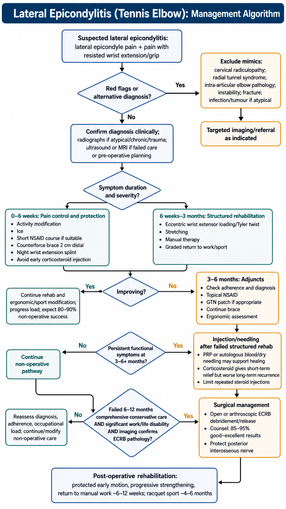
Conservative treatment is first-line for all patients, and 80-90% improve with it. The protocol is staged by time.
Structured Conservative Protocol
The goals are to reduce pain and protect healing. Avoid the provocative activities (heavy lifting, gripping); ice for 15-20 minutes several times daily; a short NSAID course of 1-2 weeks for symptom relief only; a counterforce brace worn 2cm distal to the lateral epicondyle during activities; and a wrist extension splint at night to rest ECRB. Avoid cortisone injection at this stage, as it may worsen the long-term outcome.
The goal is to restore tendon strength and endurance. The core is eccentric wrist extension with a resistance bar, lowering slowly over 3-5 seconds, the Tyler twist protocol set out in its own section below, supported by stretching of the wrist extensors, ultrasound or laser therapy, manual therapy and soft-tissue massage, and a gradual return to activities.
If the response to Phases 1-2 is inadequate: GTN (glyceryl trinitrate) patches applied to the lateral epicondyle, which may promote tendon healing; topical NSAIDs (diclofenac gel), safer than oral; continued counterforce bracing during activities; and an ergonomic assessment of work or sport. Consider injection therapy if still symptomatic at 3 months.
Only after failed structured rehabilitation. PRP may be superior to cortisone for long-term outcomes (durable to 2 years in the Peerbooms/Gosens RCT), but it has not proved superior to placebo in RCTs: much of its early effect is shared with the needling itself, so its benefit over an active needling control is modest and delayed. Consider it for the patient who wants to avoid surgery. Cortisone gives effective short-term relief for 4-8 weeks with worse long-term outcomes than PRP or placebo and increases recurrence; limit it to a maximum of 2-3 injections, and avoid it in younger athletes. Autologous blood injection and dry needling have similar efficacy to PRP, stimulate a healing response and are cheaper.
Eccentric Loading and the Tyler Twist Protocol
Why eccentric loading. Because the lesion is a failed-healing tendinosis, anti-inflammatory measures only mask symptoms. Controlled eccentric exercise, lengthening under load, applies a mechanotransduction stimulus that drives tenocyte collagen synthesis, remodels the disorganised type III collagen toward organised type I, reduces the neovascularity and its accompanying nerve ingrowth, and progressively restores the tendon's load capacity. It treats the actual pathology.
Why it is the backbone. Eccentric protocols outperform standard concentric exercise for lateral epicondylosis, and they are the single most important evidence-based conservative measure and the backbone of the conservative trial that precedes any surgery.
Tyler Twist - Step by Step
Hold a flexible resistance bar (FlexBar) vertically in the affected hand with the wrist in full extension, and grasp the top of the bar with the unaffected hand.
Twist the bar by flexing the unaffected (top) wrist while the affected wrist stays extended. This winds tension into the bar, so the concentric phase is done by the healthy side.
Move both arms forward to horizontal with the elbows straight, keeping the bar twisted and the affected wrist extended.
Slowly allow the affected wrist to move from extension into flexion against the bar's recoil over 3-4 seconds; this controlled lowering is the eccentric load on the wrist extensors and ECRB. Typically 3 sets of 15 repetitions once daily, progressing bar stiffness as tolerated; mild discomfort during the exercise is acceptable.
Surgical Technique
Open debridement and repair is the gold standard open technique.
Setup. Supine with an arm board, shoulder abducted 90 degrees, elbow flexed 90 degrees on the board and the forearm supinated for the lateral approach. General anaesthesia or a regional block (interscalene or axillary), with local anaesthetic infiltration of the site; a tourniquet gives a bloodless field but is not essential. Standard limb drape, then palpate and mark the lateral epicondyle and the radial head, the surgical landmarks.
Step-by-Step Open Technique
The Kaplan approach (modified lateral): a 4-5cm longitudinal incision centred over the lateral epicondyle, running from 2cm proximal to 2cm distal. Protect the lateral cutaneous nerve of the forearm during the subcutaneous dissection.
ECRL is the most anterior muscle of the common extensor origin. Split it longitudinally in line with its fibres and retract it anteriorly to expose ECRB beneath.
The underside of ECRB at its origin typically shows grey, friable tissue, in contrast with normal white, glistening tendon. Palpate for tears or detachment.
Excise the grey angiofibroblastic tissue from the undersurface of ECRB and extend the debridement until normal tendon is seen, preserving the normal anterior and posterior margins. Send the specimen for histology to confirm the diagnosis.
Lightly decorticate the lateral epicondyle with a curette or burr to create a bleeding bone surface for healing. Drilling 2-3 small holes in the epicondyle is optional, for an anchor if needed.
Repair ECRB to the lateral epicondyle with non-absorbable suture and side-to-side to the adjacent normal tendon, with the wrist in neutral to avoid excessive tension.
Close the ECRL split and the subcutaneous layer with absorbable suture and the skin with subcuticular or interrupted sutures, then a sterile dressing and a hinged elbow brace.
Nirschl described the pathological tissue as "grey, friable, oedematous tissue resembling crabmeat". Complete removal of it is essential for surgical success.
Pitfalls. Whichever technique is used:
- Do not detach the entire common extensor origin; preserve normal tendon anterior and posterior
- Do not release the lateral collateral ligament, which lies posterior to ECRB and is essential for elbow stability
- Do not debride too deep; the joint capsule and the PIN lie beneath
- Do not tension the repair; the wrist stays neutral to avoid re-rupture
- Do not immobilise for long; early motion prevents stiffness
The posterior interosseous nerve runs deep to the common extensor origin and supinator, at the level of the radial neck, and is at significant risk during arthroscopic lateral elbow surgery and during deep dissection or aggressive debridement in the open procedure. Keep dissection superficial, and keep the forearm supinated during portal placement and debridement to move the nerve away from the working zone.
Complications
- Incidence
- 10-20% at 1 year
- Risk Factors
- Poor rehabilitation compliance, return to activity too soon
- Management
- Repeat physiotherapy, consider injection or surgery
- Incidence
- 5-10% (skin atrophy, depigmentation)
- Risk Factors
- Superficial injection, multiple injections
- Management
- Usually cosmetic only, counsel patient pre-injection
- Incidence
- 5-15%
- Risk Factors
- Inadequate debridement, incomplete conservative trial
- Management
- Revision surgery, assess for other pathology (PIN, LCL)
- Incidence
- Less than 5% (temporary), less than 1% (permanent)
- Risk Factors
- Deep dissection, arthroscopic technique, forearm pronation
- Management
- Most resolve spontaneously in 3-6 months; permanent injury rare
- Incidence
- 5-10%
- Risk Factors
- Prolonged immobilisation, extensive dissection
- Management
- Early motion protocol, physiotherapy, manipulation if severe
- Incidence
- Less than 5%
- Risk Factors
- Excessive debridement, LCL damage
- Management
- Rare; may require LCL reconstruction
- Incidence
- Less than 1%
- Risk Factors
- Standard surgical risk
- Management
- Antibiotics; rarely requires debridement
Recurrent pain after surgery. The causes are inadequate debridement of the pathological tissue, coexisting pathology that was never addressed (PIN compression, radial tunnel syndrome, a plica), poor rehabilitation compliance, and a return to provocative activities too soon. Reassess the diagnosis with MRI, and with EMG if PIN compression is suspected; trial injection therapy; and revise only for clear residual ECRB pathology.
PIN injury. The nerve is at risk during deep dissection through supinator, arthroscopic debridement (especially from the mid-anterolateral portal) and excessive retraction with the forearm pronated, so limit the depth of dissection to superficial to the joint capsule, and identify and protect the nerve if it is encountered. Injury presents as weakness of finger and thumb extension with no sensory loss; it is usually a temporary neuropraxia.
Stiffness. The risk rises with immobilisation beyond 2 weeks, extensive capsular damage and, rarely, heterotopic ossification. Prevent it with an early motion protocol in a hinged elbow brace that allows movement, and avoid rigid splinting.
Postoperative Care and Rehabilitation
The principle. Begin gentle active motion within the first week, from day 5-7: the repaired ECRB is secure enough to tolerate motion, though not gripping, and prolonged immobilisation leads to a stiffness that is harder to treat than the original condition. Loading is then gradual, with eccentric work from 6 weeks, progressive resistance and no pain with exercise. Patients should expect 3-6 months to full recovery, and a gradual rather than premature return to work and sport.
Rehabilitation Timeline
A bulky dressing comes off at 48 hours and a hinged brace is applied at 90 degrees, allowing 90-120 degrees of motion. Gentle active elbow flexion and extension and forearm rotation begin, with no gripping or wrist exercises yet; ice and elevation control swelling, and sutures come out at 10-14 days.
Range progresses to full extension and flexion by 6 weeks. Light grip exercises begin (putty, stress ball) and the wrist works through active range only, without resistance. The brace stays on during activities and is weaned from 6 weeks; heavy lifting and forceful gripping are avoided.
The brace is discontinued at 6-8 weeks. Eccentric wrist extension against resistance (Tyler twist) begins and the load is increased gradually; office work and light manual tasks resume. The goal is full range and 70% strength by 12 weeks.
Progressive resistance continues and sport-specific training begins, with tennis players returning to play gradually. Manual work resumes at 3-4 months and racquet sports at 4-6 months; expect 90% of the final result by 6 months.
Red flags after surgery.
- Increasing pain: infection or re-injury
- Weakness: PIN injury
- Severe stiffness: needs aggressive therapy
- No improvement by 3 months: consider revision
Outcomes and Prognosis
After conservative treatment. The 80-90% who improve with structured rehabilitation (eccentric exercises, bracing, activity modification) do so within 12 months, and the natural history is self-limiting regardless of treatment.
After surgery. Open and arthroscopic release have similar success rates; the difference between them is speed of return, and the complications are mostly minor.
- Open Release
- 85-95%
- Arthroscopic Release
- 85-95%
- Notes
- No significant difference between techniques
- Open Release
- 2-3 weeks
- Arthroscopic Release
- 1-2 weeks
- Notes
- Arthroscopic slightly faster
- Open Release
- 3-4 months
- Arthroscopic Release
- 6-8 weeks
- Notes
- Significant difference favouring arthroscopic
- Open Release
- 4-6 months
- Arthroscopic Release
- 8-12 weeks
- Notes
- Gradual return important for both
- Open Release
- 10-15%
- Arthroscopic Release
- 10-15%
- Notes
- Mostly minor (stiffness, recurrence)
Predictors of a poor outcome. On the patient's side:
- Workers' compensation claim
- Secondary gain issues
- Poor rehabilitation compliance
- Smoking, which impairs healing
And on the surgeon's:
- Inadequate debridement, with grey tissue left behind
- Damage to the LCL, causing instability
- PIN injury, causing weakness
- Prolonged immobilisation, causing stiffness
- Early return to activity, causing re-injury
The six-month rule. Patients who undergo surgery after less than 6 months of conservative treatment have worse outcomes than those who complete an adequate trial. The reasons are multifactorial: the natural history means many would have improved anyway; the patient may not be fully committed to recovery; the rehabilitation attempt was inadequate; and the group is selected for more demanding patients. Always complete 6-12 months of conservative treatment before considering surgery.
Guidelines, Registries & Global Practice
Global epidemiology (source-cited):
- Figure
- 1.3%
- Source population
- Finnish general population aged 30-64 (n=4783)
- Reference
- Shiri, Am J Epidemiol 2006 (PMID 16968862)
- Figure
- 45-54 years; no sex difference
- Source population
- Same Finnish population study
- Reference
- Shiri 2006 (PMID 16968862)
- Figure
- 4.9% new LE in symptom-free workers
- Source population
- US multi-industry worker cohort (n=699 followed)
- Reference
- Descatha, Occup Environ Med 2013 (PMID 23825198)
- Figure
- Current OR 3.4; former OR 3.0
- Source population
- Finnish population study
- Reference
- Shiri 2006 (PMID 16968862)
The often-quoted "1-3% annual" figure is a range across studies. The best population estimate of definite lateral epicondylitis is 1.3% point prevalence, peaking at 45-54 years with no sex difference (Shiri 2006). Repetitive/forceful wrist and forearm activity, smoking and obesity are the strongest determinants.
Major guidelines and evidence syntheses, side by side:
- Core recommendation
- Reassure (self-limiting), analgesia/topical NSAIDs, activity modification; corticosteroid injection only for short-term relief and not routinely; consider physiotherapy if not settling
- Evidence level / basis
- Guideline summary of RCT evidence
- Core recommendation
- Avoid corticosteroid injection: worse 1-year recovery (83% vs 96%) and higher recurrence (54% vs 12%) vs placebo
- Evidence level / basis
- Level I RCT (PMID 23385272)
- Core recommendation
- PRP superior to corticosteroid at 1 and 2 years for chronic LE
- Evidence level / basis
- Level I RCT (PMID 20448192, 21422467)
- Core recommendation
- No single modality clearly best for refractory disease; combine evidence-based and patient-centred care across PRP, tenotomy, open and arthroscopic options
- Evidence level / basis
- Level IV systematic review (PMID 39106325)
- Core recommendation
- Open, percutaneous and arthroscopic release give comparable results; choose by surgeon experience and concomitant pathology
- Evidence level / basis
- Level III systematic review (PMID 17632419)
Across UK, European and North American sources the message is consistent: lateral epicondylitis is largely self-limiting, corticosteroid injection harms long-term outcomes, and no surgical technique is proven superior. Differences are mainly in how readily PRP and surgery are offered, not in the underlying evidence.
Practice variation by setting:
- Ready access to ultrasound, PRP and arthroscopic facilities
- Greater use of PRP despite cost, given Level I support over corticosteroid
- Both open and arthroscopic release available; choice driven by surgeon preference
- Emphasis on reassurance, activity modification, counterforce bracing and eccentric loading (low-cost, evidence-based)
- Corticosteroid still widely used for short-term relief despite long-term harm
- Open release favoured where arthroscopy and PRP are unavailable
Key medicolegal points for lateral epicondylitis (applicable to any health system):
- Adequate conservative trial: Document structured physiotherapy, eccentric exercises, bracing and injection therapy
- Timing: Minimum 6-12 months before surgery
- Surgical consent: Inform about PIN injury risk (less than 5%), recurrence (5-15%) and stiffness (5-10%)
- Compensable injury / secondary gain: A recognised poor prognostic factor - document carefully
- Realistic expectations: Surgery is not 100% successful; 3-6 months recovery
Common litigation themes: surgery without adequate conservative trial, unrecognised or undocumented PIN injury, failure to warn about recurrence, and unrealistic expectations.
Return-to-work principles (any setting):
- Manual workers often need extended time off (3-4 months after open release)
- Ergonomic assessment and graduated return with modified duties
- Occupational therapy input where available
MCQ Practice Points
Q: What is the characteristic histological finding in lateral epicondylitis? A: Angiofibroblastic degeneration (or angiofibroblastic hyperplasia) - disorganized type III collagen, increased fibroblasts, neovascularization, and absence of inflammatory cells. This is tendinosis, not tendinitis.
Q: Which tendon is most commonly affected in lateral epicondylitis? A: Extensor Carpi Radialis Brevis (ECRB) - the undersurface of the ECRB at its origin on the lateral epicondyle is the site of pathology in 90% of cases. The grey, friable tissue described by Nirschl is found at this location.
Q: What is Cozen's test and what does a positive test indicate? A: Resisted wrist extension with the elbow extended and forearm pronated. A positive test (pain at lateral epicondyle) indicates lateral epicondylitis. High sensitivity (80-90%) for the diagnosis.
Q: What percentage of patients with lateral epicondylitis respond to conservative management? A: 80-90% of patients improve with conservative treatment including activity modification, physiotherapy, eccentric exercises, and bracing. Natural history is self-limiting with resolution in 12-18 months in most cases.
Q: What does the evidence show regarding PRP injection for lateral epicondylitis? A: PRP is superior to corticosteroid for long-term outcomes, durable to 2 years (Peerbooms 2010 / Gosens 2011). Against an active needling control, PRP showed no benefit at 12 weeks but was superior by 24 weeks (Mishra 2014) - much of the early effect comes from needling/fenestration itself. Corticosteroid provides short-term relief but worsens long-term outcomes and increases recurrence (Coombes 2013).
Q: What is the minimum duration of conservative treatment before considering surgery for lateral epicondylitis? A: 6-12 months of failed conservative treatment including structured physiotherapy with eccentric exercises, bracing, activity modification, and at least one trial of injection therapy. Surgery without adequate conservative trial leads to worse outcomes.
Q: What nerve is at risk during surgical release for lateral epicondylitis and where is it located? A: Posterior interosseous nerve (PIN) - a purely motor branch of the radial nerve located approximately 4-5cm distal to the lateral epicondyle, deep to the supinator muscle at the level of the radial neck. Injury causes finger/thumb extension weakness without sensory loss.
Q: What is the 'Nirschl lesion' and what should be done with it? A: The grey, friable angiofibroblastic tissue at the undersurface of the ECRB origin, described by Nirschl as resembling "crabmeat." This pathological tissue should be completely excised during surgical debridement until normal white, glistening tendon is visualized.
Q: What is the success rate of surgical debridement for lateral epicondylitis after failed conservative treatment? A: 85-95% good-to-excellent results with open or arthroscopic release in appropriately selected patients (those who have failed adequate conservative trial). Recurrence/failure rate is 5-15%.
Clinical Decision Scenarios
Practise clinical reasoning and management decisions out loud
“A 42-year-old carpenter presents with 3 months of lateral elbow pain. It started gradually, worsened with work (hammering, gripping), and now affects his ability to work. He has been taking ibuprofen with minimal relief. On examination, he has tenderness 1cm distal to the lateral epicondyle and a positive Cozen's test. What is your assessment and management?”
“A 38-year-old tennis coach has had lateral elbow pain for 14 months. She has completed 6 months of structured physiotherapy with eccentric exercises, tried a counterforce brace, had two PRP injections (at 6 and 9 months), and modified her coaching activities. Despite this, she continues to have significant pain affecting her ability to coach and play. MRI shows ECRB tendinosis with partial-thickness tearing. She is requesting surgical intervention. Walk me through your surgical planning and technique.”
“You are performing an open release for lateral epicondylitis. During debridement of the ECRB, you notice the patient develops weakness of finger extension in recovery. The anesthesiologist mentions they saw some twitching of the fingers during your dissection. What has happened and how do you manage this?”
Key Anatomy
- ECRB origin at lateral epicondyle = primary pathology (90%)
- Undersurface of ECRB shows grey, friable tissue (Nirschl lesion)
- PIN located 4-5cm distal to lateral epicondyle (surgical danger)
- Common extensor origin = ECRB, ECRL, EDC, ECU
Pathophysiology
- Angiofibroblastic degeneration = tendinosis (NOT tendinitis)
- Disorganized type III collagen, neovascularization, increased fibroblasts
- No significant inflammatory cells (poor response to NSAIDs/cortisone)
- Failed healing response to repetitive microtrauma
Clinical Diagnosis
- Tenderness 1cm distal/anterior to lateral epicondyle
- Cozen's test = resisted wrist extension (high sensitivity)
- Mill's test = passive wrist flexion with forearm pronated
- Imaging NOT required (clinical diagnosis)
Conservative Management (80-90% Success)
- Activity modification + counterforce brace + ice
- Eccentric exercises (Tyler twist protocol) from 6 weeks
- Avoid cortisone early (worsens long-term outcomes)
- PRP at 3-6 months if refractory (superior to corticosteroid, durable to 2 years)
- 6-12 months trial mandatory before surgery
Surgical Technique
- Open Nirschl = split ECRL, debride ECRB (remove grey tissue), repair to bone
- Arthroscopic = portals, debride ECRB, faster recovery (same success rate)
- Protect PIN = forearm supinated, superficial dissection
- Early motion at 5-7 days, eccentric exercises at 6 weeks
- 85-95% success with appropriate patient selection
Evidence Base and Key Studies
Peerbooms et al. PRP vs Corticosteroid Injection (1-Year RCT)
- RCT of 100 patients with chronic lateral epicondylitis (PRP n=51 vs corticosteroid n=49), titled as double-blind
- Both injected via a peppering needling technique into the ECRB origin
- At 1 year, 73% of the PRP group were successful (greater than 25% reduction in VAS/DASH) versus 49% with corticosteroid (P less than 0.001)
- Corticosteroid improved early then declined, whereas PRP progressively improved
Mishra et al. Leukocyte-Enriched PRP vs Active Control (Multicentre RCT)
- Double-blind, multicentre RCT of 230 patients with chronic tennis elbow failing conservative therapy
- Leukocyte-enriched PRP needling versus active control (needling with local anaesthetic alone)
- No significant difference at 12 weeks (success 75.2% PRP vs 65.9% control, P=0.10)
- At 24 weeks PRP was superior (success 83.9% vs 68.3%, P=0.037; less residual tenderness, P=0.009)
Nirschl and Pettrone. Surgical Treatment of Lateral Epicondylitis
- Landmark case series: 88 elbows in 82 patients undergoing open ECRB debridement and repair
- Consistently identified immature fibroblastic and vascular infiltration of the ECRB origin
- Results rated excellent in 66, good in 9, fair in 11, failed in 2 (overall 97.7% improvement)
- 85.2% of patients returned to full activity including rigorous sport
Lo and Safran. Surgical Treatment of Lateral Epicondylitis - Systematic Review
- Systematic review grouping surgery into open, percutaneous and arthroscopic techniques
- No single technique was superior for pain relief, strength restoration or return to work
- Insufficient controlled trials to perform a meta-analysis (2002 Cochrane review reached the same conclusion)
- Each approach has distinct advantages and disadvantages
Gosens et al. PRP vs Corticosteroid - 2-Year Follow-up of the Peerbooms RCT
- Two-year follow-up of the same 100-patient double-blind RCT (PRP vs corticosteroid)
- PRP group significantly more often successfully treated than corticosteroid (P less than 0.0001)
- Corticosteroid DASH scores returned to baseline; PRP scores continued to improve
- No complications related to PRP
Coombes et al. Corticosteroid Injection, Physiotherapy, or Both (Factorial RCT)
- 2x2 factorial, placebo-controlled RCT of 165 patients with unilateral lateral epicondylalgia
- At 1 year, corticosteroid gave LOWER complete recovery/much improvement than placebo injection (83% vs 96%, RR 0.86)
- Corticosteroid markedly increased 1-year recurrence (54% vs 12% with placebo, RR 0.23)
- Adding physiotherapy gave no significant 1-year benefit over no physiotherapy (91% vs 88%)
- THE INTERACTION MOST OFTEN MISSED: physiotherapy DID help early, but only in patients who had not been injected - 39% vs 10% complete recovery at 4 weeks with placebo injection plus physiotherapy (p = 0.004). With a corticosteroid on board the benefit vanished (68% vs 71%, no difference). The steroid does not merely fail; it cancels the rehabilitation.