A finding, not a diagnosis — 'tennis elbow' is the commonest label and the commonest error
- Lateral epicondylitis is angiofibroblastic tendinosis of the extensor carpi radialis brevis origin — it is degenerative, not inflammatory; 'epicondylalgia' or 'tendinopathy' is the correct term.
- Tenderness is at or just anterodistal to the lateral epicondyle. Tenderness 3–5 cm distal, over the mobile wad, is radial tunnel until proven otherwise.
- Posterolateral rotatory instability (PLRI) presents as lateral pain plus clicking or giving way pushing up from a chair — always ask about previous dislocation, radial head fracture or prior lateral epicondylar surgery/steroid injection.
- Radiocapitellar arthritis and osteochondritis dissecans of the capitellum both give lateral pain with mechanical symptoms; OCD is the adolescent thrower or gymnast aged 11–15.
- Pain reproduced by neck movement, or a positive Spurling test, points to C6 radiculopathy — the elbow is innocent.
- Failure of 'tennis elbow' to settle by 6–12 months of genuine load-based rehabilitation demands a re-think, not another injection.
- “Mill test, Cozen test and resisted middle finger extension (Maudsley) — Maudsley is positive in BOTH lateral epicondylitis and radial tunnel syndrome, so it does not discriminate.
- “Radial tunnel syndrome causes pain without motor loss; posterior interosseous nerve palsy causes finger drop with preserved wrist extension in radial deviation.
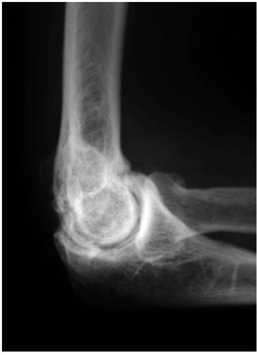
- “Calcification at the lateral epicondyle on radiograph is present in a minority of chronic cases and does not change management.
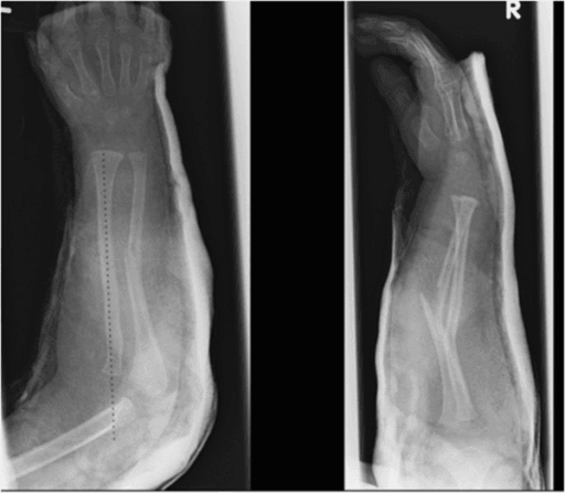
- “In a child with lateral elbow pain and effusion, exclude occult radial neck fracture and Panner disease before labelling overuse.
Up to a fifth of refractory "tennis elbow" has a second diagnosis — radial tunnel, plica, radiocapitellar chondral disease or PLRI. If tenderness is not maximal at the epicondyle, the label is wrong.
Short-term relief, worse outcomes at one year than wait-and-see, and risk of subcutaneous atrophy, depigmentation and attenuation of the lateral collateral ligament complex — a recognised iatrogenic route to PLRI.
C6 radiculopathy refers to the lateral epicondyle and radial forearm. Screen cervical range, Spurling test and brachioradialis reflex in every case that is not textbook.
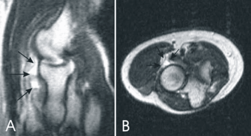
Lateral pain in a 12-year-old pitcher or gymnast with locking is capitellar osteochondritis dissecans. Plain films may be normal early — MRI is required, and continuing to throw risks an unstable fragment and permanent radiocapitellar arthrosis.
Recognising the Pattern
Definition. Pain localised to the lateral column of the elbow — the region bounded by the supracondylar ridge proximally, the radiocapitellar joint medially and the proximal mobile wad distally. It is a regional pattern, not a diagnosis, and at least eight structures within a 5 cm radius can generate it.
Confirming the pattern is genuinely lateral.
- Ask the patient to point with one finger. Diffuse whole-elbow pain is not a lateral pattern and suggests referred, inflammatory or neuropathic cause.
- Map three landmarks with the elbow flexed 90 degrees: the tip of the lateral epicondyle, the radiocapitellar joint line (palpable 1–2 cm distal, rotate the forearm to feel the radial head turn), and the mobile wad 3–5 cm distal over the supinator arcade.
- Reproduce it with load: resisted wrist extension with the elbow extended (Cozen), passive wrist flexion with forearm pronated (Mill), resisted middle finger extension (Maudsley).
Saying it out loud in a viva. "This is a pattern of lateral elbow pain. My differential is anatomical: tendon — ECRB tendinopathy; nerve — radial tunnel or posterior interosseous compression, and referred C6 radiculopathy; joint — radiocapitellar chondral injury, osteochondritis dissecans, arthritis or synovial plica; ligament — posterolateral rotatory instability from lateral ulnar collateral ligament insufficiency; and bone — occult radial head or neck fracture, and rarely tumour or infection. I would localise the point of maximal tenderness, test stability, and screen the cervical spine before committing to a diagnosis."
Mimics and false positives.
- Referred C6 radiculopathy — tenderness is poorly localised, symptoms extend to the thumb, night pain, neck-dependent.
- Cervicothoracic myofascial referral from trapezius or infraspinatus trigger points.
- Distal biceps partial tear — pain feels anterolateral but tenderness is anterior in the cubital fossa and hook test is abnormal.
- Referred wrist pathology — de Quervain or intersection syndrome patients sometimes indicate the proximal forearm.
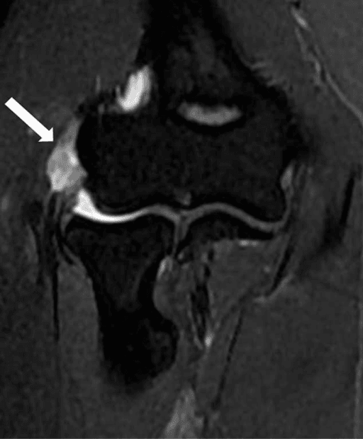
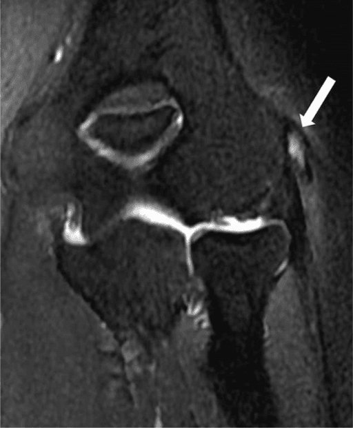
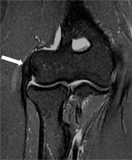
- Radiographic mimic: an accessory ossicle or the normal capitellar pseudodefect on MRI (posterolateral capitellum) misread as osteochondral lesion — the pseudodefect lies posterior to the articulating zone.
Next Investigation
The Differential
- Typical age / setting
- Any age; prior elbow dislocation, radial head fracture, or previous lateral release / repeated steroid
- Discriminating feature
- Apprehension and clicking pushing up out of a chair with forearm supinated; painful clunk in mid-flexion, NOT point tenderness at the epicondyle
- What confirms it
- Lateral pivot-shift test under anaesthesia; chair push-up and table-top relocation tests; MRI showing lateral ulnar collateral ligament attenuation
- Typical age / setting
- Any age; immunosuppressed, IVDU, recent injection or wound
- Discriminating feature
- Pain at rest with global loss of rotation and a hot swollen joint — the elbow is held at 70–80 degrees and resists ALL movement, not just resisted extension
- What confirms it
- Aspiration for cell count, Gram stain and culture; raised CRP; urgent washout
- Typical age / setting
- Osteosarcoma 10–20; metastasis or myeloma greater than 50
- Discriminating feature
- Night pain unrelated to activity, constant and progressive, with local swelling; pain does not respect resisted testing
- What confirms it
- Radiographs showing aggressive periosteal reaction or lysis; MRI whole bone plus staging and biopsy at a tumour unit
- Typical age / setting
- Adults after fall on outstretched hand; children with radial neck buckle
- Discriminating feature
- Acute onset with a defined injury, effusion and painful pronosupination — resisted wrist extension is comparatively comfortable
- What confirms it
- Anterior and posterior fat pad signs on true lateral; radiocapitellar view; CT or repeat films at 10–14 days
- Typical age / setting
- 35–55; manual workers, racquet sports, plumbers, keyboard use
- Discriminating feature
- Point tenderness within 1 cm of the epicondyle at the ECRB origin, pain on gripping with elbow extended and relief with elbow flexed
- What confirms it
- Clinical — Cozen and Mill tests positive; ultrasound shows hypoechoic thickened ECRB with neovascularity if imaging needed
- Typical age / setting
- 30–50; repetitive forceful pronation-supination
- Discriminating feature
- Maximal tenderness 3–5 cm DISTAL to the epicondyle over supinator; aching night pain; NO weakness; pain on resisted supination with elbow extended
- What confirms it
- Diagnostic local anaesthetic block into the radial tunnel abolishing pain; electrodiagnostics usually normal — a normal EMG does not exclude it
- Typical age / setting
- 40–60; degenerative cervical spondylosis
- Discriminating feature
- Pain extends proximally to neck and distally to thumb; reproduced by neck extension and ipsilateral rotation, not by resisted wrist extension
- What confirms it
- Positive Spurling test, reduced brachioradialis reflex; cervical MRI showing C5/6 foraminal stenosis
- Typical age / setting
- Greater than 50; prior radial head fracture, heavy manual work, or post-traumatic
- Discriminating feature
- Pain and crepitus specifically on pronosupination with the elbow extended; terminal extension block; tenderness at the joint line, not the epicondyle
- What confirms it
- AP and lateral radiographs showing radiocapitellar joint space loss and osteophytes; radiocapitellar oblique view
- Typical age / setting
- 11–15; overhead throwers, gymnasts (dominant arm)
- Discriminating feature
- Lateral pain with catching, locking and a 15–20 degree extension block in an adolescent athlete — mechanical symptoms are the giveaway
- What confirms it
- AP in 45 degrees flexion showing capitellar lucency; MRI grades stability and cartilage integrity
- Typical age / setting
- 5–10; younger than OCD, often non-athletic
- Discriminating feature
- Diffuse lateral ache without locking; self-limiting; the whole capitellar ossific nucleus fragments and later reconstitutes
- What confirms it
- Radiograph showing fragmented sclerotic capitellar epiphysis with normal contour restoration on follow-up films
- Typical age / setting
- 20–40; throwers and racquet players
- Discriminating feature
- Reproducible painful snap in the posterolateral gutter at 90–110 degrees of flexion during pronation; tenderness posterior to the radiocapitellar joint
- What confirms it
- MR arthrogram showing a thickened posterolateral plica greater than 3 mm; arthroscopic excision is diagnostic and therapeutic
- Typical age / setting
- Any; mass lesion, lipoma, rheumatoid synovitis, penetrating injury
- Discriminating feature
- MOTOR loss — finger and thumb extension weakness with preserved radially deviated wrist extension and normal sensation
- What confirms it
- EMG showing PIN denervation; MRI or ultrasound to find a compressive mass
- Typical age / setting
- Adults, repetitive rotation
- Discriminating feature
- Palpable snap of a thickened annular ligament over the radial head in rotation, with focal swelling but no extensor origin tenderness
- What confirms it
- Dynamic ultrasound during pronosupination
- Typical age / setting
- 30–60; polyarticular, morning stiffness greater than 30 minutes
- Discriminating feature
- Boggy synovial swelling in the lateral gutter with symmetrical small joint involvement — the elbow is one of several joints
- What confirms it
- Rheumatoid factor / anti-CCP, raised CRP, ultrasound synovitis with power Doppler
Narrowing It Down
- 1Step 1 — Is this acute trauma or insidious overuse?
Take the onset history first, and radiograph any defined injury before examining further.
A fall or dislocation mandates radiographs: fat pad signs, radial head or neck fracture, coronoid fracture. Insidious onset over weeks to months moves you to tendon, nerve and joint causes. Constant progressive night pain with no mechanical pattern buys a tumour and infection workup regardless of history.
- 2Step 2 — How old is the patient?
Let age set the differential before the examination refines it.
Age 5 to 10 with lateral ache: Panner disease. Age 11 to 15 thrower or gymnast: capitellar OCD. Age 35 to 55 manual or racquet: ECRB tendinopathy and radial tunnel dominate. Over 55 with rotation pain and stiffness: radiocapitellar arthritis. Age extremes with rest pain: neoplasm.
- 3Step 3 — Where exactly is the point of maximal tenderness?
Localise with one finger and measure the distance from the epicondyle, rather than accepting 'lateral elbow'.
At or within 1 cm anterodistal to the epicondyle is ECRB tendinopathy. Three to five centimetres distal in the mobile wad is radial tunnel. Over the radiocapitellar joint line is chondral, arthritic or plica pathology. Posterior to the joint line with a snap is posterolateral plica. Poorly localised is referred cervical.
- 4Step 4 — Are there mechanical symptoms?
Ask specifically about locking, catching, a fixed block to extension, and about a clunk on pushing up out of a chair.
Locking, catching or an extension block shifts you intra-articular: OCD, loose body, plica, arthritis. A painful clunk on chair push-up or apprehension in supination shifts you to posterolateral rotatory instability. Pure activity-related ache with no mechanical event stays extra-articular.
- 5Step 5 — Is there any neurological deficit?
Test finger extension separately from wrist extension, and check the brachioradialis reflex.
Weak finger extension with preserved wrist extension is PIN palsy - image for a mass. Sensory change in thumb and index with a reduced brachioradialis reflex is C6 radiculopathy. Pain with entirely normal neurology in the mobile wad is radial tunnel, which is a pain syndrome and not a palsy.
- 6Step 6 — Test the ligament before you accept a tendon diagnosis
Perform the chair push-up test, the table-top relocation test and the lateral pivot shift, and ask directly about previous steroid injections and previous lateral surgery.
Undiagnosed posterolateral rotatory instability is the classic reason a tennis elbow release fails and leaves the patient worse. Both repeated steroid and open lateral release compromise the lateral ulnar collateral ligament, so the history of prior treatment is itself a risk factor for the diagnosis you are looking for.
- 7Step 7 — Are there systemic features?
Screen for fever, rest pain, morning stiffness, other joints, weight loss and prior malignancy.
Fever, rest pain and global movement loss are sepsis until aspirated. Morning stiffness, multiple joints and a family history are inflammatory arthropathy. Weight loss, prior malignancy or bone pain elsewhere are metastatic disease and warrant staging.
MCQ Practice Points
Q: Which structure is the primary site of pathology in lateral epicondylitis?
A: The origin of extensor carpi radialis brevis on the anterior aspect of the lateral epicondyle. Nirschl and Pettrone found the lesion consistently at operation in 88 elbows and described it as immature fibroblastic and vascular infiltration — angiofibroblastic change, with no acute inflammatory cells. Extensor digitorum communis is involved in roughly a third of cases. The "-itis" in the name is a misnomer, and it is the reason an anti-inflammatory injection was always a mechanistic mismatch.
Q: Which single clinical feature best distinguishes radial tunnel syndrome from lateral epicondylitis?
A: The location of maximal tenderness — 3 to 5 cm distal to the lateral epicondyle over the supinator, rather than at the epicondyle itself. Night pain and failure of an epicondylar injection support it; a diagnostic radial tunnel anaesthetic block that abolishes the pain, when an epicondylar injection does not, confirms it.
Q: What is the primary restraint lost in posterolateral rotatory instability?
A: The lateral ulnar collateral ligament, running from the lateral epicondyle to the supinator crest of the ulna. Loss allows radius and ulna to rotate externally as a unit away from the humerus, producing the pivot shift and the chair push-up apprehension sign. Both repeated steroid injection and open lateral release can compromise it — which is why the failed tennis elbow release is the classic presentation.
Q: How does Panner disease differ from capitellar osteochondritis dissecans?
A: Age, extent and outcome. Panner disease occurs at 5 to 10 years, involves the entire capitellar ossific nucleus, produces no loose bodies or locking, and reconstitutes spontaneously. OCD occurs at 11 to 15 years in throwers and gymnasts, is a focal anterolateral lesion, may fragment into loose bodies and can lead to radiocapitellar arthrosis.
Q: What is the evidence-based effect of corticosteroid injection in lateral epicondylitis?
A: Better at six weeks and WORSE at a year — and worse than placebo, not merely worse than the alternatives. Smidt's three-arm trial: 92 per cent success at 6 weeks falling to 69 per cent at 52 weeks, against 83 per cent for wait-and-see and 91 per cent for physiotherapy. Bisset: 47 of 65 injection successes subsequently regressed. Coombes, the only placebo-controlled, injection-blinded trial: recovery at 1 year 83 per cent with steroid versus 96 per cent with placebo injection, and recurrence 54 versus 12 per cent — and adding a steroid abolished the early benefit of physiotherapy. Local risks include skin atrophy, depigmentation and attenuation of the lateral collateral ligament complex.
Exam Viva Scenarios
Practise clinical reasoning and management decisions out loud
“You are shown radiographs of a 46-year-old plumber, 14 months after open lateral epicondylar debridement and three prior corticosteroid injections. Films look unremarkable. He reports ongoing lateral pain and now describes his elbow 'giving way' when he pushes himself up out of a chair.”
“You are shown an AP radiograph in 45 degrees of flexion of a 13-year-old competitive gymnast with six months of lateral elbow pain, intermittent locking and a 20 degree loss of terminal extension. There is a lucency in the capitellum with surrounding sclerosis.”
“You are shown normal radiographs of a 38-year-old assembly line worker with nine months of lateral elbow ache, worse at night, unresponsive to two epicondylar steroid injections. Grip is strong. Tenderness is maximal about 4 cm distal to the lateral epicondyle.”
Localise by centimetres
- At the epicondyle (within 1 cm): ECRB tendinopathy
- 3–5 cm distal, mobile wad: radial tunnel syndrome
- Radiocapitellar joint line: arthritis, OCD, loose body
- Posterolateral gutter with a snap: synovial plica
- Poorly localised, neck-dependent: C6 radiculopathy
Red flags
- Rest and night pain unrelated to activity — tumour
- Hot swollen elbow with global movement loss — sepsis
- Giving way on chair push-up — PLRI
- Finger drop with preserved wrist extension — PIN palsy
- Locking with extension block in an adolescent — unstable OCD
Tests worth knowing
- Cozen: resisted wrist extension, elbow extended
- Mill: passive wrist flexion, forearm pronated
- Maudsley: resisted middle finger extension — positive in BOTH epicondylitis and radial tunnel
- Chair push-up and table-top relocation: PLRI
- Lateral pivot shift: PLRI, best under anaesthesia
- Spurling: cervical radiculopathy
Imaging strategy
- Typical tendinopathy under 6 weeks — no imaging
- Radiographs for trauma, mechanical symptoms, restricted rotation, age under 16 or over 55
- Ultrasound for tendon tear, dynamic snapping, guided injection
- MRI for suspected PLRI, OCD staging, mass or 6 months of failure
- Local anaesthetic block to confirm radial tunnel
Management anchors
- Eccentric loading programme is first-line for tendinopathy; most settle within 12 months
- Counterforce bracing and activity modification for load reduction
- Avoid repeated corticosteroid — worse 12-month outcomes and LUCL risk
- Surgery for tendinopathy only after 6–12 months of genuine rehabilitation and after excluding PLRI, radial tunnel and intra-articular causes
- PLRI is treated by LUCL reconstruction, not debridement
Evidence Base
Effect of Corticosteroid Injection, Physiotherapy, or Both on Clinical Outcomes in Patients with Unilateral Lateral Epicondylalgia - A Randomized Controlled Trial
- 165 patients, 2x2 factorial, injection-BLINDED and PLACEBO-controlled - corticosteroid, placebo injection, and each with or without multimodal physiotherapy; one-year follow-up
- At 1 year, corticosteroid gave WORSE complete recovery or much improvement than placebo injection: 83 versus 96 per cent (RR 0.86, 99 per cent CI 0.75-0.99, p = 0.01)
- One-year RECURRENCE was 54 per cent after corticosteroid against 12 per cent after placebo (RR 0.23, 99 per cent CI 0.10-0.51, p less than 0.001)
- The gap was already open at 26 weeks: 55 versus 85 per cent recovered (p less than 0.001)
- Physiotherapy made NO difference at 1 year - 91 versus 88 per cent recovered (p = 0.56); its only benefit was at 4 weeks, and only in patients who had NOT had a steroid (39 versus 10 per cent, p = 0.004)
- Adding physiotherapy to corticosteroid changed nothing at 4 weeks (68 versus 71 per cent) - the steroid abolished the physiotherapy benefit
Corticosteroid Injections, Physiotherapy, or a Wait-and-See Policy for Lateral Epicondylitis - A Randomised Controlled Trial
- 185 primary-care patients with at least six weeks of symptoms randomised to six weeks of corticosteroid injection, physiotherapy, or an explicit WAIT-AND-SEE policy
- At 6 weeks injection was better than everything: success 92 per cent, against 47 per cent for physiotherapy and 32 per cent for wait-and-see
- At 52 weeks the ranking had INVERTED: 69 per cent for injection, 91 per cent for physiotherapy, 83 per cent for wait-and-see
- Recurrence in the injection group was high; long-term differences significantly favoured physiotherapy
- Physiotherapy beat wait-and-see at a year but NOT significantly - the authors note the relative gain of physiotherapy is small
Mobilisation with Movement and Exercise, Corticosteroid Injection, or Wait and See for Tennis Elbow - Randomised Trial
- 198 participants aged 18 to 65 with at least six weeks of tennis elbow, randomised to eight physiotherapy sessions, corticosteroid injection, or wait and see; 52-week follow-up
- Corticosteroid was significantly better at 6 weeks - and then 47 OF 65 SUCCESSES SUBSEQUENTLY REGRESSED
- Long-term outcomes were significantly POORER after injection than after physiotherapy
- Physiotherapy beat wait and see in the short term; by 52 weeks there was no difference, with most participants in both groups reporting success
- Patients who had physiotherapy sought less additional treatment, including fewer NSAIDs, than either other group
- The authors' own phrasing: the short-term benefits of injection are 'paradoxically reversed' after six weeks