Pure Motor, Pure Trouble
- The radial nerve divides into superficial (sensory) and deep (motor) branches at the level of the radiocapitellar joint, between brachialis and brachioradialis.
- The deep branch becomes the posterior interosseous nerve as it passes beneath the arcade of Frohse into the supinator.
- It carries NO cutaneous sensory fibres - only articular branches ending in a pseudoganglion on the dorsal wrist capsule.
- PIN palsy gives finger drop with preserved wrist extension in radial deviation, because extensor carpi radialis longus is spared and extensor carpi ulnaris is not.
- Pronating the forearm carries the nerve away from the lateral radius and lengthens the safe zone from roughly 3.3 cm to 5.2 cm.
- “Sensation is normal in PIN palsy - if the first dorsal web is numb, the lesion is proximal to the division.
- “The tenodesis test separates PIN palsy from extensor tendon rupture in rheumatoid disease.
- “The arcade of Frohse is tendinous in about 30 per cent of adults and is never tendinous in the fetus - it is acquired.
- “Radial tunnel syndrome is a pain syndrome with no reliable electrodiagnostic signature; PIN syndrome is a motor palsy.
PIN palsy or ruptured tendons?
- Both give loss of active metacarpophalangeal extension.
- Test: Tenodesis. Passively flex the wrist. If the fingers extend, the tendons are intact and the problem is the nerve.
- Also test: Passively extend the metacarpophalangeal joints and ask the patient to hold - a sagittal band rupture holds, a torn tendon does not.
Radial tunnel or tennis elbow?
- Tennis elbow is tender AT the lateral epicondyle.
- Radial tunnel is tender 3 to 5 cm DISTAL to it, over the mobile wad and supinator.
- The two coexist frequently - releasing only one leaves a dissatisfied patient.
- Location
- Between brachialis and brachioradialis
- Supplies
- Brachioradialis, ECRL, often ECRB
- What Puts It At Risk
- Distal humeral fracture, plating
- Location
- Radial tunnel, anterior to radial head
- Supplies
- Supinator, variably ECRB
- What Puts It At Risk
- Radial head fracture, leash of Henry, ECRB edge
- Location
- Between the two heads of supinator
- Supplies
- Nothing (transit segment)
- What Puts It At Risk
- Arcade of Frohse, supinator synovitis
- Location
- On abductor pollicis longus, deep extensor plane
- Supplies
- ECU, EDC, EDM, APL, EPB, EPL, EI
- What Puts It At Risk
- Proximal radius plating, distal supinator edge
- Location
- Floor of fourth dorsal compartment
- Supplies
- Wrist capsule (articular only)
- What Puts It At Risk
- Harvested deliberately for denervation or graft
FREASThe Five Sites of PIN Compression
Hook:FREAS runs proximal to distal down the radial tunnel - release all five or the patient comes back.
Superficial then DeepWhat the PIN Supplies After the Supinator
Hook:The superficial group (ECU, EDC, EDM) leaves first; the deep group (APL, EPB, EPL, EI) is reached by a longer descending branch.
WAD OUTSIDE, JOINT INSIDERadial Tunnel Boundaries
Hook:A 5 cm corridor with a joint for a floor - synovitis in that joint compresses the nerve.
Overview
The posterior interosseous nerve (PIN) is the terminal motor continuation of the radial nerve in the forearm. Everything that makes it examinable follows from two facts: it is almost purely motor, and it crosses the proximal radius at the exact level surgeons need to expose for radial head and proximal shaft work.
The radial nerve (C5 to T1) enters the anterior compartment of the arm through the lateral intermuscular septum and lies in the groove between brachialis medially and brachioradialis laterally. At the level of the radiocapitellar joint it divides into a superficial branch (sensory, running under brachioradialis) and a deep branch (motor). The deep branch is renamed the posterior interosseous nerve once it passes under the arcade of Frohse into the substance of supinator. Its dominant root contribution is C7 with C8, with a variable C6 component.
Because it carries no cutaneous fibres, a complete PIN lesion produces a dramatic motor picture with an entirely normal sensory examination. Candidates who test only sensation to screen for radial nerve injury will miss it completely.
The wrist has three prime extensors. Extensor carpi radialis longus (ECRL) is supplied by the radial nerve proper, before the division, and is therefore spared. Extensor carpi radialis brevis (ECRB) has variable innervation - from the radial nerve proper, from the superficial branch, or from the PIN. Extensor carpi ulnaris (ECU) is always PIN.
So in a complete PIN palsy the patient still extends the wrist, but the unopposed ECRL pulls it into radial deviation. That radially deviated wrist extension with finger drop is pathognomonic, and it is the single most commonly examined sign on this nerve. A true wrist drop means the lesion is proximal to the division - a radial nerve lesion in the spiral groove, not a PIN lesion.
The nerve also matters in reverse: it is a useful structure to sacrifice. Its terminal articular branch is routinely divided in dorsal wrist denervation for painful degenerative wrists, and the terminal segment in the floor of the fourth dorsal compartment is a recognised short interpositional graft for digital nerve gaps.
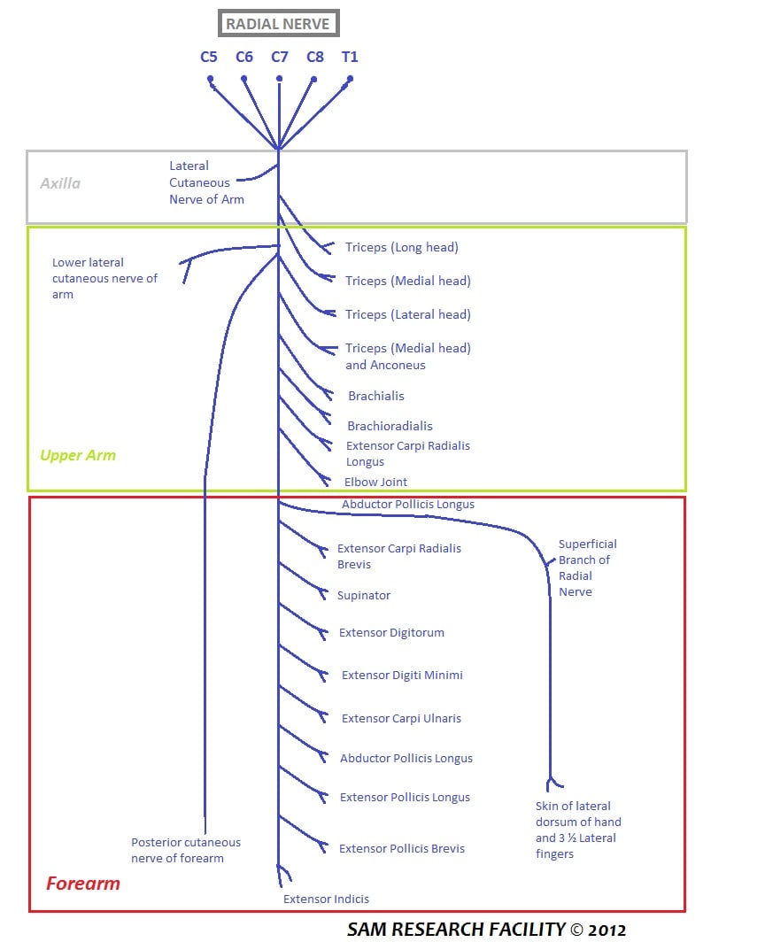
Course, Motor Supply and Relations
Division of the radial nerve
- The radial nerve divides at the level of the radiocapitellar joint, roughly at the lateral epicondyle, deep in the interval between brachialis and brachioradialis.
- The superficial branch continues distally on the deep surface of brachioradialis and remains sensory.
- The deep branch turns laterally and posteriorly around the radial neck to reach the supinator.
The radial tunnel
- A fibro-osseous and muscular corridor roughly 5 cm long, running from the level of the radiocapitellar joint to the distal edge of the supinator.
- Lateral wall and roof: brachioradialis, extensor carpi radialis longus, extensor carpi radialis brevis (the mobile wad).
- Medial wall: brachialis proximally, biceps tendon distally.
- Floor: the anterior capsule of the radiocapitellar joint, then the deep head of supinator.
- Radiocapitellar synovitis, a radial head fracture haematoma, or a proximal radioulnar joint ganglion all raise pressure inside this corridor.
Through the supinator
- The nerve enters between the superficial and deep heads of supinator, passing under the arcade of Frohse - the arched proximal border of the superficial head.
- It winds around the radial neck from anterior to posterior within the muscle, crossing the shaft obliquely.
- It emerges from the distal border of supinator onto the deep extensor plane, typically 4 to 8 cm distal to the radiocapitellar joint depending on forearm rotation.
Terminal division
- Superficial (recurrent) descending branches: to extensor carpi ulnaris, extensor digitorum communis and extensor digiti minimi.
- Deep descending branch: runs on the interosseous membrane, on the surface of abductor pollicis longus, supplying abductor pollicis longus, extensor pollicis brevis, extensor pollicis longus and extensor indicis.
- The terminal fibres run in the floor of the fourth dorsal compartment, expand into a pseudoganglion, and end as articular branches to the wrist.
The parent trunk is developed in radial nerve anatomy, and the injuries that most often threaten this segment in radial head fractures and Monteggia fractures.
Surface Anatomy and Examination
Locating the nerve on the surface
- Mark the lateral epicondyle and the radial head (palpable, and confirmed by rotating the forearm under your thumb).
- The radial tunnel lies in the interval just anterior to the radial head, deep to the mobile wad.
- The maximal tenderness point in radial tunnel syndrome is over the supinator, 3 to 5 cm distal to the lateral epicondyle - distinctly distal to the tender point of lateral epicondylitis.
Motor examination sequence
- Wrist extension: present, but observe the direction. Radial deviation implies loss of extensor carpi ulnaris.
- Finger metacarpophalangeal extension: the primary deficit. Test with the interphalangeal joints flexed to remove the intrinsic contribution.
- Thumb retropulsion: palm flat on the table, lift the thumb. Tests extensor pollicis longus.
- Thumb radial abduction: abductor pollicis longus.
- Supination with the elbow extended: removes the biceps contribution and isolates supinator, but is often preserved because the supinator branches leave above the arcade.
- Sensation everywhere, including the first dorsal web space.
- Triceps power and reflex.
- Brachioradialis contraction on resisted elbow flexion in mid-pronation - visible and palpable.
- Wrist extension power (weak but present).
- Any abnormality here relocates the lesion proximally, to the radial nerve or the C7 root.
The tenodesis test
The examination that decides the operation in a rheumatoid hand.
- Method: support the forearm, passively flex the wrist, and observe the fingers.
- Tendons intact (nerve lesion): the metacarpophalangeal joints extend passively through the tenodesis effect.
- Tendons ruptured: the affected digits stay flexed while the intact ones extend.
- Caveat: long-standing PIN palsy with fixed metacarpophalangeal contracture blunts the response, and a bulky dorsal tenosynovitis can restrict excursion. Assess passive metacarpophalangeal extension first.
Named tests around the radial tunnel
- How to Perform
- Elbow extended, resist extension of the middle finger metacarpophalangeal joint
- Positive Finding
- Pain over the supinator, 3-5 cm distal to the epicondyle
- False Positives
- Also positive in lateral epicondylitis - judge the SITE of pain, not its presence
- How to Perform
- Elbow extended, forearm pronated, resist supination
- Positive Finding
- Pain in the proximal extensor mass
- False Positives
- Low specificity; painful in any lateral elbow tendinopathy
- How to Perform
- Palpate over supinator with the forearm rotated
- Positive Finding
- Point tenderness distal to the epicondyle
- False Positives
- Deep pressure is uncomfortable in normal people - always compare sides
- How to Perform
- Small volume placed accurately in the radial tunnel
- Positive Finding
- Relief of pain, often with transient finger drop confirming the nerve was reached
- False Positives
- A large volume spreads to the epicondyle and gives a false positive
- How to Perform
- Passively flex the wrist and watch the fingers
- Positive Finding
- Fingers extend - extensor tendons in continuity, so the lesion is neurological
- False Positives
- Fixed metacarpophalangeal contracture or bulky tenosynovitis blunts the response
- How to Perform
- Palpate a nine-square grid over the proximal forearm
- Positive Finding
- Tenderness confined to the proximal lateral squares
- False Positives
- Descriptive mapping only - never validated as diagnostic
- In PIN syndrome (motor palsy), needle electromyography shows denervation confined to PIN-supplied muscles with normal brachioradialis and extensor carpi radialis longus - that sparing pattern is the localising evidence.
- In radial tunnel syndrome (pain only), nerve conduction studies and electromyography are typically normal. A normal study does not exclude it, and an abnormal study should prompt a search for a different diagnosis. This is why radial tunnel syndrome remains a clinical diagnosis and why its surgical results are the least predictable of the compression neuropathies.
Complications
- Mechanism
- Hohmann levered around the radial neck
- How to Avoid It
- Blunt retractors, subperiosteal, forearm pronated
- What to Do
- Usually neuropraxia - observe with electromyography at 3-4 weeks
- Mechanism
- Exposure carried beyond the safe zone blind
- How to Avoid It
- Identify the nerve before extending past 3 cm from the radiocapitellar joint
- What to Do
- Immediate exploration and repair
- Mechanism
- Distal supinator edge or ECRB edge left unreleased
- How to Avoid It
- Release all five FREAS sites every time
- What to Do
- Revision release under direct vision
- Mechanism
- Fibrosis after direct decompression - around 17 per cent
- How to Avoid It
- Meticulous haemostasis, early mobilisation
- What to Do
- Consider indirect supinator-origin release at revision
- Mechanism
- Coexisting lateral epicondylitis not addressed
- How to Avoid It
- Examine and treat both if both are present
- What to Do
- Address the epicondyle; counsel on realistic outcome
- Mechanism
- Chronic radial head dislocation not recognised
- How to Avoid It
- Check the radiocapitellar line on every elbow radiograph
- What to Do
- Correct the deformity as well as decompressing
- Mechanism
- Two-incision biceps repair, or extensive dissection off the interosseous membrane
- How to Avoid It
- Avoid stripping the ulna, limit muscle splitting
- What to Do
- Excision with interposition once mature
Denervation consequences if recovery fails
- Finger drop is functionally disabling in a way that pure wrist drop is not - the hand cannot open to grasp.
- Reliable reconstruction exists: pronator teres to extensor carpi radialis brevis (wrist extension), flexor carpi radialis or flexor carpi ulnaris to extensor digitorum communis (finger extension), and palmaris longus to extensor pollicis longus (thumb extension) - the standard radial nerve palsy transfer set. Because ECRL and brachioradialis are spared in a PIN lesion, wrist extension usually does not need reconstruction and only the finger and thumb transfers are required.
- Time the transfer for the point at which spontaneous recovery is no longer plausible, applying the roughly 1 mm per day regeneration principle from the level of the lesion, and remembering that motor end plates degenerate irreversibly after roughly 12 to 18 months.
Clinical Relevance
The clinical picture
- Finger drop: loss of active metacarpophalangeal extension of all digits, often asymmetric between digits.
- Thumb: loss of extension and retropulsion.
- Wrist: extends, but in radial deviation.
- Sensation: entirely normal.
- Pain: often a prodrome of deep aching in the proximal forearm for weeks before the weakness.
Causes
- Compressive: any of the five FREAS sites, most commonly the arcade of Frohse.
- Space-occupying: lipoma, ganglion of the proximal radioulnar joint, synovial cyst, nerve sheath tumour. A mass is found in a meaningful minority of spontaneous palsies, which is why imaging is worthwhile before labelling a palsy idiopathic.
- Inflammatory: rheumatoid synovitis of the radiocapitellar joint compressing the nerve in the tunnel - the reason PIN palsy and extensor tendon rupture occur in the same patient population.
- Traumatic: radial head and neck fracture, Monteggia injury, proximal radius fracture.
- Iatrogenic: proximal radius plating, radial head arthroplasty, bicipital tuberosity exposure, elbow arthroscopy portals.
- Neuralgic amyotrophy: the hourglass fascicular constriction described in neuralgic amyotrophy causes a painful, spontaneous, often partial palsy in an otherwise healthy person. Severe pain preceding the weakness is the clue.
Imaging
- Ultrasound: first line. Shows nerve calibre change at the arcade, a mass lesion, or an hourglass constriction, and allows dynamic assessment during pronation and supination.
- MRI: denervation oedema in supinator and the extensor group in the acute phase; fatty atrophy when chronic. Isolated supinator oedema without extensor changes is a recognised incidental finding and should not by itself be reported as PIN syndrome.
- Radiographs: always look at the radial head and the radiocapitellar relationship. A missed Monteggia is a diagnosis made on the plain film.
Surgical Relevance
Pronate the forearm. Pronation rolls the posterior interosseous nerve medially and distally, away from the lateral surface of the radius, and flattens the angle at which it crosses the shaft. Diliberti and colleagues measured this directly in 32 cadaveric specimens through a posterolateral approach: pronation gave an average safe zone of 52.0 plus or minus 7.8 mm distal to the radiocapitellar joint, and allowed safe exposure of at least the proximal 38 mm in every specimen. Supination shortened this to an average of 33.4 plus or minus 5.7 mm, and to as little as 22 mm in the worst specimen. The nerve-to-shaft angle fell from 47.4 degrees in supination to 27.8 degrees in pronation.
Posterolateral (Thompson) approach to the proximal radius
- Superficial interval: extensor carpi radialis brevis and extensor digitorum communis proximally; abductor pollicis longus and extensor carpi radialis brevis distally.
- Deep: the supinator is encountered with the PIN inside it. The nerve must be identified if the exposure extends beyond the safe zone.
- Technique: pronate fully, identify the distal border of supinator, trace the nerve proximally under direct vision, and elevate the supinator subperiosteally from the radius with the nerve protected within the muscle.
Extensor digitorum communis splitting approach
- A direct lateral approach that gives better proximal radius exposure than the standard posterolateral interval.
- Schimizzi and colleagues measured the nerve in 15 cadaveric arms: mean radiocapitellar-joint-to-nerve distance 48.2 plus or minus 7.9 mm in pronation, 44.5 mm in neutral, 40.8 mm in supination. Mean lateral-epicondyle-to-nerve distance was 64.7 mm in pronation.
- Crucially, the shortest distances observed were 29 mm from the radiocapitellar joint and 42 mm from the lateral epicondyle in pronation. Those minima, not the means, define the safe zone.
- The nerve lay a mean 10.2 plus or minus 2.4 mm deep to the most superficial aspect of extensor digitorum communis - shallow enough that an aggressive muscle split reaches it.
- Put the two studies side by side and the trade-off appears. Diliberti's posterolateral approach was safe for at least 38 mm in every one of 32 specimens in pronation. Schimizzi's EDC-splitting approach came within 29 mm of the joint in its closest specimen. So the approach that gives the better view of the proximal radius is also the one whose worst case sits 9 mm nearer the nerve, and its nerve lies only about a centimetre below the muscle surface. Exposure and safety move in opposite directions here; choose the split when you need the view, and identify the nerve rather than trusting a number.
Kocher approach (anconeus and extensor carpi ulnaris)
- Used for radial head fracture and arthroplasty. It is the posterior-most of the lateral intervals and therefore the furthest from the nerve at the level of the radial head.
- The trade-off is the lateral ulnar collateral ligament: keep the capsular incision anterior to the midaxial line of the radial head to avoid creating posterolateral rotatory instability.
Anterior (Henry) approach
- The PIN is protected by full supination, which brings the supinator insertion onto the anterolateral radius into view and rotates the nerve posteriorly, away from the incision.
- Elevate the supinator strictly subperiosteally from its radial insertion, staying on bone, with the nerve carried away inside the muscle. Never dissect into the substance of the supinator from the front.
- Note the deliberate opposite of the posterolateral rule: the anterior approach uses supination, the posterolateral approach uses pronation. The principle is identical - rotate the nerve away from the side you are working on.
A posterior interosseous nerve palsy that is present before operation, in a closed injury, may be observed. A palsy that is new after operation, or that appears after manipulation of a Monteggia lesion, implies transection, suture entrapment, retractor crush or entrapment in the fracture. It should be explored early - typically within days - not observed for three months. The default assumption for an iatrogenic palsy is that something reversible is compressing the nerve and can be undone.
Guidelines, Registries & Global Practice
Anatomical variation across populations
- The arcade of Frohse is membranous in fetal specimens and becomes a distinct tendinous arch in around 30 per cent of adults. Reported prevalence varies widely between cadaveric series and populations, from under a fifth to over half, depending on how a tendinous arch is defined. That variability, rather than any single true figure, is the examinable point.
- Innervation of extensor carpi radialis brevis is genuinely inconsistent across studies, arising from the radial nerve proper, the superficial branch or the deep branch in different specimens. No single pattern predominates strongly enough to be used as a localising sign.
- The level at which the nerve emerges from the supinator varies with forearm length and with rotation. Cadaveric measurements referenced to the radiocapitellar joint rather than to the lateral epicondyle are more reproducible, because the epicondyle-to-joint distance itself varies.
- Accessory and bifid patterns of the deep branch are described but uncommon; a surgeon who has identified one branch should not assume there is not a second.
Differences in described technique
- Emphasis
- Proximal radius exposure with the forearm pronated; identify the nerve before extending distally
- Practical Point
- Anterior (Henry) approach uses supination; posterolateral uses pronation - rotate the nerve away from your side
- Emphasis
- Complete five-site release for radial tunnel syndrome, including the distal supinator edge
- Practical Point
- Incomplete release is the leading avoidable cause of failure
- Emphasis
- Indirect decompression by dividing the superficial supinator origin, to reduce perineural scarring
- Practical Point
- An alternative where recompression rates after direct release are a concern
- Emphasis
- Clinical diagnosis with a diagnostic block; non-operative care first and honest consent
- Practical Point
- Manage expectations before offering release in an occupational injury
- Emphasis
- In Monteggia lesions, restore ulnar length and radiocapitellar alignment first
- Practical Point
- Nerve palsy usually resolves without exploration once the radial head is reduced
Registry, service and resource considerations
- There is no dedicated peripheral nerve injury registry equivalent to the arthroplasty registries. Outcome data for posterior interosseous nerve decompression come almost entirely from single-centre retrospective series, which is why the reported success rate for radial tunnel release spans such a wide range.
- High-resource settings have ready access to high-resolution nerve ultrasound and magnetic resonance neurography, which increasingly detect hourglass fascicular constrictions in spontaneous palsies and can change the decision from observation to early exploration.
- Limited-resource settings rely on careful serial clinical examination. The key discriminators here - radially deviated wrist extension, an entirely normal sensory examination, and the tenodesis test - require no equipment at all and are more informative than most investigations.
- Occupational context matters everywhere. Radial tunnel syndrome presenting in the context of a workplace claim has consistently poorer surgical outcomes across health systems, and this should shape both counselling and the threshold for operating.
- Timing principle, universally applied: a closed traumatic palsy is observed with electrodiagnostic reassessment at 3 to 4 weeks and again at 3 months; an iatrogenic or open palsy is explored early; and reconstruction by tendon transfer should not be deferred past the point at which motor end plates are irrecoverable.
MCQ Practice Points
Q: At what level does the radial nerve divide into its superficial and deep branches? A: At the level of the radiocapitellar joint, in the interval between brachialis and brachioradialis, roughly at the lateral epicondyle.
Q: Which wrist extensor is always spared in a posterior interosseous nerve palsy, and why does that matter? A: Extensor carpi radialis longus, because it is supplied by the radial nerve proper above the division. Its preservation, combined with denervated extensor carpi ulnaris, produces wrist extension in radial deviation.
Q: What is the cutaneous sensory territory of the posterior interosseous nerve? A: None. It supplies articular branches only, terminating in a pseudoganglion on the dorsal wrist capsule. Numbness in the first dorsal web space means the lesion is proximal to the radial nerve division.
Q: Which of the five compression sites is most commonly implicated? A: The arcade of Frohse, the arched proximal border of the superficial head of supinator - tendinous in roughly 30 per cent of adults and never tendinous in the fetus.
Q: Which forearm position protects the nerve during a posterolateral approach to the proximal radius? A: Pronation. It increases the average safe zone distal to the radiocapitellar joint from about 3.3 cm in supination to about 5.2 cm, and guaranteed at least 3.8 cm in cadaveric measurement.
Q: What is the leash of Henry? A: The radial recurrent vessels fanning across the deep branch of the radial nerve within the radial tunnel - one of the five recognised compression sites and a source of troublesome bleeding during decompression.
Q: Which single bedside test separates posterior interosseous nerve palsy from extensor tendon rupture? A: The tenodesis test. Passive wrist flexion extends the fingers if the tendons are intact.
Q: Which nerve is most commonly injured in a Monteggia fracture dislocation? A: The posterior interosseous nerve, stretched across the arcade of Frohse by the anteriorly or laterally displaced radial head. Most recover once the radial head is reduced.
Q: What are the length and the floor of the radial tunnel? A: Approximately 5 cm, from the radiocapitellar joint to the distal border of supinator. The floor is the radiocapitellar joint capsule proximally - which is why radiocapitellar synovitis compresses the nerve.
Q: Which tendon transfers are needed for an established, unrecoverable posterior interosseous nerve palsy? A: Finger and thumb extension only - typically flexor carpi radialis or flexor carpi ulnaris to extensor digitorum communis and palmaris longus to extensor pollicis longus. Wrist extension does not usually need reconstruction because extensor carpi radialis longus is intact.
Exam Viva Scenarios
Practise clinical reasoning and management decisions out loud
“A 62-year-old woman with 20 years of seropositive rheumatoid arthritis presents unable to extend the fingers of her right hand at the metacarpophalangeal joints. She can extend her wrist. Sensation is normal. How do you work this out?”
“You are called to review a 45-year-old man on the first postoperative day after a radial head arthroplasty performed through a Kocher approach for a comminuted radial head fracture. He cannot extend his fingers or thumb. Wrist extension is present but radially deviated. Sensation is intact. What is your assessment and plan?”
“A 38-year-old carpenter has had 14 months of lateral elbow and proximal forearm pain. He has had two corticosteroid injections at the lateral epicondyle with brief relief, and a previous lateral epicondylar debridement elsewhere which did not help. Motor examination is normal. Nerve conduction studies and electromyography are normal. He asks you to operate.”
Anatomy High Yield
- Roots: C7, C8 (variable C6)
- Division: at the radiocapitellar joint
- Tunnel: radial tunnel, about 5 cm long
- Entry: arcade of Frohse into supinator
- Ends: pseudoganglion on the dorsal wrist capsule
Five Compression Sites (FREAS)
- Fibrous bands at the radial head
- Radial recurrent vessels - leash of Henry
- ECRB tendinous medial edge
- Arcade of Frohse - commonest
- Supinator distal edge - most often missed
Clinical Signs
- Finger drop at the metacarpophalangeal joints
- Wrist extends but deviates radially (ECU out, ECRL in)
- Thumb retropulsion lost
- Sensation entirely NORMAL
- Tenodesis test separates palsy from tendon rupture
Key Numbers
- 5.2 cm: average safe zone in pronation
- 3.8 cm: minimum safe zone in pronation
- 3.3 cm: average safe zone in supination
- 2.9 cm: shortest measured joint-to-nerve distance in pronation
- 1 cm: depth of the nerve deep to superficial EDC
- 17 per cent: recompression by scar after direct release
Differentials
- High radial nerve palsy (wrist drop plus sensory loss)
- Extensor tendon rupture (Vaughan-Jackson)
- Sagittal band rupture (holds after passive extension)
- C7 radiculopathy (triceps involved)
- Neuralgic amyotrophy (severe pain then weakness)
Evidence Base
Safe Zone of the Proximal Radius in Pronation and Supination
- Posterolateral approach between anconeus and extensor carpi ulnaris performed in 32 cadaveric specimens with direct exposure of the posterior interosseous nerve
- Pronation allowed safe exposure of at least the proximal 38 mm of the lateral radius, with an average safe zone of 52.0 plus or minus 7.8 mm
- Supination reduced the safe zone to an average of 33.4 plus or minus 5.7 mm and to as little as 22 mm; the nerve-to-shaft angle fell from 47.4 degrees in supination to 27.8 degrees in pronation
Safe Zone During the Extensor Digitorum Communis Splitting Approach
- Fifteen cadaveric arms dissected through an extensor digitorum communis splitting approach with the nerve measured in neutral, supination and pronation
- Mean radiocapitellar-joint-to-nerve distances were 44.5, 40.8 and 48.2 mm in neutral, supination and pronation respectively; lateral-epicondyle-to-nerve distances were 61.7, 57.6 and 64.7 mm
- The shortest distances in pronation were 29 mm from the radiocapitellar joint and 42 mm from the lateral epicondyle; the nerve lay a mean 10.2 plus or minus 2.4 mm deep to the superficial surface of extensor digitorum communis
The Arcade of Frohse and Posterior Interosseous Nerve Paralysis
- Anatomical study of the proximal border of the superficial head of supinator in fetal and adult specimens
- The arcade is membranous in the fetus and becomes tendinous in a substantial proportion of adults, indicating that the fibrous arch is acquired rather than congenital
- Established the arcade as the principal anatomical site of posterior interosseous nerve compression
Long-term Results of Posterior Interosseous Nerve Decompression
- 110 patients operated for posterior interosseous nerve compression, with 69 reviewed at a mean of 41 months
- Only 60 per cent achieved an excellent or good result by the Roles and Maudsley criteria; the mean DASH score was 32
- Recompression of the nerve by scarring occurred in 17 per cent, and 52 per cent of patients also had lateral epicondylitis, leading the authors to regard radial tunnel syndrome as a specific form of tennis elbow
- BECAUSE OF THAT SCARRING RATE THE AUTHORS ABANDONED DIRECT DECOMPRESSION and moved to Wilhelm's denervation procedure, which relaxes the arcade of Frohse indirectly by dividing the superficial origin of supinator - their results with it are described only as preliminary and promising
Radial Tunnel Syndrome - Contemporary Review
- Radial tunnel syndrome is a pain syndrome without specific radiological or electrodiagnostic findings
- Treatment should begin non-operatively; surgery requires release of all potential entrapment sites including complete division of the superficial head of supinator
- Outcomes are generally good but are poorer in patients with associated lateral epicondylitis and in those with compensation claims
Tardy Posterior Interosseous Nerve Palsy - Mechanism
- Partial posterior interosseous nerve palsy developed more than two decades after childhood osteomyelitis of the proximal radial metaphysis
- Previous reports of tardy palsy had all involved a dislocated radial head, frequently from an unrecognised Monteggia fracture dislocation, causing compression at the arcade of Frohse
- In this case the radial head was located, and compression at the arcade combined with traction against inflamed periosteum produced the late presentation
Superficial Radial Nerve Anatomy and the Danger of the Anatomic Snuffbox
- The study's actual purpose was K-wire safety: 40 wires were placed into the anatomic snuffbox of 10 cadaveric forearms, then assessed radiologically and by dissection
- A branch of the superficial radial nerve was injured in 22 per cent (2 of 9) and the cephalic vein in 33 per cent (3 of 9); the radial artery was never hit but lay within 1 mm of a wire in 22 per cent
- The nerve emerged between brachioradialis and extensor carpi radialis longus in 9 of 10 specimens, a mean 7.8 cm proximal to the radial styloid
- A mean of 5.1 branches crossed the level of the radial styloid, with the first and second divisions a mean of 4.5 cm and 3.0 cm proximal to it
- The authors concluded that despite relatively consistent proximal anatomy the snuffbox remains dangerous, and that an OPEN approach is preferred to percutaneous wiring