Quadrangular Space, Triangular Space and Triangular Interval
- Teres minor is the muscle that separates the quadrangular space from the triangular space; the long head of triceps is medial to the quadrangular space and lateral to the triangular space.
- The triangular interval lies BELOW teres major - it is the only one of the three that is inferior to that muscle, and it delivers the radial nerve from the axilla into the spiral groove.
- Seen from the front the superior boundary of the quadrangular space is the subscapularis and the inferior capsule, not teres minor - the same space has different superior walls from the two sides.
- The axillary nerve emerges from the quadrangular space at the inferior border of teres minor and runs around the surgical neck 5 to 7 cm distal to the lateral edge of the acromion - the limit of a safe deltoid split.
- Quadrilateral space syndrome is a compressive lesion of the axillary nerve, its posterior branch, or the posterior circumflex humeral artery, classically in an overhead athlete, and the MRI clue is isolated teres minor fatty atrophy.
- “Isolated teres minor atrophy on MRI is more often idiopathic or due to a paralabral cyst at the inferior glenoid than due to true quadrilateral space syndrome - it is a clue, not a diagnosis.
- “The vascular form of quadrilateral space syndrome causes posterior circumflex humeral artery aneurysm or thrombosis with distal digital embolisation in throwers and volleyball players - a cold, mottled hand in an athlete with posterior shoulder pain.
- “The infraspinatus-teres minor interval is a true internervous plane: suprascapular nerve above, axillary nerve below. It is the working interval for posterior glenoid exposure.
- “The radial nerve is found reliably at the inferior border of teres major as it leaves the triangular interval - the fixed landmark for the posterior approach to the humerus.
Overview
The posterior wall of the axilla is crossed by two muscles running laterally from the scapula to the humerus - teres minor above and teres major below - and by the long head of triceps, which descends between them from the infraglenoid tubercle. Where these three cross one another and the humerus, they create three named gaps, and each gap transmits exactly one important neurovascular structure.
That regularity is why examiners love this region and why the surgeon needs it. Every posterior approach to the shoulder, every posterior arthroscopic portal, every latissimus dorsi transfer and every posterior exposure of the humerus is defined by where these three spaces lie and what comes out of them.
Start with a single rectangle bounded by teres minor above, teres major below, the long head of triceps medially and the humerus laterally. That is the quadrangular space, and the axillary nerve with the posterior circumflex humeral artery runs through it.
Now note that the long head of triceps runs obliquely, from the infraglenoid tubercle downward and laterally. It therefore crosses behind teres minor and in front of teres major, and in doing so it splits the region into three:
- Medial to the long head of triceps, between the two teres muscles is the triangular space - three sides only, because there is no humerus involved. It transmits the circumflex scapular artery, which winds around the lateral scapular border into the infraspinous fossa.
- Lateral to the long head of triceps, between the two teres muscles and the humerus is the quadrangular space - four sides, because the humerus makes the fourth. It transmits the axillary nerve and the posterior circumflex humeral artery.
- Below teres major, between the long head of triceps medially and the humerus (with the lateral head of triceps) laterally, is the triangular interval - the only space inferior to teres major. It transmits the radial nerve and the profunda brachii artery on their way from the axilla into the spiral groove.
One further point that catches candidates: the quadrangular space looks different from the front. Approached anteriorly through the axilla, its superior boundary is the subscapularis and the inferior glenohumeral joint capsule, not teres minor. The same aperture, two different roofs, depending on which side of it you stand.
QSA - TSC - TIRContents of the Three Spaces
Hook:Four sides equals axillary nerve. Three sides above teres major equals circumflex scapular artery. Three sides below teres major equals radial nerve.
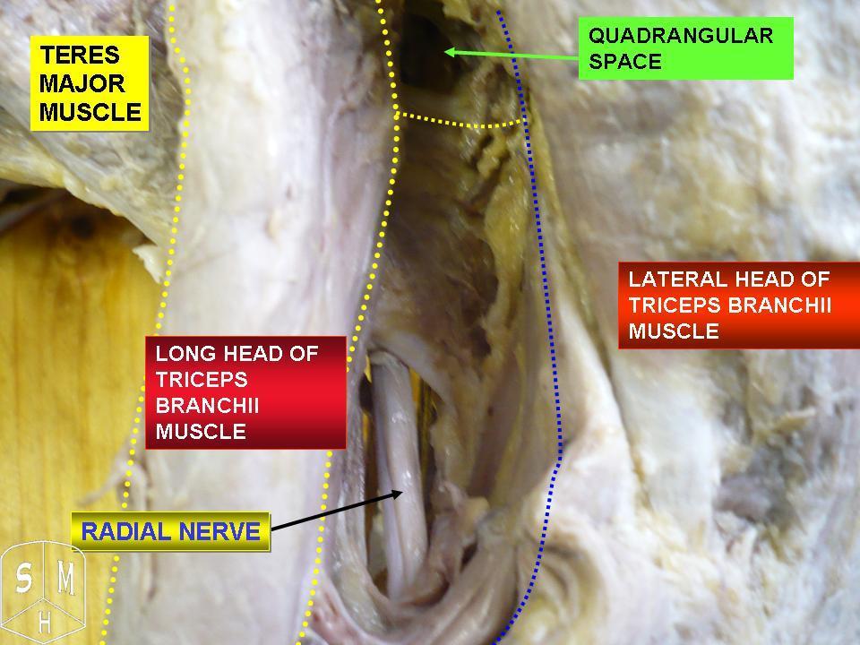
Boundaries, Contents and Relations
Boundaries seen from BEHIND
- Superior: teres minor (and above it the inferior border of the glenohumeral joint capsule).
- Inferior: teres major.
- Medial: long head of triceps brachii.
- Lateral: surgical neck of the humerus.
Boundaries seen from the FRONT (through the axilla)
- Superior: subscapularis and the inferior glenohumeral joint capsule.
- Inferior: teres major.
- Medial: long head of triceps.
- Lateral: surgical neck of the humerus.
The discrepancy is real and is a favourite question. Teres minor and subscapularis both attach around the joint; from behind you see teres minor, from in front you see subscapularis.
Contents
- Axillary nerve (C5, C6) - from the posterior cord. It passes through the space and immediately divides into:
- an anterior (deltoid) branch that winds around the surgical neck deep to the deltoid,
- a posterior branch supplying teres minor, the posterior deltoid, and continuing as the superior lateral cutaneous nerve of the arm over the posterolateral deltoid,
- an articular branch to the inferior glenohumeral joint capsule.
- Posterior circumflex humeral artery and its venae comitantes, arising from the third part of the axillary artery and anastomosing around the surgical neck with the anterior circumflex humeral artery.
Key distances
- The axillary nerve lies approximately 5 to 7 cm distal to the lateral edge of the acromion on the humeral shaft - the classical limit of a lateral deltoid-splitting approach. Treat this as a rule of thumb rather than a guarantee: in a cadaveric series of 102 shoulders the nerve was less than 5 cm from the palpable acromial edge in nearly 20 per cent, was as close as 3.1 cm in one small female specimen, and abduction of the shoulder brought it nearly 30 per cent closer.
- The inferior glenoid rim to axillary nerve distance is of the order of 10 to 25 mm, decreasing with adduction and increasing with abduction - the reason posteroinferior capsular work is done with the arm abducted.
- The posterior branch and the teres minor motor branch leave within the space itself, which is why a compressive lesion there can produce isolated teres minor denervation with an intact deltoid.
Commit these numbers; they are the ones you will be asked for and the ones that prevent an iatrogenic palsy:
- Axillary nerve: approximately 5 to 7 cm distal to the lateral edge of the acromion - but 5 cm is NOT an absolute safe zone. It was under 5 cm in nearly 20 per cent of cadaveric shoulders and as little as 3.1 cm in a short-armed specimen, and abduction reduces the distance by nearly 30 per cent. Never extend a lateral deltoid split beyond 5 cm without identifying and protecting the nerve, and place a stay suture at the distal apex of the split.
- Axillary nerve to inferior glenoid rim: of the order of 10 to 25 mm, closest in adduction. Abduct the arm before working in the axillary pouch.
- Suprascapular nerve at the spinoglenoid notch: approximately 1.5 to 2 cm medial to the posterior glenoid rim. Do not place a posterior anchor or a retractor medial to that line.
- Suprascapular nerve at the suprascapular notch: approximately 2 to 3 cm medial to the base of the coracoid.
- Circumflex scapular artery: about 2 to 3 cm distal to the glenoid on the lateral scapular border.
- Radial nerve: at the inferior border of teres major proximally, and piercing the lateral intermuscular septum about 10 cm proximal to the lateral epicondyle distally.
Dynamic Anatomy and Biomechanics
The spaces change size with arm position
These are not fixed apertures. Their dimensions vary with the position of the humerus, and that variation is the mechanism of both the compressive syndromes and the surgical safe zones.
- Effect on the Quadrangular Space
- Space is at its most open; axillary nerve slack
- Clinical Consequence
- The position in which the nerve is least at risk from stretch
- Effect on the Quadrangular Space
- Teres minor and teres major approximate and the space narrows; the nerve is drawn taut against the surgical neck
- Clinical Consequence
- The provocative position for quadrilateral space syndrome, and the position that reproduces symptoms in the thrower
- Effect on the Quadrangular Space
- Axillary nerve moves further from the inferior glenoid rim
- Clinical Consequence
- Abduct the arm before posteroinferior capsular release or plication
- Effect on the Quadrangular Space
- Axillary nerve is closest to the inferior glenoid rim
- Clinical Consequence
- The most dangerous position for inferior capsular work and for thermal or radiofrequency devices
- Effect on the Quadrangular Space
- Repetitive narrowing with each throw
- Clinical Consequence
- The load cycle that produces fibrous band formation and arterial injury in the overhead athlete
Why the axillary nerve is mechanically vulnerable
- It is tethered at both ends - proximally at the posterior cord and distally where it wraps the surgical neck and enters the deltoid. A tethered nerve cannot glide away from a narrowing aperture.
- It runs directly on bone at the surgical neck, so a displaced fracture or a retractor placed inferiorly compresses it against a hard surface.
- Its posterior branch and the teres minor motor branch arise within the space itself, so a lesion inside the space can denervate teres minor while sparing the deltoid entirely - the anatomical basis of isolated teres minor atrophy.
Why the posterior circumflex humeral artery is vulnerable
- It shares the aperture with the nerve and is subjected to the same repetitive narrowing.
- In throwers and volleyball players it can develop intimal injury, thrombosis or aneurysm, and thrombus embolises distally to the digital arteries. A young athlete with cold, mottled fingers and posterior shoulder pain has this diagnosis until imaging in the abducted, externally rotated position excludes it. Note what you are looking for: a structural lesion — thrombus, aneurysm, distal embolus. Mere positional narrowing of the vessel is not the finding, because that occurs in asymptomatic shoulders too.
Functional role of the muscles that bound the spaces
- Teres minor is the only cuff muscle supplied by the axillary nerve; it is a pure external rotator and, with infraspinatus, forms the posterior arm of the transverse force couple. Its selective loss reduces external rotation power in adduction and, in a cuff-deficient shoulder, contributes to the hornblower sign.
- Teres major is an internal rotator, adductor and extensor - the "latissimus dorsi's little helper" - and is transferred with latissimus dorsi in the bipolar L'Episcopo-type transfer for external rotation deficit.
- Long head of triceps originates from the infraglenoid tubercle, so it crosses the glenohumeral joint and contributes to inferior stability with the arm adducted.
Surface Anatomy and Examination
Locating the spaces on the surface
- Identify the posterolateral corner of the acromion. Two centimetres inferior and one centimetre medial to it is the posterior "soft spot" - the standard posterior arthroscopic portal, entering the joint through the infraspinatus-teres minor interval.
- Drop the finger 3 to 4 cm further inferiorly and slightly laterally, and press deeply just lateral to the palpable lateral border of the long head of triceps: this is the surface projection of the quadrangular space. Deep, localised tenderness here in a thrower is the single most useful physical sign of quadrilateral space syndrome.
- The posterior axillary fold is formed by latissimus dorsi and teres major; the triangular interval lies just above and lateral to its lower border, deep to the fold.
Examination for quadrilateral space syndrome
The most useful physical sign.
- With the patient prone or seated and the arm relaxed, press deeply in the quadrangular space 3 to 4 cm inferior to the posterolateral acromion, lateral to the long head of triceps.
- Positive: exquisite, focal, deep tenderness reproducing the patient's posterior shoulder pain.
- Means: supports quadrilateral space syndrome. Compare with the asymptomatic side, which is usually mildly tender too.
Reproduces the mechanism.
- Hold the arm in 90 degrees of abduction with maximal external rotation for one minute.
- Positive: reproduction of the deep posterior ache and of paraesthesia over the lateral arm, or blanching of the hand in the vascular form.
- Means: dynamic narrowing of the quadrangular space. Compare with the contralateral side and time the onset.
Detects selective denervation.
- Arm abducted to 90 degrees in the scapular plane with the elbow flexed to 90 degrees; the patient externally rotates against resistance.
- Positive: inability to hold the hand away from the mouth, or measurable weakness of external rotation in abduction compared with the other side.
- Means: teres minor insufficiency. In quadrilateral space syndrome this may be present with a completely normal deltoid, because the teres minor branch leaves within the space.
Never omit it in a thrower.
- Inspect the hand for splinter haemorrhages, digital pallor, mottling and cool fingertips; check capillary refill and the radial and ulnar pulses in neutral and in abduction with external rotation.
- Positive: digital ischaemic changes, or pulse diminution reproducing hand symptoms in the provocative position.
- Means: the vascular form - posterior circumflex humeral artery aneurysm or thrombosis with distal embolisation. This is urgent.
Pitfalls
- Deltoid strength is usually normal in quadrilateral space syndrome, because the anterior branch escapes. Do not exclude the diagnosis because the deltoid is strong.
- The sensory disturbance is vague and non-dermatomal over the lateral shoulder, not a crisp regimental badge patch. Expecting a textbook sensory deficit leads to under-diagnosis.
- Isolated teres minor atrophy on MRI is not synonymous with the syndrome. In a prospective review of 217 consecutive shoulder MRI studies it was present in 5.5 per cent, 92 per cent of those patients had a rotator cuff or labroligamentous tear, none had a mass in the quadrilateral space, and the mean age was 60 - not the profile of quadrilateral space syndrome, which the authors concluded is a very rare cause of the finding.
- Exclude the mimics: posterior labral tear, internal (posterosuperior) impingement, suprascapular neuropathy at the spinoglenoid notch (infraspinatus atrophy, not teres minor), cervical radiculopathy, and thoracic outlet syndrome.
Complications
Iatrogenic axillary nerve injury
- Where: a deltoid split extended beyond 5 cm; inferior capsular release or plication with the arm adducted; an inferiorly placed retractor in a Latarjet or open Bankart; thermal capsular treatment at the 6 o'clock position; reverse shoulder arthroplasty; posterior approaches dissecting below the inferior border of teres minor.
- Consequence: deltoid palsy with loss of abduction power, teres minor weakness, and lateral shoulder numbness. A flail deltoid in a young patient is a life-changing injury.
- Avoidance: respect the 5 cm rule, place a stay suture at the distal apex of any deltoid split, abduct the arm for inferior work, palpate the nerve inside the joint through the axillary pouch before instrumenting it, and use blunt retractors placed under vision.
Iatrogenic suprascapular nerve injury
- From medial retraction of infraspinatus in the posterior approach, from anchors placed medial to the spinoglenoid notch, or from splitting the infraspinatus raphe instead of the true interval.
- Consequence: infraspinatus wasting and loss of external rotation power.
Radial nerve injury
- From posterior humeral exposure without identifying the nerve, from plate application across the spiral groove, from mobilisation of the teres major tendon in a transfer, and from a retractor placed blindly in the triangular interval.
- Consequence: wrist drop, loss of digital extension, and loss of thumb retropulsion. Post-plating radial nerve palsy is one of the most frequently litigated complications in upper limb trauma.
Circumflex scapular artery haemorrhage
- Encountered on the lateral border of the scapula about 2 to 3 cm distal to the glenoid during posterior scapular exposure. Bleeds briskly and retracts into the triangular space. Identify and ligate it before mobilising the lateral border.
Failure of quadrangular space decompression
- Usually reflects the wrong diagnosis: internal impingement, a posterior labral tear, suprascapular neuropathy or a cervical radiculopathy treated as quadrilateral space syndrome. Objective findings and a considered exclusion of the mimics before surgery are the only protection.
- Recurrence from re-formation of scar within the space is described.
Vascular complications in the athlete
- Missed posterior circumflex humeral artery thrombosis or aneurysm with ongoing digital embolisation. Any thrower with a cold, mottled or painful hand needs arteriography with the arm in the provocative position - not reassurance.
Fluid extravasation in arthroscopy
- Prolonged posterior arthroscopy with high pump pressures extravasates fluid into the deltoid and into the quadrangular space, producing a transient compressive axillary neuropathy. Keep pressures low and operating times short.
Clinical Relevance
Definition and forms
Compression of the axillary nerve, its posterior branch, or the posterior circumflex humeral artery within the quadrangular space. Two clinically distinct forms:
- Neurogenic - the commoner presentation: poorly localised posterior shoulder pain with non-dermatomal paraesthesia over the lateral arm, worse with abduction and external rotation, and sometimes with isolated teres minor weakness or atrophy.
- Vascular - posterior circumflex humeral artery thrombosis, occlusion or aneurysm with distal digital embolisation. Presents as a cold, mottled or painful hand with splinter haemorrhages in an overhead athlete. Uncommon but limb-threatening.
Who gets it
- Overhead and throwing athletes - baseball pitchers, volleyball players (particularly the hitting arm), swimmers, javelin throwers.
- After trauma - proximal humeral fracture, glenohumeral dislocation, or a direct blow with subsequent scarring.
- Space-occupying lesions - paralabral (spinoglenoid or inferior) cyst, lipoma, ganglion, or an axillary nerve schwannoma.
- Fibrous bands within the space are the commonest structural finding at decompression, exactly as in thoracic outlet syndrome.
Investigation
- MRI of the shoulder - looking for isolated teres minor fatty atrophy (with a normal deltoid and infraspinatus), for a paralabral cyst, and for a mass. Interpret atrophy cautiously: it appears in about 5.5 per cent of consecutive shoulder MRI studies, and in 92 per cent of those cases it accompanies a cuff or labroligamentous tear rather than quadrilateral space compression.
- MR neurography of the axillary nerve where available.
- EMG and nerve conduction studies - may be normal in a purely dynamic compression, so a normal study does not exclude the diagnosis. Useful chiefly to exclude a cervical radiculopathy or a wider plexopathy.
- Subclavian and brachial arteriography with the arm in abduction and external rotation. This is the test that identifies an arterial lesion, and conventional imaging with the arm at the side can be entirely normal — but positional occlusion on its own is not diagnostic, because it happens in asymptomatic people too. Mochizuki imaged six healthy volunteers by MR angiography and found the artery occluded on abduction in 80 per cent of them, concluding bluntly that MR angiography "has no value in the diagnosis of quadrilateral space syndrome". Robinson, using colour Doppler in the abducted and externally rotated position across 93 asymptomatic shoulders, found occlusion or significant stenosis in a much lower 16 per cent. The two disagree on the rate and the reason is methodological — six volunteers versus 93 shoulders, MR angiography versus Doppler, abduction alone versus full ABER — but they agree on the point that matters: a positive study does not make the diagnosis.
- Use the vascular study to rule OUT, not to rule in. That is Robinson's own conclusion: axillary neurovascular compression is unlikely if Doppler shows no compromise during ABER. What converts a positive study into a diagnosis is a structural arterial lesion — thrombosis, aneurysm, distal embolus — together with the clinical picture, not the fact that the vessel narrows when the arm goes overhead.
- Duplex ultrasound with provocative positioning as the screening test, on the same logic: its value is in the negative.
- Venous varicosity at the quadrilateral space is a newer and more specific MRI sign. Ying's blinded comparative study of 31 patients against 119 controls found maximum venous diameter 3.0 plus or minus 0.7 mm versus 2.2 plus or minus 0.5 mm (p less than 0.001), with good inter-observer reliability. It is a retrospective level 3 study from a single centre and no diagnostic threshold has been validated prospectively, so treat it as supporting evidence rather than a rule.
Management
- Non-operative first in the neurogenic form: relative rest from the provocative activity, correction of throwing or serving mechanics, posterior capsular stretching, scapular and cuff rehabilitation, and time. The majority of athletes improve.
- Surgical decompression through a posterior approach for the patient who fails 3 to 6 months of rehabilitation with a convincing clinical picture: release of the fibrous bands within the space, excision of any cyst or mass, and neurolysis of the axillary nerve and its posterior branch.
- Vascular form: this is not managed conservatively. Ligation or excision of the thrombosed or aneurysmal posterior circumflex humeral artery, decompression, and management of the distal emboli, with reconstruction where indicated. Return to overhead sport is realistic after treatment of the arterial lesion.
- Nerve
- Suprascapular
- Site of Lesion
- Suprascapular notch
- Typical Cause
- Ganglion, transverse scapular ligament, traction
- Nerve
- Suprascapular (distal)
- Site of Lesion
- Spinoglenoid notch
- Typical Cause
- Paralabral cyst from a posterosuperior labral tear
- Nerve
- Axillary (posterior branch)
- Site of Lesion
- Quadrangular space
- Typical Cause
- Usually incidental - 92 per cent have a cuff or labral tear; true quadrilateral space compression is rare
- Nerve
- Axillary (main trunk)
- Site of Lesion
- Proximal to or within the quadrangular space
- Typical Cause
- Dislocation, fracture, iatrogenic injury
- Nerve
- Posterior cord
- Site of Lesion
- Infraclavicular plexus
- Typical Cause
- Plexus injury, subcoracoid compression
Surgical Relevance
Posterior approaches that use these intervals
The interval
The infraspinatus-teres minor interval is a true internervous plane: infraspinatus is supplied by the suprascapular nerve and teres minor by the axillary nerve. It is the working interval for exposure of the posterior glenoid, the posterior capsule and the posterior humeral head.
Indications
Posterior glenoid rim fracture, posterior bone block or glenoid osteotomy for posterior instability, open posterior capsulolabral repair, reverse Hill-Sachs lesion, and posterior fracture-dislocation.
Technique
- Lateral decubitus or beach-chair with the arm draped free.
- Incision along the scapular spine, curving distally over the posterior corner of the acromion.
- Deltoid either split (limited exposure) or, more usefully, detached from the scapular spine and reflected laterally.
- The infraspinatus-teres minor interval is developed. A practical tip: infraspinatus is bipennate with a visible raphe that is often mistaken for the interval - developing that raphe splits the infraspinatus and risks the branch of the suprascapular nerve within it. Find the true interval by following teres minor from its inferior border upward.
- The posterior capsule is now exposed and can be opened for glenoid access.
Structures at risk with distances
- Where
- Emerging from the quadrangular space at the inferior border of teres minor
- Distance
- 5-7 cm distal to the posterolateral acromion; 10-25 mm from the inferior glenoid rim
- Avoidance
- Do not dissect below the inferior border of teres minor; abduct the arm for inferior work
- Where
- Spinoglenoid notch, wrapping the base of the scapular spine
- Distance
- About 1.5-2 cm medial to the posterior glenoid rim
- Avoidance
- Do not retract infraspinatus medially with force; no anchors medial to this line
- Where
- Triangular space, lateral scapular border
- Distance
- About 2-3 cm distal to the glenoid rim
- Avoidance
- Identify and ligate before mobilising the lateral border
- Where
- Within the infraspinatus raphe
- Distance
- Variable
- Avoidance
- Do not mistake the raphe for the interval
Guidelines, Registries & Global Practice
Anatomical variation
- Fibrous bands within the quadrangular space are the commonest structural finding at decompression and are present in a proportion of asymptomatic shoulders, which is why anatomy alone does not make the diagnosis.
- The axillary nerve's division point varies: it may divide before entering, within, or after leaving the quadrangular space, which explains the variable combinations of deltoid and teres minor involvement.
- Accessory subscapularis and teres slips crossing the space are described and can produce a congenital predisposition to compression.
- The distance from the acromion to the axillary nerve scales with patient height, so the 5 cm rule is a conservative minimum rather than a fixed anatomical constant - in a tall patient the nerve is further away, and in a short patient it is closer.
- Isolated teres minor atrophy is found in a small but consistent minority of routine shoulder MRI studies, frequently without any clinical syndrome.
Practice framing across bodies
- Position Relevant to the Posterior Shoulder Spaces
- Emphasises identification and protection of the axillary nerve in the deltoid-splitting approach and of the radial nerve before plating the humerus posteriorly.
- Position Relevant to the Posterior Shoulder Spaces
- Recognise quadrilateral space syndrome as a diagnosis of exclusion requiring failure of non-operative treatment before decompression, with urgent management of the vascular form.
- Position Relevant to the Posterior Shoulder Spaces
- Posteroinferior portals should be established under direct arthroscopic visualisation; thermal treatment of the inferior capsule is discouraged because of axillary nerve risk.
- Position Relevant to the Posterior Shoulder Spaces
- Digital ischaemia in an overhead athlete warrants arteriography in the provocative position, and the arterial lesion is treated surgically.
- Position Relevant to the Posterior Shoulder Spaces
- The circumflex scapular artery emerging through the triangular space is the pedicle of the scapular and parascapular flaps and part of the subscapular chimeric system.
Resource-dependent practice
- Well-resourced settings: MR neurography of the axillary nerve, dynamic angiography with the arm in the provocative position, and endoscopic quadrangular space decompression.
- Limited-resource settings: the anatomy itself is the tool. Deep point tenderness in the quadrangular space, the provocative abduction-external rotation test, and careful examination of the hand identify the patients who matter; the 5 cm deltoid rule, identification of the radial nerve at the inferior border of teres major, and the 1.5 to 2 cm spinoglenoid rule require no equipment at all and prevent most of the avoidable injuries in this region.
Global framing
- Quadrilateral space syndrome remains an uncommon and somewhat contested diagnosis, reported almost exclusively in case series from sports medicine centres. Its neurogenic form is over-diagnosed on MRI atrophy and under-diagnosed clinically; its vascular form is genuinely rare and genuinely dangerous.
- Iatrogenic axillary and radial nerve injury, by contrast, is common, well documented internationally, and largely preventable by respecting distances that have been consistent across cadaveric studies for decades. That is the reason to memorise them.
MCQ Practice Points
Q: Name the four boundaries of the quadrangular space from behind. A: Teres minor above, teres major below, long head of triceps medially, surgical neck of the humerus laterally.
Q: What is the superior boundary of the quadrangular space seen from the front? A: Subscapularis and the inferior glenohumeral joint capsule - not teres minor.
Q: What passes through the triangular space? A: The circumflex scapular artery only - a branch of the subscapular artery. No named nerve.
Q: Is the long head of triceps medial or lateral to the triangular space? A: Lateral. It is medial to the quadrangular space and lateral to the triangular space.
Q: What passes through the triangular interval and where is it? A: The radial nerve and profunda brachii artery, below teres major, between the long head of triceps medially and the humerus laterally.
Q: How far distal to the lateral acromion can a deltoid split safely extend? A: 5 cm as a working limit - but it is not an absolute safe zone. The nerve was less than 5 cm from the acromial edge in nearly 20 per cent of cadaveric shoulders, as little as 3.1 cm in one, and abduction brings it nearly 30 per cent closer. Place a stay suture at the distal apex.
Q: How far medial to the posterior glenoid rim is the suprascapular nerve at the spinoglenoid notch? A: Approximately 1.5 to 2 cm. No anchor or retractor medial to that line.
Q: Why is the infraspinatus-teres minor interval a true internervous plane? A: Infraspinatus is supplied by the suprascapular nerve and teres minor by the axillary nerve.
Q: Give two fixed landmarks for the radial nerve in the arm. A: The inferior border of teres major proximally, and where it pierces the lateral intermuscular septum about 10 cm proximal to the lateral epicondyle distally.
Q: Which circumflex humeral artery contributes most to humeral head perfusion? A: The posterior circumflex humeral artery, which runs in the quadrangular space - contrary to older teaching that emphasised the anterior vessel.
Exam Viva Scenarios
Practise clinical reasoning and management decisions out loud
“Point to the posterior shoulder on this specimen and describe the three spaces, their boundaries and their contents. Which is which, and how do you avoid confusing them?”
“A 23-year-old volleyball hitter has six months of vague, deep pain in the back of the dominant shoulder. In the last two weeks his fingertips have become cold and mottled, with a small splinter haemorrhage under one nail. Deltoid power is normal. What is going on?”
“You are exposing the posterior glenoid to fix a displaced posterior rim fracture. Describe your approach, your interval, and everything you must protect - with distances.”
Quadrangular Space
- Teres minor above, teres major below
- Long head of triceps medial, surgical neck of humerus lateral
- Contents: axillary nerve and posterior circumflex humeral artery
- From the front the roof is subscapularis and the inferior capsule
Triangular Space and Interval
- Triangular space: teres minor above, teres major below, triceps LATERAL
- Contains the circumflex scapular artery only
- Triangular interval: teres major above, triceps medial, humerus lateral
- Contains the radial nerve and profunda brachii artery
Distances
- Axillary nerve about 5-7 cm distal to the lateral acromion - under 5 cm in nearly 20 per cent, and closer in abduction
- Axillary nerve 10-25 mm from the inferior glenoid rim, closest in adduction
- Suprascapular nerve 1.5-2 cm medial to the posterior glenoid rim
- Circumflex scapular artery 2-3 cm distal to the glenoid on the lateral border
Clinical
- Quadrilateral space syndrome: point tenderness, abduction-external rotation provocation
- Isolated teres minor atrophy: 5.5 per cent of shoulder MRIs, 92 per cent with cuff or labral tears - a clue, not a diagnosis
- Vascular form: PCHA thrombosis or aneurysm with digital emboli - urgent
- Infraspinatus-teres minor is a true internervous plane (suprascapular / axillary)
Evidence Base
Quadrilateral Space Syndrome
- The syndrome is caused by compression of the posterior humeral circumflex artery and the axillary nerve or one of its major branches within the quadrilateral space
- Forward flexion and/or abduction with external rotation of the humerus aggravate the symptoms
- Discrete point tenderness is ALWAYS found posteriorly in the quadrilateral space
- A positive subclavian arteriogram shows occlusion of the posterior humeral circumflex artery with the arm in abduction and external rotation
- Of 18 patients operated on through a posterior approach, eight had dramatic and complete relief, eight improved and two showed no improvement
Isolated Teres Minor Atrophy - Quadrilateral Space Syndrome or Traction Injury to the Axillary Nerve?
- 217 consecutive shoulder MRI examinations prospectively reviewed over a 3-month period
- Twelve patients (5.5 per cent) had non-compressive isolated teres minor atrophy
- Eleven of the twelve (92 per cent) had rotator cuff or labroligamentous tears, and NO patient had a mass within the quadrilateral space
- Mean age was 60 years and 11 of 12 were male - not the expected clinical profile of quadrilateral space syndrome
- The authors concluded quadrilateral space syndrome appears to be a VERY RARE cause of isolated teres minor atrophy, raising humeral decentering as an alternative mechanism
Surgical Anatomy of the Axillary Nerve
- 102 shoulders from 51 embalmed cadavers dissected to measure the axillary nerve relative to the palpable acromial edge
- In nearly 20 per cent of cadavers the nerve lay LESS than 5 cm from the palpable acromial edge at some point in its course
- This was especially true of female cadavers with short arm spans; in one the nerve was only 3.1 cm from the acromial edge
- Abducting the shoulder decreased the nerve-to-acromion distance by nearly 30 per cent
- The authors concluded that 5 cm does NOT describe an absolute safe zone
The 7-o'clock Posteroinferior Portal for Shoulder Arthroscopy
- Six paired cadaveric shoulders used to develop and test a 7-o'clock posteroinferior portal, with distances measured in six arm positions
- The portal lay 39 plus or minus 4 mm from the axillary nerve and 28 plus or minus 2 mm from the suprascapular nerve
- There was no significant change in these distances with flexion, extension, abduction or adduction
- An inside-to-outside technique placed the portal approximately 5 mm further from both nerves than an outside-to-inside technique
- The portal gives safe, direct working access to the inferior capsular recess
Quantitative Assessment of the Vascularity of the Proximal Part of the Humerus
- Twenty-four fresh-frozen cadaver shoulders in twelve matched pairs, with gadolinium-enhanced MRI quantification after selective vessel ligation
- The posterior humeral circumflex artery provided 64 per cent of the blood supply to the humeral head; the anterior humeral circumflex artery provided 36 per cent
- The posterior vessel supplied significantly more of three of the four quadrants of the humeral head
- This challenges the prior consensus that the anterolateral branch of the anterior humeral circumflex artery is the main supply, and may explain the low rate of osteonecrosis despite disruption of that vessel in 80 per cent of proximal humeral fractures
An Anatomical Study of the Suprascapular Nerve
- The course of the suprascapular nerve and its distance from fixed scapular landmarks measured in 90 cadaveric shoulders
- In 15 further cadavers, pins were passed through the middle of the glenoid neck just inferior and lateral to the coracoid base, and the distance from the posterior exit site to the nerve was recorded
- Inferiorly directed pins were furthest from the nerve, averaging 16 mm
- A relative safe zone in the posterior glenoid neck is described on the basis of these measurements
Occlusion of the Posterior Humeral Circumflex Artery: MR Angiography in Healthy Volunteers
- Two-dimensional MR angiography of both shoulders in one symptomatic patient and SIX asymptomatic volunteers - ten posterior humeral circumflex arteries in the control group
- With the arm in neutral, every artery in every subject appeared normal
- With the arm ABDUCTED, occlusion was seen in the symptomatic patient AND in 80 per cent of the asymptomatic volunteers
- The authors' stated conclusion: occlusion of the posterior humeral circumflex artery is common in asymptomatic volunteers, and MR angiography therefore HAS NO VALUE in the diagnosis of quadrilateral space syndrome
Occlusion and Stenosis of the Posterior Circumflex Humeral Artery: Ultrasound in a Normal Population
- The paper's premise is that angiographic occlusion on abduction and external rotation had been 'accepted as evidence of mechanical compression' and 'considered pathognomonic' of quadrilateral space syndrome, with almost no normal-population data behind that belief
- Colour Doppler of both shoulders in healthy volunteers with no shoulder complaints, each shoulder treated separately, in neutral and in the abducted, externally rotated position
- 15 of 93 shoulders (16 per cent) showed occlusion or significant stenosis despite being asymptomatic
- The authors' conclusion is framed as a RULE-OUT: axillary neurovascular compression is unlikely if colour Doppler shows no posterior circumflex humeral artery compromise during ABER
MRI Venous Varicosity as a Sign of Quadrilateral Space Syndrome
- Retrospective comparative study, the authors' own stated level of evidence 3: 31 patients with quadrilateral space syndrome against 119 controls drawn from normal subjects and rotator cuff tear patients
- Two blinded independent observers scored venous tortuosity, engorgement and maximum venous diameter on coronal and sagittal slices; tortuous plus engorged equals varicosity
- Maximum venous diameter was 3.0 plus or minus 0.7 mm in the syndrome group versus 2.2 plus or minus 0.5 mm in controls (p less than 0.001)
- Inter-rater reliability was good - ICC 0.85 for tortuosity and 0.82 for engorgement; intra-rater ICCs ranged 0.85 to 0.96
- The authors propose venous varicosity posterior to the quadrilateral space as a simple and specific supporting sign