Subscapularis Attachment | Posterior Dislocation Association | Medial Displacement
ISOLATED VS ASSOCIATED
Critical Must-Knows
- Subscapularis attachment: The only rotator cuff tendon on the lesser tuberosity
- Medial displacement: Deforming force pulls fragment medially
- Posterior dislocation: Always rule out associated posterior dislocation (seizures/electrocution)
- Axillary view essential: Shows lesser tuberosity profile anteriorly
- Biceps tendon: Medial to GT, lateral to LT (in groove) - at risk during fixation
Clinical Pearls
- "Modified Axillary view helps visualize lesser tuberosity profile
- "Chronic malunion can cause mechanical block to internal rotation
- "Open reduction requires deltopectoral approach
- "Hardware must avoid the bicipital groove
Clinical Imaging
Imaging Gallery


Critical Lesser Tuberosity Exam Points
Posterior Dislocation
Mandatory Check: Any lesser tuberosity fracture should raise high suspicion for a posterior shoulder dislocation until proven otherwise. Check axillary view carefully.
Subscapularis Function
Internal Rotation: The lesser tuberosity is the insertion for the subscapularis. Avulsion leads to loss of internal rotation strength (Lift-off test, Belly press).
Biceps Tendon
Bicipital Groove: The long head of biceps runs in the groove lateral to the lesser tuberosity. It is at risk during injury and surgical fixation.
Imaging Pitfall
AP View Miss: Isolated lesser tuberosity fractures can be easily missed on standard AP views as the fragment overlaps the humeral head. Axillary view is diagnostic.
At a Glance - Management Decision
| Pattern | Displacement | Symptoms | Treatment |
|---|---|---|---|
| Minimally displaced | Less than 5mm | Minimal weakness | Non-operative (sling) |
| Displaced | Greater than 5mm | Weakness / Block | Surgical fixation |
| With Posterior Dislocation | Variable | Locked shoulder | Reduce dislocation, then reassess |
| Chronic Malunion | Healed medial | Internal rotation block | Excision or Osteotomy |
LESSERLesser Tuberosity Features
| L | Lift-off test Tests subscapularis function |
| E | Electric shock/Epilepsy Common causes (posterior dislocation) |
| S | Subscapularis The attached tendon |
| S | Small tuberosity Medial to the groove |
| E | Early motion If stable, to prevent stiffness |
| R | Rotation block Malunion blocks internal rotation |
| L | Lift-off test Tests subscapularis function | S | Subscapularis The attached tendon | E | Early motion If stable, to prevent stiffness |
| E | Electric shock/Epilepsy Common causes (posterior dislocation) | S | Small tuberosity Medial to the groove | R | Rotation block Malunion blocks internal rotation |
Hook:LESSER tuberosity fractures are tied to Subscapularis function and Posterior dislocation!
BLOCKManagement Indications
| B | Block to motion Mechanical block to internal rotation |
| L | Large displacement Greater than 5mm |
| O | Open reduction Needed for posterior dislocation |
| C | Chronic weakness Symptomatic subscapularis insufficiency |
| K | Kill the pain Unresolving pain in active patient |
| B | Block to motion Mechanical block to internal rotation | C | Chronic weakness Symptomatic subscapularis insufficiency |
| L | Large displacement Greater than 5mm | K | Kill the pain Unresolving pain in active patient |
| O | Open reduction Needed for posterior dislocation |
Hook:Remember BLOCK when deciding to operate on a lesser tuberosity fracture.
LIGHTPosterior Dislocation Signs
| L | Lightbulb sign Humeral head looks like lightbulb on AP |
| I | Internal rotation locked Arm held in internal rotation |
| G | Glenoid rim empty Positive vacancy sign anteriorly |
| H | History of seizure Or shock/trauma |
| T | Trough line Reverse Hill-Sachs lesion visible |
| L | Lightbulb sign Humeral head looks like lightbulb on AP | H | History of seizure Or shock/trauma |
| I | Internal rotation locked Arm held in internal rotation | T | Trough line Reverse Hill-Sachs lesion visible |
| G | Glenoid rim empty Positive vacancy sign anteriorly |
Hook:Look for the LIGHT to diagnose the associated posterior dislocation.
Overview
Lesser tuberosity fractures are rare isolated injuries but significant due to their association with posterior shoulder dislocations and subscapularis function. The lesser tuberosity is situated on the anterior aspect of the proximal humerus and serves as the insertion site for the subscapularis tendon.
Epidemiology
Incidence:
- Rare as isolated injury (2-5% of proximal humerus fractures)
- Commonly associated with posterior shoulder dislocation (15-30%)
- Often missed on initial presentation
Mechanism of Injury
Acute:
- Posterior glenohumeral dislocation (avulsion)
- Seizures or electric shock (violent muscle contraction)
- Forced external rotation of adducted arm
Direct:
- Direct blow to anterior shoulder (rare)
Anatomy and Pathophysiology
Anatomical Considerations
Lesser Tuberosity:
- Anterior projection of proximal humerus.
- Medial border of bicipital groove.
- Distal to anatomical neck.
- Smaller than greater tuberosity.
Relationships:
- Lateral: Bicipital groove (Biceps Long Head).
- Lateral to groove: Greater Tuberosity.
- Medial: Articular surface.
Review normal anatomy to identify subtle displacements.
Classification
Classification
Based on Fragment Size & Displacement:
- Type I: Minimally displaced (less than 5mm), small avulsion.
- Type II: Displaced (greater than 5mm), large fragment involving articular surface.
- Type III: Comminuted fracture.
- Type IV: Associated with posterior dislocation.
This descriptive system aids in surgical planning.
Quick Classification Guide
| Type | Description | Key Feature | Treatment |
|---|---|---|---|
| Isolated Avulsion | Small fragment | Subscapularis intact/avulsed | Fix if greater than 5mm |
| Associated with Dislocation | Posterior dislocation | Locked head possible | Reduce first |
| Non-displaced | Anatomic position | Stable | Non-operative |
| Comminuted | Fragmented | Poor bone stock | Suture anchors |
Clinical Pearl
There is no widely used specific alphanumeric classification for isolated lesser tuberosity fractures equivalent to Neer's for GT. They are generally described by fragment size and displacement (less than or greater than 5mm).
Clinical Assessment
History and Physical Examination
History
Mechanism:
- History of seizure? (Must ask).
- Electric shock?
- Trauma with arm in adduction/internal rotation.
- Sensation of "pop" or instability.
Symptoms:
- Anterior shoulder pain.
- Weakness in internal rotation.
- Pain with overhead activity.
Mechanism of injury is a strong predictor of this fracture pattern.
Examination
Inspection:
- Anterior swelling/bruising.
- Posterior prominence (if dislocated) - flattening of anterior shoulder.
Range of Motion:
- Limited external rotation (painful stretch of subscap).
- Limited internal rotation (weakness or block).
- Locked internal rotation suggests posterior dislocation.
Special Tests:
- Lift-off test: Positive (unable to lift hand off back).
- Belly press test: Positive (wrist flexion/elbow drop).
- Bear hug test: Sensitive for upper subscapularis.
Physical exam must confirm joint reduction first.
Investigations
Imaging Studies
Standard Series:
- True AP (Grashey).
- Scapular Y.
- Axillary View (Essential).
Findings:
- AP: Fragment often superimposed on head (double density) or seen medially.
- Axillary: Shows profile of lesser tuberosity anteriorly. Confirms glenohumeral reduction.
- Scapular Y: Helps rule out dislocation.
Standard series is usually sufficient for initial screening.
Management Algorithm
Treatment Decision Making
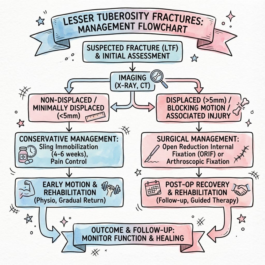
Indications:
- Displacement less than 5mm.
- Minimally displaced.
- Low demand patient.
- Fragment does not block motion.
Protocol:
- Sling immobilization for 4-6 weeks.
- Passive external rotation restricted (protect subscap) usually to neutral.
- Active internal rotation avoided for 6 weeks.
- Elbow/wrist/hand ROM immediately.
- Progressive strengthening after 6-8 weeks.
Close radiographic follow-up is required to ensure no late displacement.
Surgical Technique
Fixation Techniques
Deltopectoral Approach:
- Standard approach for lesser tuberosity.
- Position: Beach chair.
- Incision: Coracoid to axillary fold.
- Plane: Pectoralis major (medial) and Deltoid (lateral).
- Cephalic vein: Retract laterally with deltoid.
- Expose clavipectoral fascia, identify conjoined tendon.
Adequate exposure is critical for anatomical reduction.
Complications
Potential Complications
Subscapularis Deficiency
Weakness/Insufficiency: Failure of healing or non-union leads to weak internal rotation and anterior instability. Positive lift-off/belly press tests. This is the most common reason for revision if non-op fails.
Biceps Pathology
Ten.donitis/Subluxation: The biceps tendon runs adjacent to the fracture. Callus formation or hardware can cause tenosynovitis or rupture. Incarceration blocks reduction.
Posterior Instability
Recurrent Dislocation: If the lesser tuberosity (anterior stabilizer) fails to heal, the humeral head may subluxate posteriorly.
Malunion
Mechanical Block: Medial malunion is tolerated well, but prominent anterior malunion can block internal rotation or impinge on coracoid. This requires excision.
Postoperative Care
Rehabilitation Protocol
- Sling immobilization.
- No active internal rotation.
- Passive external rotation limited (usually to 0 degrees or neutral).
- Elbow/wrist/hand ROM.
- Pendulum exercises started early.
- Wean from sling.
- Progressive passive ROM.
- Gentle active-assisted Grade 1-2.
- Avoid forceful ER (puts tension on subscapularis repair).
- Forward elevation active-assisted.
- Active ROM allowed.
- Isometrics to Isotonics.
- Internal rotation strengthening initiated.
- Scapular stabilization focus.
- Hydrotherapy can be useful.
- Return to sport/heavy labor.
- Full ROM goal.
- Maintenance of cuff strength.
- Return to contact sports only when strength is 90% of contralateral side.
Outcomes
Prognosis
- Union Rates: High, generally excellent healing potential due to cancellous bed. Non-union is rare but symptomatic.
- Function: Good to excellent in 85-90% of surgically treated cases.
- Missed Diagnosis: Leads to chronic pain and weakness. Chronic posterior dislocation has poor prognosis if missed greater than 3 weeks (often requires arthroplasty).
- Subscapularis Strength: Often recovers to near normal, but some residual weakness in lift-off is common even with successful repair.
Differential Diagnosis
Distinguishing the Lesser Tuberosity Fracture
Anterior Shoulder Pain with Internal Rotation Weakness
| Diagnosis | Key Distinguishing Feature | Best Test |
|---|---|---|
| Lesser tuberosity fracture | Bony fragment medial to bicipital groove; avulsion mechanism | Axillary radiograph and CT |
| Isolated subscapularis tendon tear | Tendon retraction without a bony fragment; older or overhead athlete | MRI; lift-off and belly-press |
| Posterior fracture-dislocation | Locked internal rotation, empty glenoid, reverse Hill-Sachs | Axillary view and CT |
| Anterior dislocation with greater tuberosity fracture | Fragment lateral to groove; arm in external rotation | AP and axillary radiograph |
| Biceps long-head pathology | Pain over groove, no bony fragment; positive Speed/Yergason | MRI or ultrasound |
| Adhesive capsulitis | Global passive restriction, no trauma or fragment | Clinical; normal radiograph |
Clinical Pearl
The single most useful discriminator on imaging is the position of the fragment relative to the bicipital groove: a lesser tuberosity fragment sits medial to the groove, a greater tuberosity fragment lateral to it.
Controversies and Areas of Uncertainty
Where the Evidence Is Thin
Displacement Threshold
The 5 mm operative threshold is expert consensus extrapolated from small series, not a validated cut-off. Some advocate fixation at 3 mm in overhead athletes; others tolerate larger displacement in low-demand patients.
Screw vs Suture Anchor
No comparative trial exists. Large bony fragments favour lag screws; small, comminuted, or apophyseal fragments favour an anchor lasso. Choice remains surgeon preference guided by fragment size and bone quality.
Role of Arthroscopy
Arthroscopic-assisted repair is increasingly described but evidence is limited to case series; open deltopectoral fixation remains the reference standard, especially when a posterior dislocation must also be addressed.
Reverse Hill-Sachs Cut-offs
Defect-size thresholds guiding McLaughlin transfer (roughly 25-40%) versus rotational osteotomy or arthroplasty (greater than 40-50%) are derived from small cohorts and vary between authors.
Evidence Base
Key Studies
Ogawa & Takahashi - Long-Term Outcome of Isolated Lesser Tuberosity Fractures
- Series of 10 isolated lesser tuberosity fractures (6 acute, 4 chronic), mean age 30 years
- ORIF most often recommended for displaced acute injuries; all 3 operated acute cases excellent/satisfactory
- In chronic cases conservative (muscle strengthening) is first line, with ORIF reserved for failures
Garrigues et al. - Subscapularis Avulsion of the Lesser Tuberosity in Adolescents
- 6 skeletally immature patients plus pooled literature review; mean follow-up over 4 years
- Prototype: 13-year-old male, abduction-extension sporting injury, positive belly-press and lift-off
- Suture-anchor lasso technique gave predictably good results (mean ASES 97, WOSI 94)
Levine et al. - Avulsion Fractures of the Lesser Tuberosity in Adolescents
- Case report plus literature review of lesser tuberosity apophyseal avulsion in adolescents
- Often present late as chronic shoulder pain after a missed acute diagnosis
- ORIF of the displaced apophyseal fragment restored full motion and strength at 4 months
Gerber & Krushell - Isolated Rupture of the Subscapularis Tendon
- 16 men with isolated subscapularis rupture from forced hyperextension or external rotation of the adducted arm
- Loss of internal-rotation strength and increased passive external rotation without instability
- Introduced and validated the lift-off test for clinically relevant subscapularis lesions
Xiong et al. - Modified McLaughlin for Locked Chronic Posterior Dislocation
- 5 locked chronic posterior dislocations with reverse Hill-Sachs defects of 30-40% of the head
- Lesser tuberosity plus artificial bone transferred into the defect, fixed with two lag screws and sutures
- Constant-Murley score improved from 46.0 to 85.8 with no recurrent instability at mean 19.8 months
Court-Brown et al. - Epidemiology of Proximal Humeral Fractures
- Prospective 5-year study of 1,027 proximal humeral fractures
- Unimodal age distribution peaking in women aged 80-89 years
- AO classification more comprehensive than Neer; isolated tuberosity patterns are uncommon
Viva Scenarios
Use these scenarios to practise clinical reasoning and management decisions
"A 40-year-old male presents with shoulder pain after a seizure. X-rays show a lesser tuberosity fracture. What is your immediate concern and how do you investigate it?"
"What are the surgical indications for a lesser tuberosity fracture?"
"Describe the Deltopectoral Approach for fixing a lesser tuberosity fracture."
MCQ Practice
Self-Assessment Questions
Q1: Anatomy
Q: Which muscle attaches to the lesser tuberosity of the humerus?
- A) Supraspinatus
- B) Infraspinatus
- C) Teres Minor
- D) Subscapularis
- E) Pectoralis Major
A: D - The subscapularis is the only rotator cuff muscle that attaches to the lesser tuberosity. Supraspinatus, Infraspinatus, and Teres Minor attach to the Greater Tuberosity. Pectoralis Major attaches to the lateral lip of the bicipital groove.
Q2: Associated Injury
Q: An isolated lesser tuberosity fracture following a seizure should raise highest suspicion for:
- A) Anterior dislocation
- B) Posterior dislocation
- C) Axillary nerve injury
- D) Biceps rupture
- E) Rotator cuff tear
A: B - Seizures cause violent muscle contractions leading to posterior dislocation. The lesser tuberosity fracture is often an avulsion injury associated with this mechanism.
Q3: Imaging
Q: What is the most sensitive radiographic view for diagnosing a lesser tuberosity fracture profile?
- A) AP Internal Rotation
- B) AP External Rotation
- C) Scapular Y
- D) Axillary Lateral
- E) Outlet View
A: D - The Axillary Lateral view projects the lesser tuberosity anteriorly, allowing assessment of its profile and displacement. It is also diagnostic for posterior dislocation.
Q4: Surgical Threshold
Q: What is the generally accepted displacement threshold for surgical fixation of lesser tuberosity fractures in active patients?
- A) 1mm
- B) 3mm
- C) 5mm
- D) 10mm
- E) Any displacement requires surgery
A: C - 5mm is the commonly cited threshold (Ogawa et al.) where surgical fixation is recommended to restore subscapularis function and prevent mechanical block.
Q5: Approach
Q: Which surgical approach is most appropriate for open reduction internal fixation of a lesser tuberosity fracture?
- A) Deltoid Splitting
- B) Deltopectoral
- C) Posterior
- D) Mackenzie
- E) Trans-acromial
A: B - The Deltopectoral approach utilizes the interval between the deltoid and pectoralis major to provide direct anterior access to the lesser tuberosity and subscapularis.
Guidelines, Registries & Global Practice
Global Epidemiology
- Isolated lesser tuberosity fracture is rare — under 0.5% of all fractures and a small minority of proximal humeral fractures (Court-Brown 2001, N=1027 prospective series).
- Two distinct populations: adolescent males with an apophyseal avulsion during sport (abduction-extension mechanism), and adults sustaining avulsion during a posterior dislocation from seizure, electric shock, or trauma.
- Posterior glenohumeral dislocation itself accounts for only 2-4% of all shoulder dislocations and is missed at first presentation in roughly half of cases.
Side-by-Side Guidance
How Major Bodies Frame the Injury
| Body | Region | Emphasis |
|---|---|---|
| AO Foundation | Global | Anatomical reduction of the subscapularis footprint; screw or suture-anchor fixation; protect the long head of biceps |
| AAOS (proximal humerus CPG) | US | Most proximal humeral fractures non-operative; shared decision-making and early supervised motion |
| BOA / BOAST (shoulder trauma) | UK | Senior review of fracture-dislocations; CT for surgical planning; urgent reduction of locked dislocations |
| EFORT / European consensus | Europe | Recognise posterior dislocation early; reverse Hill-Sachs size drives reconstruction strategy |
No society publishes a lesser-tuberosity-specific guideline; recommendations are extrapolated from proximal humerus and posterior instability guidance. The widely cited 5 mm displacement threshold is expert-consensus, not guideline-mandated.
Registry Note
- Proximal humerus registries (e.g. national arthroplasty registries) capture this injury only when it progresses to arthroplasty for a chronic locked fracture-dislocation; isolated tuberosity fixation is not separately tracked, so the evidence base remains case series rather than registry data.
High- vs Limited-Resource Practice
- Well-resourced settings: Cross-sectional CT for fragment sizing, MRI for tendon integrity, arthroscopic-assisted or open anchor repair.
- Limited-resource settings: Diagnosis rests on a good axillary radiograph and clinical subscapularis testing; transosseous suture repair through drill holes substitutes for anchors, with comparable healing in cancellous bone.
- Universal priority: Early reduction of any associated locked posterior dislocation — delay beyond 3 weeks markedly worsens the outcome regardless of setting.
Lesser Tuberosity Fractures - Exam Quick Reference
Clinical summary
Key Facts
- •Attachment: Subscapularis
- •Threshold: 5mm displacement
- •Assoc: Posterior Dislocation (Seizures)
- •Nerve at risk: Axillary (inf), Musculocutaneous (medial retraction)
- •Structure at risk: Biceps Tendon (lateral)
Surgical Steps
- •Deltopectoral Approach
- •Identify LHB and LT
- •Reduce fragment (medial to lateral)
- •Screw fixation (large) or Suture Anchor (small)
- •Protect Biceps
Common Pitfalls
- •Missing posterior dislocation
- •Missing associated reverse Hill-Sachs
- •Hardware in bicipital groove
- •Failure to recognize subscapularis weakness
Examiner Favorites
- •What muscle attaches here?
- •Mechanism of injury?
- •How do you test clinical function? (Lift-off)
- •Surgical approach anatomy?