White and Panjabi | Dynamic Imaging | Fusion Decisions
- White & Panjabi: loss of ability to maintain normal motion pattern
- Radiographic: greater than 4mm translation OR greater than 10-15° angular motion
- Clinical diagnosis supported by imaging, not purely radiographic
- Bilateral greater than 50% facetectomy OR complete unilateral = iatrogenic instability
- Add fusion if extensive decompression creates instability
- “Instability is CLINICAL with radiographic support
- “Flexion-extension films must be standing/weight-bearing
- “Degenerative cascade: dysfunction → instability → restabilization
- “Three-column theory: 2+ columns = unstable
Overview and Epidemiology
The definition. White and Panjabi defined instability as the loss of the spine's ability to maintain its normal pattern of motion under physiological loads without neurological deficit, major deformity or incapacitating pain. Learn it verbatim, because the whole topic turns on one word in it: the diagnosis is clinical.
Clinical versus radiographic. Clinical instability is a symptomatic motion disorder, assessed by history, examination and function, and its treatment trigger is failed conservative care. Radiographic instability is excessive motion on flexion-extension films, translation of more than 4 mm or angular motion of more than 10-15° at a single segment, and it is treated only if it is symptomatic. The clinical diagnosis is primary; imaging supports it but does not define it, and symptoms and function guide the treatment decisions.
How common. The exact prevalence is uncertain because of diagnostic variability, but an estimated 10-25% of chronic low back pain has an instability component, and the proportion increases with age as the degenerative cascade progresses. Fusion for degenerative instability is among the most common spine procedures; the condition is a significant contributor to chronic disability and lost working days, and the cost of its conservative and surgical care is substantial.
Who. Degenerative instability peaks at 40-65 years. Women are affected 2:1, attributed to ligamentous laxity and hormonal factors; heavy manual labour increases the risk, and familial clustering is observed in degenerative spine disease.
Where.
- L4-L5 - most commonly affected, 50-60% of cases
- L5-S1 - second, 20-30%
- Multilevel involvement in approximately 20%
- Upper lumbar levels - less common; seen with trauma or iatrogenic causes
Pathophysiology and Mechanisms
Three columns. Denis divided the spine into an anterior column (anterior longitudinal ligament, anterior vertebral body and disc, which take the compression loads), a middle column (posterior longitudinal ligament, posterior body and disc, the column critical for stability) and a posterior column (pedicles, facets, lamina and ligaments, which take tension and rotation). Injury to two or more columns indicates mechanical instability requiring stabilisation.
The facets. In the neutral position the facet joints resist 20% of the axial load, and 40-50% in extension. Remove more than 50% of both facets and flexion-extension motion increases significantly; remove one facet completely and axial rotation increases. Those two observations are the working rule that decides fusion during a decompression, listed under Management.
What Abumi actually showed. The cadaveric study usually cited for that rule (Abumi and colleagues, 1990) never reports a percentage of facet resected. Its categories were anatomical: medial (partial) facetectomy and division of the supraspinous and interspinous ligaments did not significantly affect stability, whereas total facetectomy destabilised the segment even when unilateral, and the mode affected was axial rotation. Extension and lateral bending were unaffected even after bilateral total facetectomy, so the instability it describes is direction-specific, and the 50% figure is conventional teaching rather than Abumi's finding. Asked for the evidence, answer in his terms: medial facetectomy is safe, total facetectomy destabilises even unilaterally.
The disc. An intact annulus provides torsional stiffness. Discectomy reduces that stiffness by approximately 30%, and large annular defects increase the risk of instability.
Panjabi's three subsystems. Stability is the product of three interacting subsystems, and failure of any one of them can produce instability, which is why structural findings do not always correlate with symptoms.
- Passive - the osteoligamentous structures: vertebrae, discs, facet joints and ligaments. They provide intrinsic mechanical resistance and function mainly at the end of the range; degenerative change here leads to structural instability
- Active - the musculature: paraspinals (multifidus, erector spinae) and core (transversus abdominis, obliques). They stabilise dynamically during movement; weakness or dysfunction causes functional instability
- Neural control - proprioceptive feedback from mechanoreceptors, coordinated by the central nervous system into anticipatory and reactive muscle activation. It is impaired in chronic low back pain
Rehabilitation targets the active and neural subsystems.
The neutral zone. Within a segment's total range of motion, the neutral zone is the inner region around the neutral posture where there is minimal resistance to motion, the slack of the segment, bounded by the elastic zone in which ligament and disc resistance rises rapidly. Panjabi showed that the neutral zone is a more sensitive marker of instability than total range of motion. Degeneration, through annular tears and facet and capsular laxity, enlarges it: the segment becomes loose around neutral even when its end-range motion looks normal, which is why the standard flexion-extension thresholds can miss early clinical instability.
Why it ties the topic together. Anything that reduces an enlarged neutral zone reduces pain: the osteophytes of restabilisation in the degenerative cascade, fusion, which abolishes the neutral zone surgically, and active muscle activation of multifidus and transversus abdominis, which is the biomechanical reason core-stabilisation physiotherapy works. The neutral zone is a laboratory quantity, measured on cadaveric specimens under applied moments; it cannot be measured in a living patient, so it explains instability rather than diagnosing it.
The degenerative cascade (Kirkaldy-Willis). Degeneration runs through three phases, and the phase predicts the natural history and the response to treatment.
- Dysfunction (15-45 years) - circumferential and radial tears in the annulus fibrosus, disc dehydration with loss of proteoglycans, minor loss of disc height. Localised back pain and no instability yet
- Instability (35-70 years) - progressive disc collapse and height loss, facet subluxation and capsular laxity, abnormal segmental motion. Mechanical back pain with instability symptoms: a catch, a feeling of giving way
- Restabilisation (over 60 years) - osteophytes bridging the disc space, facet hypertrophy and arthrosis, ligamentous ossification. Motion decreases but stenosis develops, and the pain changes from mechanical to neurogenic
Classification Systems
Three classifications, each answering a different question: the cause guides treatment approach and prognosis, the radiographic pattern describes the abnormal motion, and the clinical severity guides the intensity of treatment and the surgical decision.
- Mechanism
- Disc and facet degeneration
- Typical Age
- 40-65 years
- Key Features
- Most common, L4-L5
- Mechanism
- Pars defect with spondylolisthesis
- Typical Age
- 15-35 years
- Key Features
- Athletes, L5-S1
- Mechanism
- Post-laminectomy, facetectomy
- Typical Age
- Post-surgery
- Key Features
- Excessive bone removal
- Mechanism
- Fracture-dislocation
- Typical Age
- Any age
- Key Features
- Three-column injury
- Mechanism
- Tumour, infection
- Typical Age
- Any age
- Key Features
- Bone destruction
Clinical Assessment
History. The pain is mechanical: worse with activity and with prolonged standing, better sitting or lying, stiff in the morning after inactivity, and not a night pain unless the degeneration is severe. The features in the history that point to instability:
- The instability catch - a sharp pain at the transition between movements
- A sensation of giving way - the back feels unstable
- Positional relief - better sitting than standing
- Activity limitation - avoids bending and twisting
- Needing to support the back with the hands
Inspection and palpation. Look for loss of the lumbar lordosis from muscle spasm, a forward-flexed posture and asymmetric paraspinal muscle bulk. Feel for paraspinal spasm, midline tenderness over the affected level and, in spondylolisthesis, a palpable step-off.
Movement. Movements are guarded, with limited flexion and extension, and the instability catch is sought during the transition from flexion back to extension.
Neurology. Often normal in pure mechanical instability; there may be a radiculopathy if there is associated stenosis or foraminal narrowing. Examine lower-limb power, sensation and reflexes.
Special tests.
- Technique
- Flex then extend spine
- Positive Finding
- Sharp catch pain during transition
- Technique
- Prone, legs off table, press spinous process
- Positive Finding
- Pain relieved with leg lift
- Technique
- Prone, PA pressure on spinous process
- Positive Finding
- Excessive motion or pain
- Technique
- Forward bend while palpating
- Positive Finding
- Excessive segmental motion felt
The prone instability test has high specificity for lumbar instability. The patient lies prone with the legs off the table and the examiner applies posteroanterior pressure to the spinous process; pain indicates instability. The patient then lifts the legs, which activates the paraspinals, and relief of the pain with that muscle activation confirms dynamic instability. The positive likelihood ratio is approximately 4.0.
Differential diagnosis. The mimics are separated by the pattern of pain and by what provokes it.
- Pain pattern
- Mechanical, worse with activity/transition, relief with sitting
- Discriminating feature
- Instability catch, giving-way; positive prone instability test
- Key test
- Standing flexion-extension radiographs (greater than 4mm / greater than 10-15°)
- Pain pattern
- Neurogenic claudication, worse standing/walking, relief with flexion
- Discriminating feature
- Symptoms positional and neurogenic, not motion-triggered
- Key test
- MRI showing canal/lateral recess stenosis
- Pain pattern
- Axial pain worse with flexion/sitting
- Discriminating feature
- Provoked by sustained flexion, not by motion transition
- Key test
- MRI HIZ/Modic changes; no abnormal dynamic motion
- Pain pattern
- Axial pain worse with extension/rotation
- Discriminating feature
- Relief with medial branch block
- Key test
- Diagnostic facet/medial branch block (greater than 50% relief)
- Pain pattern
- Mechanical back pain ± radiculopathy in young athlete
- Discriminating feature
- Pars defect; step-off; commonest L5-S1
- Key test
- Oblique radiograph / CT pars; lateral standing film
- Pain pattern
- Buttock pain below L5, often unilateral
- Discriminating feature
- Provocation cluster positive; pain over PSIS
- Key test
- SIJ provocation tests / image-guided block
Investigations
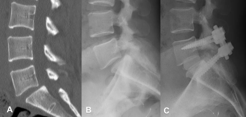
Standing films first. The AP and lateral radiographs must be weight-bearing, because a supine film misses instability. Assess disc height, alignment and osteophytes, and measure the slip if there is a spondylolisthesis. A traction spur, a horizontal osteophyte, indicates chronic instability.
Flexion-extension laterals are the conventional dynamic study, the one on which the thresholds were defined, and they are essential because static films miss functional instability. The patient performs maximal safe flexion and extension, and translation and angular motion are measured between the two films against the radiographic criteria above (more than 4 mm of sagittal translation, or more than 10-15° of segmental motion):
- Translation - the posterior body offset between adjacent vertebrae
- Angular motion - the change in segmental Cobb angle from flexion to extension
- Use the same anatomical landmarks on both views
The thresholds are conventional working values, not validated cut-offs. They derive from the White and Panjabi textbook rather than from a diagnostic-accuracy study, published values vary between sources (4 mm and 4.5 mm; 10 per cent and 15 per cent of vertebral body width both appear), and no threshold has an established sensitivity or specificity for a clinical outcome. Treat a number near the cut-off as one input alongside the history and examination, never as the diagnosis.
Flexion-extension is often not the pair that shows the most motion. Tarpada and colleagues (PMID 29432395) added a supine lateral to the standard series in 59 patients with single-level degenerative spondylolisthesis. Mean mobility on flexion-extension was 5.53%, against 7.83% on flexion-to-supine (P=0.001), and maximal mobility appeared on flexion-extension in only 11 of 59 patients, against 37 on flexion-to-supine. The relaxed supine position reduces an anterolisthesis more completely than active extension does, because extension is limited by pain and by the same arthritic facets you are interrogating. The practical consequence is that flexion-extension alone will underestimate translation, so a slip measuring under 4 mm on that pair has not been excluded; consider a supine lateral in place of, or in addition to, the extension film.
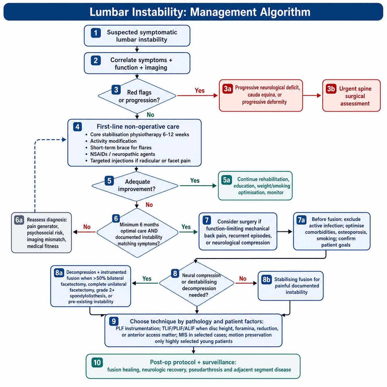
Management Algorithm
Who. Mild to moderate symptoms, no progressive deformity, no neurological deficit, the patient who prefers it, and the patient whose medical comorbidities preclude surgery. Approximately 30-40% improve adequately with conservative care, with better outcomes from a structured physiotherapy programme; the predictors of success are mild symptoms, good compliance and no significant deformity.
Core stabilisation physiotherapy is the most important non-surgical intervention. A structured 6-12 week programme targets transversus abdominis and multifidus, adds proprioceptive training and postural education, and aims to enhance compensation by the active subsystem.
Activity modification. Avoid repetitive bending and twisting, teach proper lifting mechanics, assess the workplace ergonomically, and lose weight if obese.
Bracing. A lumbosacral corset provides external support for an acute flare, for 2-4 weeks at most. Prolonged use causes muscle deconditioning, so wean it as core strength improves.
Medication. NSAIDs for inflammation and pain, a short course of muscle relaxant for spasm, and a neuropathic agent if there is a radicular component. Avoid opioids for chronic mechanical pain.
Injections. Epidural steroid injection for radicular symptoms; facet joint injections, diagnostic and therapeutic; medial branch blocks; and radiofrequency ablation for facet-mediated pain.
How long. A minimum of 6 months of conservative treatment before surgery is considered, unless there is progressive neurology.
Smoking significantly increases pseudarthrosis risk. Nicotine impairs bone healing and fusion rates. Non-smokers: 90-95% fusion. Smokers: 70-80% fusion. Strongly encourage smoking cessation for a minimum of 4 weeks pre-operatively and throughout healing. Consider bone morphogenetic protein (BMP) in smokers.
Complications
In theatre.
- Dural tear (5-10%) - primary repair, bed rest, avoid Valsalva
- Neural injury (1-2%) - nerve root or cauda equina, from retraction or instrumentation
- Vascular injury (under 1%) - aorta, vena cava or iliac vessels; the risk is higher in ALIF
- Excessive bleeding - from the epidural venous plexus and from bone
In the first six weeks.
- Wound infection (2-5%) - superficial or deep, higher with multilevel surgery and obesity
- Haematoma - epidural or in the wound, and may cause neurological compression
- CSF leak - from an unrecognised or inadequately repaired dural tear
- Medical - DVT and PE, myocardial infarction, pneumonia, urinary tract infection
Prevention is meticulous haemostasis, prophylactic antibiotics, DVT prophylaxis, and careful retraction and handling of neural tissue. Early recognition and management are critical to the outcome.
Guidelines, Registries & Global Practice
Global Epidemiology
Lumbar instability is most often a manifestation of degenerative disc and facet disease, the leading structural driver of low back pain (LBP). A systematic review of 165 studies from 54 countries estimated the global mean point prevalence of LBP at approximately 11.9% and the 1-month prevalence at approximately 23.2%, with the highest burden in women and those aged 40-80 years (Hoy et al. 2012, DOI). LBP is consistently ranked the single leading cause of years lived with disability worldwide, and the absolute number affected is rising with population ageing.
- Most affected segment: L4-L5 (degenerative spondylolisthesis), then L5-S1
- Sex: Degenerative spondylolisthesis is markedly more common in women
- Trend: Lumbar fusion volumes have risen faster than decompression in high-income systems, without a fall in reoperation rates (Martin et al. 2007, DOI)
Guideline & Society Positions
- Position on fusion for instability
- Do NOT offer fusion for non-specific LBP outside a trial; reserve for deformity/instability with clear indication
- Evidence basis
- RCT-informed, conservative
- Position on fusion for instability
- Decompression PLUS fusion supported for symptomatic degenerative spondylolisthesis with instability
- Evidence basis
- Level I/II (SPORT, Herkowitz, Fischgrund)
- Position on fusion for instability
- MDT assessment; fusion for documented instability or iatrogenic destabilisation after wide decompression
- Evidence basis
- Consensus + RCT
- Position on fusion for instability
- Reserve fusion for true instability, deformity, or post-decompression destabilisation; favour selective single-level constructs
- Evidence basis
- Consensus + biomechanical
Registry & Trial Evidence
- SPORT (NEJM 2007): as-treated benefit of surgery over non-operative care for degenerative spondylolisthesis at 2 years (Weinstein et al., DOI).
- Fischgrund 1997 Volvo Award: instrumentation raised fusion rate (82% vs 45%) without improving 2-year clinical outcome (DOI) — a recurring theme in registry analyses.
- National spine registries (e.g. Swespine, the British Spine Registry, and the Norwegian/Danish spine registries) collect PROMs (Oswestry Disability Index, EQ-5D) to benchmark fusion outcomes; routine reporting of reoperation and adjacent-segment surgery is now standard.
Practice Variation
Marked international and intra-national variation exists in fusion rates for degenerative lumbar disease, driven by reimbursement, surgeon training, and differing thresholds for diagnosing "instability." Conservative-first pathways (physiotherapy-led core stabilisation, activity modification, judicious injections) are universally endorsed before elective fusion, except for progressive neurological deficit, cauda equina syndrome, or progressive deformity. Minimally invasive and navigation/robotic-assisted fusion techniques (TLIF/PLIF) are increasingly available at tertiary centres internationally, and structured multidisciplinary post-operative rehabilitation, including occupational therapy and vocational return-to-work coordination, supports recovery and reintegration.
MCQ Practice Points
Q: What radiographic findings on flexion-extension X-rays define lumbar instability? A: Translation greater than 4mm (or greater than 10% of vertebral body width) OR angular motion greater than 10-15 degrees between adjacent segments. These are the classic White-Panjabi criteria for clinical instability.
Q: How much facet resection causes iatrogenic instability requiring fusion? A: Greater than 50% bilateral facetectomy or complete unilateral facetectomy. The facet joints contribute 40-50% of torsional stability. Partial medial facetectomy (less than 50% per side) typically preserves stability.
Q: What is the difference between clinical and mechanical instability? A: Clinical instability produces symptoms (pain, neurological signs) with motion; mechanical instability is radiographic abnormal motion that may be asymptomatic. Surgical fusion addresses mechanical instability but is only indicated when clinically symptomatic.
Q: When should fusion be added to decompression for degenerative conditions? A: When pre-existing instability exists (greater than 4mm translation), when decompression creates iatrogenic instability (extensive facetectomy), or when deformity correction is required. SPORT trial showed no benefit of routine fusion for stable stenosis.
Exam Viva Scenarios
Practise clinical reasoning and management decisions out loud
“A 58-year-old woman underwent L4-L5 laminectomy for stenosis 18 months ago with initial good relief of leg symptoms. She now presents with mechanical back pain worse with activity. Flexion-extension X-rays show 6mm translation at L4-L5. How would you manage this?”
“You are planning L4-L5 decompression for severe central and lateral recess stenosis. Pre-operative flexion-extension films show no instability. Intraoperatively, you find you need bilateral 60% facetectomy to adequately decompress. What do you do?”
“A 52-year-old manual laborer has chronic mechanical low back pain worse with activity. MRI shows severe L4-L5 disc degeneration with facet arthropathy. Flexion-extension films show 3mm translation and 8 degrees angular motion - below instability thresholds. He has failed 12 months of physiotherapy. Does he have instability? Would you offer surgery?”
Key Definitions
- White & Panjabi: Loss of spine's ability to maintain normal motion pattern under physiologic loads without neurological deficit, major deformity, or incapacitating pain
- Clinical instability: CLINICAL diagnosis supported by imaging, not purely radiographic
- Radiographic thresholds: Greater than 4mm translation OR greater than 10-15° angular motion on flexion-extension films
- Panjabi subsystems: Passive (osteoligamentous), Active (muscular), Neural (proprioceptive control)
Classification
- Etiological: Degenerative (most common), isthmic (pars defect), iatrogenic (post-laminectomy), traumatic, pathological
- Three columns (Denis): Anterior (ALL, anterior VB/disc), Middle (PLL, posterior VB/disc - KEY), Posterior (facets, ligaments)
- Degenerative cascade: Dysfunction (15-45y) → Instability (35-70y) → Restabilization (60+y)
- Instability rule: Injury to 2 or more columns = mechanical instability
Clinical Assessment
- History: Mechanical back pain worse with activity, instability catch, giving way sensation, relief with sitting
- Prone instability test: PA pressure on spinous process causes pain, relieved with leg lift (muscle activation)
- Flexion-extension radiographs: the conventional dynamic study and must be weight-bearing - but NOT the most sensitive pair. Maximal mobility appeared on flexion-extension in only 11 of 59 patients (Tarpada, PMID 29432395); flexion-to-supine showed more (7.83% vs 5.53%), so flexion-extension alone UNDERESTIMATES translation
- MRI findings: Disc degeneration, facet effusion, Modic Type II changes, high-intensity zone in annulus. Remember MRI is SUPINE and missed the slip in 83% of patients who had one on standing films (Caterini, PMID 21597993) - facet effusion may be its only trace
Surgical Indications
- Failed conservative management minimum 6 months (unless progressive neurology)
- Documented instability on imaging with symptom correlation
- Iatrogenic: Bilateral greater than 50% facetectomy OR complete unilateral facetectomy
- Progressive deformity with functional impairment
- Add fusion to decompression if creating instability
Surgical Techniques
- Posterolateral fusion with instrumentation: the conventional default. Fusion rate 82% instrumented vs 45% non-instrumented in degenerative spondylolisthesis (Fischgrund) - the commonly quoted 85-95% is higher than the randomised figure, and instrumentation did NOT improve clinical outcome
- TLIF: Unilateral approach, less neural retraction, excellent foraminal decompression
- PLIF: Bilateral approach, direct neural decompression, higher dural tear risk
- ALIF: Anterior approach, large graft area, useful for spondylolisthesis reduction
Complications
- Pseudarthrosis: 5-10% instrumented PLF. Risk factors: smoking, obesity, multilevel, diabetes
- Adjacent segment disease: 2-3% per year, 15-20% require surgery at 10 years
- Dural tear: 5-10%. Management: primary repair, bed rest, avoid Valsalva
- Smoking increases pseudarthrosis risk: 90-95% fusion non-smokers vs 70-80% smokers
Evidence Pearls
- White & Panjabi (1990): Foundation definition and radiographic thresholds
- Panjabi three subsystem model: Explains variable clinical presentation
- Abumi facetectomy study: Bilateral greater than 50% = significant motion increase
- Kirkaldy-Willis cascade: Dysfunction → Instability → Restabilization phases
- Conservative success 30-40%, surgical 70-80% good/excellent outcomes
Evidence Base
Panjabi: Clinical Spinal Instability and Low Back Pain
- Clinical instability defined as loss of the normal pattern of spinal motion causing pain and/or neurological dysfunction
- Stabilising system comprises three subsystems: spinal column, spinal muscles, and the neural control unit
- The neutral zone is a more sensitive parameter than range of motion for detecting destabilisation and restabilisation
- Reduction of an enlarged neutral zone (by fusion, osteophytes, or muscle activation) correlates with pain relief
Panjabi Three Subsystem Model
- Described spinal stability as interaction of passive (osteoligamentous), active (muscular), and neural control subsystems
- Explained how dysfunction of any subsystem can be compensated, adapted to, or lead to injury and clinical instability
- Provided framework for understanding why structural findings do not always correlate with symptoms
- Supported role of rehabilitation targeting the active and neural subsystems
Kirkaldy-Willis Degenerative Cascade
- Described three phases: dysfunction, instability, and restabilisation
- Instability phase characterised by abnormal motion and mechanical pain
- Restabilisation phase shows decreased motion but stenosis develops
- Natural history shows transition from mechanical to neurogenic symptoms
Abumi: Graded Facetectomies and Lumbar Stability
- In vitro study of fresh human functional spinal units under six load modes (range of motion and neutral zone measured)
- Medial facetectomy (partial) and division of supraspinous/interspinous ligaments did NOT significantly affect stability
- Total facetectomy, even when created UNILATERALLY, rendered the segment unstable - the mode affected was AXIAL ROTATION (right axial rotation range of motion increased after left unilateral total facetectomy)
- The flexion increase belonged to unilateral MEDIAL facetectomy and was described as slight; range of motion was NOT affected even by BILATERAL TOTAL facetectomy in extension or lateral bending
- Division of the supraspinous and interspinous ligaments did not affect range of motion in any load mode
- The discriminator is anatomical - medial versus total - and the paper offers NO percentage of facet resection
Martin: Reoperation Rates After Lumbar Surgery
- Washington State discharge registry; adults having lumbar surgery for degenerative disease in 1990-1993 (n=24,882)
- Cumulative incidence of reoperation was 19% over the subsequent 11 years
- For spondylolisthesis, reoperation was LESS likely after fusion than after decompression alone (17.1% vs 28.0%, P=0.002)
- For other degenerative diagnoses, reoperation was HIGHER after fusion than decompression alone (21.5% vs 18.8%, P=0.008); 62.5% of post-fusion reoperations involved device complication or pseudarthrosis
Herkowitz & Kurz: Decompression vs Decompression + Arthrodesis
- 50 patients with spinal stenosis and degenerative spondylolisthesis prospectively studied (mean follow-up 3 years)
- Concomitant intertransverse-process arthrodesis gave significantly better relief of back and leg pain than decompression alone
- Established that an unstable spondylolisthetic segment should be fused at the time of decompression
- Landmark evidence behind fusing the listhetic level during decompression
Fischgrund (1997 Volvo Award): Instrumentation and Fusion
- 76 patients randomised to instrumented versus non-instrumented posterolateral fusion after decompression; 67 available at 2-year follow-up
- Successful arthrodesis: 82% instrumented vs 45% non-instrumented (P=0.0015)
- Clinical outcome excellent/good in 76% instrumented vs 85% non-instrumented (P=0.45, not significant)
- Pedicle-screw instrumentation improves fusion rate but did not improve 2-year clinical pain outcome
- Overall, successful fusion did not influence patient outcome (P=0.435) - union itself did not track with how the patient did
SPORT: Surgical vs Nonsurgical for Degenerative Spondylolisthesis
- 304 randomised plus 303 observational patients (607 total) across 13 centres in 11 US states; one-year crossover approximately 40% in EACH direction in the randomised cohort, and 17% to surgery with 3% to nonsurgical care in the observational cohort
- THE INTENTION-TO-TREAT ANALYSIS OF THE RANDOMISED COHORT SHOWED NO STATISTICALLY SIGNIFICANT EFFECT on any primary outcome
- The as-treated analysis of both cohorts combined favoured surgery at 2 years: bodily pain 18.1 (95% CI 14.5 to 21.7), physical function 18.3 (95% CI 14.6 to 21.9), Oswestry -16.7 (95% CI -19.5 to -13.9)
- Little evidence of harm from either treatment
Tarpada: Supine Lateral Radiographs Reveal More Instability than Flexion-Extension
- 59 patients (51 women, 8 men), mean age 63.0 years, single-level degenerative spondylolisthesis; supine lateral added to standing neutral/flexion/extension series
- Mean mobility on flexion-extension 5.53% versus 7.83% on flexion-to-supine (P=0.00133), independent of age and BMI
- MAXIMAL mobility appeared on flexion-extension in only 11 of 59 patients, against 37 of 59 on flexion-to-supine and 11 on neutral-to-supine
- The relaxed supine position reduces an anterolisthesis more completely than active extension does
Caterini: Supine MRI Misses Most Degenerative Slips, and Facet Effusion is the Tell
- 52 patients (mean age 64.7) had weight-bearing flexion-extension radiographs and supine MRI prospectively compared
- Degenerative spondylolisthesis was seen radiographically in 12 patients (23.1%) - and was NOT evident on the supine sagittal MRI in 10 of those 12 (83.3%)
- Exaggerated facet fluid on axial T2 was present at the corresponding level in 8 of the 12 patients with a slip (66%)
- Facet fluid was also present in 7 of 52 (13.4%) with NO radiographic instability, so it is not specific
References
- White AA, Panjabi MM. Clinical Biomechanics of the Spine. 2nd ed. Philadelphia: Lippincott; 1990.
- Panjabi MM. The stabilizing system of the spine. Part I. Function, dysfunction, adaptation, and enhancement. J Spinal Disord. 1992;5(4):383-389.
- Kang CH, Shin MJ, Kim SM, Lee SH, Lee CS. MRI of paraspinal muscles in lumbar degenerative kyphosis patients and control patients with chronic low back pain. Clin Radiol. 2007;62(5):479-486.
- Kirkaldy-Willis WH, Farfan HF. Instability of the lumbar spine. Clin Orthop Relat Res. 1982;(165):110-123.
- Kalichman L, Li L, Kim DH, et al. Facet joint osteoarthritis and low back pain in the community-based population. Spine. 2008;33(23):2560-2565.
- Battié MC, Videman T, Parent E. Lumbar disc degeneration: epidemiology and genetic influences. Spine. 2004;29(23):2679-2690.
- Sengupta DK, Herkowitz HN. Degenerative spondylolisthesis: review of current trends and controversies. Spine. 2005;30(6 Suppl):S71-81.
- Martin BI, Deyo RA, Mirza SK, et al. Expenditures and health status among adults with back and neck problems. JAMA. 2008;299(6):656-664.
- Adams MA, Roughley PJ. What is intervertebral disc degeneration, and what causes it? Spine. 2006;31(18):2151-2161.
- Fujiwara A, Lim TH, An HS, et al. The effect of disc degeneration and facet joint osteoarthritis on the segmental flexibility of the lumbar spine. Spine. 2000;25(23):3036-3044.
- Ghiselli G, Wang JC, Bhatia NN, Hsu WK, Dawson EG. Adjacent segment degeneration in the lumbar spine. J Bone Joint Surg Am. 2004;86(7):1497-1503.
- Abumi K, Panjabi MM, Kramer KM, Duranceau J, Oxland T, Crisco JJ. Biomechanical evaluation of lumbar spinal stability after graded facetectomies. Spine. 1990;15(11):1142-1147. (PMID 2267608)
- Denis F. The three column spine and its significance in the classification of acute thoracolumbar spinal injuries. Spine. 1983;8(8):817-831.
- Kim DH, Vaccaro AR, Berta SC. Spinal Instrumentation: Surgical Techniques. New York: Thieme; 2005.
- Meyerding HW. Spondylolisthesis. Surg Gynecol Obstet. 1932;54:371-377.
- Kalichman L, Kim DH, Li L, Guermazi A, Hunter DJ. Computed tomography-evaluated features of spinal degeneration: prevalence, intercorrelation, and association with self-reported low back pain. Spine J. 2010;10(3):200-208.
- Panjabi MM. Clinical spinal instability and low back pain. J Electromyogr Kinesiol. 2003;13(4):371-379.
- Pearcy MJ, Tibrewal SB. Lumbar intervertebral disc and ligament deformations measured in vivo. Clin Orthop Relat Res. 1984;(191):281-286.
- Nachemson AL. Newest knowledge of low back pain. A critical look. Clin Orthop Relat Res. 1992;(279):8-20.
- Frymoyer JW, Selby DK. Segmental instability. Rationale for treatment. Spine. 1985;10(3):280-286.
- Hides JA, Richardson CA, Jull GA. Multifidus muscle recovery is not automatic after resolution of acute, first-episode low back pain. Spine. 1996;21(23):2763-2769.
- MacDonald D, Moseley GL, Hodges PW. Why do some patients keep hurting their back? Evidence of ongoing back muscle dysfunction during remission from recurrent back pain. Pain. 2009;142(3):183-188.
- O'Sullivan PB, Twomey LT, Allison GT. Evaluation of specific stabilizing exercise in the treatment of chronic low back pain with radiologic diagnosis of spondylolysis or spondylolisthesis. Spine. 1997;22(24):2959-2967.
- Weinstein JN, Lurie JD, Tosteson TD, et al. Surgical versus nonsurgical treatment for lumbar degenerative spondylolisthesis. N Engl J Med. 2007;356(22):2257-2270.
- Herkowitz HN, Kurz LT. Degenerative lumbar spondylolisthesis with spinal stenosis. A prospective study comparing decompression with decompression and intertransverse process arthrodesis. J Bone Joint Surg Am. 1991;73(6):802-808.
- Martin BI, Mirza SK, Comstock BA, Gray DT, Kreuter W, Deyo RA. Reoperation rates following lumbar spine surgery and the influence of spinal fusion procedures. Spine. 2007;32(3):382-387.
- Tarpada SP, Cho W, Chen F, Amorosa LF. Utility of supine lateral radiographs for assessment of lumbar segmental instability in degenerative lumbar spondylolisthesis. Spine. 2018;43(18):1275-1280. (PMID 29432395)
- Caterini R, Mancini F, Bisicchia S, Maglione P, Farsetti P. The correlation between exaggerated fluid in lumbar facet joints and degenerative spondylolisthesis: prospective study of 52 patients. J Orthop Traumatol. 2011;12(2):87-91. (PMID 21597993)
- Bendo JA, Ong B. Importance of correlating static and dynamic imaging studies in diagnosing degenerative lumbar spondylolisthesis. Am J Orthop (Belle Mead NJ). 2001;30(3):247-250. (PMID 11300136)