Progressive Instability | Mayfield Stages | Emergent Reduction | Ligament Reconstruction
- 25% missed initially - always look for carpal arc disruption on lateral X-ray
- Spilled teacup sign = Stage IV lunate dislocation (lunate tips volar into carpal tunnel)
- Mayfield progression: SL then capitolunate then lunotriquetral ligaments fail sequentially
- Acute median nerve symptoms common in Stage IV - urgent reduction required
- Greater arc injuries = fracture-dislocations (trans-scaphoid most common)
- “Lateral X-ray: Lunate should articulate with both radius and capitate
- “Lesser arc = pure ligamentous; Greater arc = bone fractures
- “Trans-scaphoid perilunate = scaphoid fracture + perilunate dislocation
- “Open reduction via combined volar + dorsal approach preferred
Overview
Perilunate and lunate dislocations are a spectrum of high-energy carpal injuries in which the ligaments around the lunate fail in sequence, the Mayfield progression. The two names are the last two stages of that sequence. In a perilunate dislocation (Mayfield Stage III) the lunate stays articulated with the radius while the rest of the carpus dislocates dorsally around it; in a lunate dislocation (Stage IV) the lunate itself tips volarly out of its fossa into the carpal tunnel while the carpus partially reduces.
Why they are missed. Herzberg's seven-centre series of 166 injuries found the diagnosis missed at first presentation in 41 (25%), and delayed diagnosis means poorer outcomes. The diagnosis is made on the lateral radiograph, where the lunate should articulate with both the radius and the capitate; failing to look for that colinearity is what underlies the missed-diagnosis rate.
The spectrum. Associated fractures are common, the scaphoid especially. An injury whose path passes through bone is a greater arc fracture-dislocation and one that passes through ligament alone is a lesser arc injury; both arcs are set out under Classification. Treatment is urgent reduction followed by open repair for every injury, to relieve the median nerve and to prevent progressive carpal instability.
- Lunate Dislocation
- Stage IV
- Perilunate Dislocation
- Stage III
- Lunate Dislocation
- Displaced volar into carpal tunnel
- Perilunate Dislocation
- Remains in lunate fossa
- Lunate Dislocation
- Partially reduced
- Perilunate Dislocation
- Displaced dorsally
- Lunate Dislocation
- Spilled teacup
- Perilunate Dislocation
- Lunate-capitate malalignment
- Lunate Dislocation
- High (volar lunate directly compresses nerve)
- Perilunate Dislocation
- Lower (acute median neuropathy still common across all perilunate injuries)
- Lunate Dislocation
- Mandatory
- Perilunate Dislocation
- Considered if symptoms present
- Lunate Dislocation
- Lower (50-60%)
- Perilunate Dislocation
- Higher (70-80%)
Anatomy
The lunate as keystone. The lunate is the keystone of the proximal carpal row, tied to its neighbours by intrinsic ligaments and to the radius by extrinsic ones. The perilunate injury is the sequential failure of exactly these attachments, which is why knowing them lets you read the stage from the film and predict the associated injuries.
Intrinsic (interosseous) ligaments. The scapholunate ligament is strongest dorsally and is the key stabiliser; the lunotriquetral ligament is strongest volarly. Both are critical to carpal stability.
Extrinsic ligaments. The volar radiocarpal ligaments are the radioscaphocapitate (RSC), the long radiolunate (LRL) and the short radiolunate (SRL); dorsally there is the dorsal radiocarpal (DRC) ligament. Between the RSC and the LRL lies the space of Poirier, a weak zone, and it is the path along which the lunate dislocates.
Pathophysiology
Mechanism. A fall on the outstretched hand with the wrist in dorsiflexion, typically from high-energy trauma: a motor vehicle accident, a fall from height or sport. The combination that fails the perilunate ring is hyperextension, ulnar deviation and intercarpal supination, the loading Mayfield reproduced in the cadaver.
Force transmission. The sequence runs:
- Axial load through the palm
- Ground reaction force applied to the thenar eminence
- The wrist forced into hyperextension
- Sequential ligament failure from the radial to the ulnar side
Perilunate injuries frequently cause acute median neuropathy: Wickramasinghe found it in approximately 47% across the spectrum, and acute carpal tunnel syndrome is especially associated with volar lunate dislocation (Stage IV), where the displaced lunate directly compresses the median nerve. Emergent reduction is required, because prolonged sustained nerve compression risks permanent damage.
Classification Systems
Mayfield's classification describes the sequential failure of the perilunate structures from the radial side to the ulnar as the wrist is loaded in hyperextension, ulnar deviation and intercarpal supination.
Stage I, scapholunate dissociation. The scapholunate interosseous ligament is disrupted. The scaphoid flexes and the lunate extends, the beginning of a DISI pattern, and the PA radiograph shows a widened scapholunate interval of over 3mm, the Terry Thomas sign.
Stage II, capitolunate dissociation. The space of Poirier disrupts and the capitate dislocates dorsally relative to the lunate. The lunate still articulates with the radius as the ligament failure continues.
Stage III, perilunate dislocation. The lunotriquetral ligament fails and the entire carpus dislocates dorsally around the lunate. The lunate remains in the lunate fossa of the radius, which is the key point, and no longer articulates with the capitate; the lateral radiograph shows the capitate displaced dorsally.
Stage IV, lunate dislocation. The dorsal radiocarpal ligament, the final ligament, fails. The lunate rotates and tips volarly out of the fossa into the carpal tunnel, the spilled teacup, and a Stage IV injury with nerve symptoms is a surgical emergency.
About 97% of perilunate dislocations are dorsal. In the rare volar (palmar) perilunate dislocation the capitate and distal carpus dislocate volarly relative to a lunate that stays in its fossa, the mirror image of the usual pattern; do not confuse it with a Stage IV lunate dislocation, where the lunate itself tips volar into the carpal tunnel. The force is atypical, a dorsally directed blow on the flexed wrist rather than hyperextension, ulnar deviation and supination, so the carpus is driven palmar to the lunate.
It is easily missed because it is unfamiliar. It follows the same principle of emergency reduction then operative repair, but the reduction manoeuvre is reversed, the carpus being brought from volar back to dorsal. Define the direction of carpal displacement relative to the lunate on the true lateral before planning any reduction.
Distinct from the Mayfield spectrum are the rare axial (longitudinal) carpal dislocations, in which the carpus splits longitudinally into columns rather than dislocating around the lunate. The mechanism is a severe crush or blast injury to the dorsopalmar plane of the hand, an industrial press or a run-over, which drives the metacarpals and carpus apart along the long axis and disrupts the transverse intercarpal and intermetacarpal ligaments.
Garcia-Elias classifies them by the column displaced:
- Axial-radial: the radial column (thumb and index ray with the adjacent carpus) displaces radially, peritrapezial or peri-scaphoid-trapezial
- Axial-ulnar: the ulnar column (small and ring ray, peri-pisiform or peri-hamate) displaces ulnarly
- Combined axial-radial-ulnar: a central column retained between two diverging columns
These are high-energy crush injuries with major soft-tissue, neurovascular (ulnar and median branches), tendon and skin compromise, and the soft-tissue injury, compartment syndrome and contamination dominate the prognosis far more than the bony dislocation. Management is emergency debridement, compartment release if needed, anatomic realignment of the columns with K-wire or screw fixation, and staged soft-tissue and neurovascular reconstruction, very different from the perilunate algorithm.
Clinical Assessment
History. The mechanism is typically high-energy: a fall on the outstretched hand, a motor vehicle accident, or a contact-sport or cycling injury. Patients describe severe wrist pain, rapid swelling and an inability to move the wrist, sometimes a feeling of a "pop" or "shift", and numbness in the median nerve distribution, especially in Stage IV.
Inspection and palpation. The wrist is markedly swollen with loss of its normal contour. A dorsal prominence suggests a perilunate dislocation and volar fullness a lunate dislocation, and in Stage IV the lunate itself may be palpable volarly. Tenderness is diffuse. Assess skin tension and look for an open injury or skin compromise, because these decide how urgently the wrist must be reduced.
The median nerve. This is the critical part of the examination. Test sensation in the thumb, index and middle fingers, thenar muscle strength (opponens pollicis) and two-point discrimination, and compare with the other side. Range of motion is severely limited by pain; do not force it, but document a baseline for postoperative comparison.
Red flags. Any of the following demands emergent intervention:
- Acute carpal tunnel symptoms (numbness, tingling, weakness)
- Open injury
- Skin blanching or tension
- Vascular compromise
- Signs of compartment syndrome
Differential diagnosis. The painful, swollen, deformed wrist after a fall on the outstretched hand has several mimics, and the single most important discriminator is the lateral radiograph: in a perilunate or lunate dislocation the normal radius-lunate-capitate colinearity is lost. That is the distinction that is missed.
- Distinguishing Features
- High-energy FOOSH; gross swelling; median nerve symptoms common; loss of radius-lunate-capitate colinearity on lateral; disrupted Gilula's arcs on PA
- Management Pointer
- Emergent reduction then operative repair/fixation
- Distinguishing Features
- Common in all ages; dorsal/volar angulation at the metaphysis; carpal alignment preserved; lunate stays in fossa
- Management Pointer
- Closed reduction; cast or ORIF depending on stability
- Distinguishing Features
- Anatomical snuffbox tenderness; carpus aligned; no carpal dislocation; may be radiographically occult early
- Management Pointer
- Cast; ORIF for displaced/unstable; MRI if occult
- Distinguishing Features
- Widened SL gap (Terry Thomas sign); DISI on lateral; carpus otherwise reduced; no full perilunate dislocation
- Management Pointer
- Acute repair / pinning; reconstruction if chronic
- Distinguishing Features
- Lower-energy mechanism; tenderness without bony or alignment abnormality; normal radiographs and Gilula's arcs
- Management Pointer
- Symptomatic; re-image or MRI if symptoms persist
- Distinguishing Features
- More insidious; sclerosis/collapse of lunate; no acute dislocation; chronic ulnar-negative association
- Management Pointer
- Staged management per Lichtman stage
Investigations
Radiographs. Three standard views:
- PA (posteroanterior)
- True lateral
- Oblique views
The PA view. Gilula's arcs are the landmarks, and the diagnosis on this view is a break in them:
- Arc I: proximal articular surfaces of the proximal row
- Arc II: distal articular surfaces of the proximal row
- Arc III: proximal articular surfaces of the capitate and hamate
Look for disruption of the arcs, overlap or crowding of the carpal bones, a widened scapholunate interval (the Terry Thomas sign described under Stage I), and a triangular or pie-shaped lunate, which means the lunate has rotated.
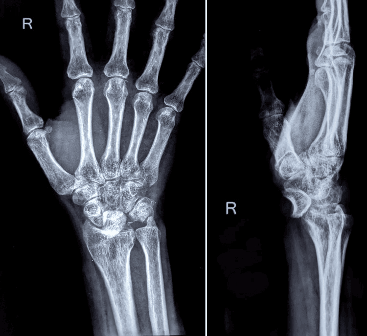
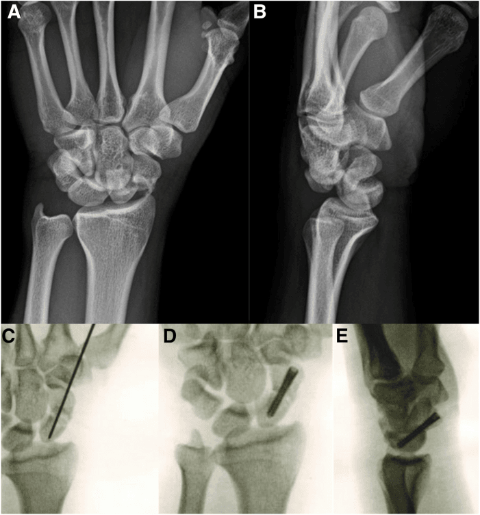
The lateral view. The radius, lunate and capitate should be colinear, the lunate cup holding the capitate ball. In a Stage III perilunate dislocation the lunate remains in the fossa and the capitate is dislocated dorsally, no longer articulating with it. In a Stage IV lunate dislocation the lunate is tilted volarly out of the fossa, and the capitate may partially reduce into the fossa it has left.
On the lateral, the normal lunate is a cup holding the capitate. In a Stage IV lunate dislocation the lunate rotates and "spills" volarly, the spilled teacup sign, pathognomonic for lunate dislocation.
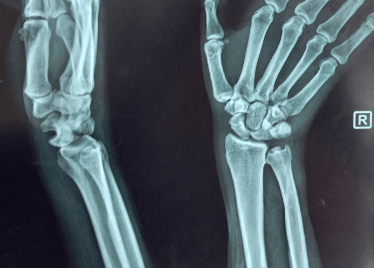
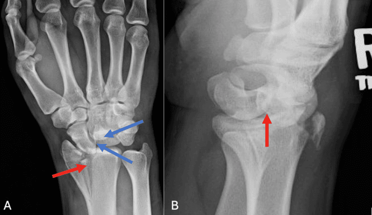
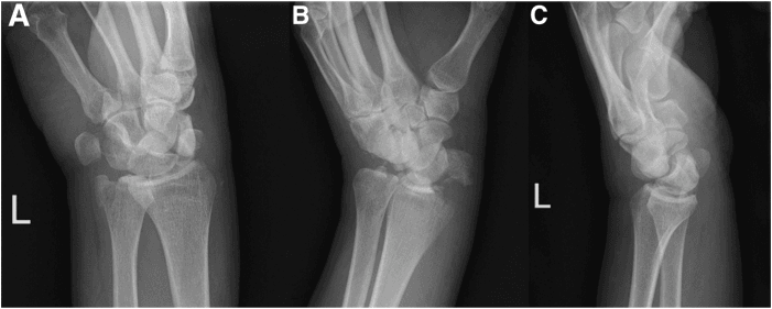
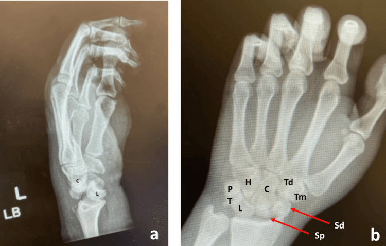
CT confirms the diagnosis and identifies the associated fractures of a greater arc injury, the fracture lines in the scaphoid, capitate, triquetrum or styloid, with the extent of articular involvement and the size and displacement of the fragments. It is the pre-operative planning study and, afterwards, the way to judge the quality of reduction.
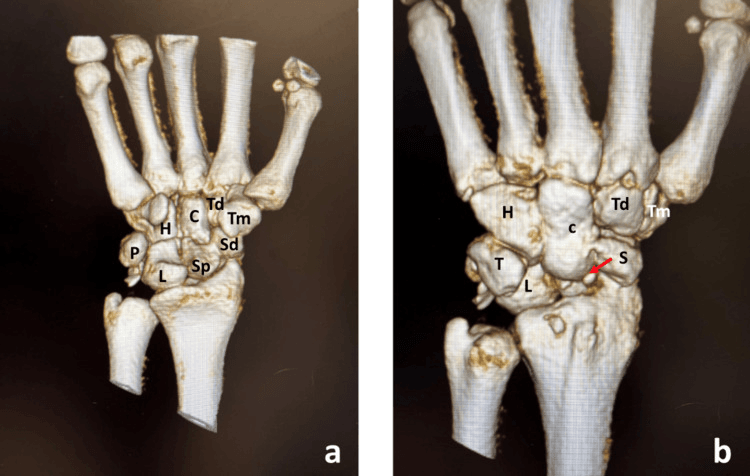
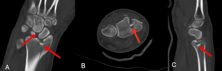
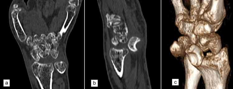
MRI is usually not needed acutely. Subacutely it may help assess ligament integrity, showing scapholunate and lunotriquetral tears, the pattern of bone bruising and any TFCC injury, and it is useful when planning a staged reconstruction.
Management Algorithm
Initial assessment. Before anything else:
- Neurovascular status immediately, the median nerve above all
- Mechanism of injury
- Skin integrity and tension
- PA, lateral and oblique radiographs
- Splint in the position of comfort initially
When to reduce emergently. Any of these means the wrist is reduced now:
- Acute carpal tunnel syndrome (Stage IV lunate dislocation)
- Skin compromise or threatened skin
- Open injury
- Vascular compromise
Closed reduction. Under a regional block or procedural sedation, the manoeuvre reverses the injury: longitudinal traction through the fingers first, then the wrist extended to disengage the carpus and recreate the deformity, thumbs over the dorsal carpus applying pressure, and the carpus pushed volarly while the wrist is flexed. Take immediate post-reduction films to verify the reduction.
TRAPClosed Reduction Steps
Hook:TRAP the lunate back in place: Traction, Recreate, Apply pressure, Push and flex
After reduction. Splint in neutral, repeat the neurovascular examination, confirm the reduction on radiographs, and arrange definitive surgery within 24-72 hours. Closed reduction is temporising only: every perilunate and lunate dislocation requires definitive surgical treatment, because closed treatment alone results in unacceptable rates of instability and arthritis.
If closed reduction fails, interposed tissue is preventing it, and the answer is emergent open reduction in theatre.
Surgical Technique
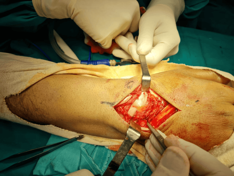
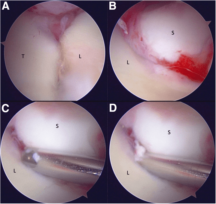
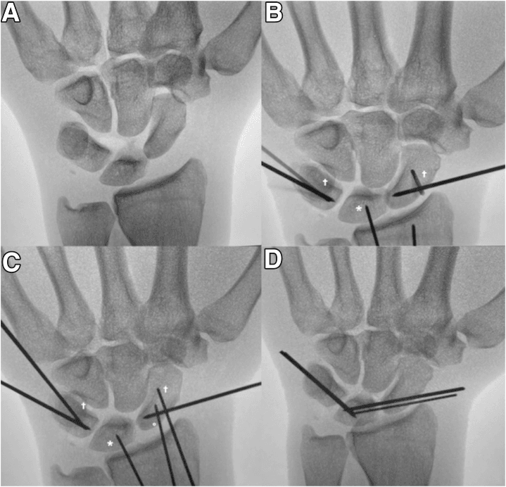
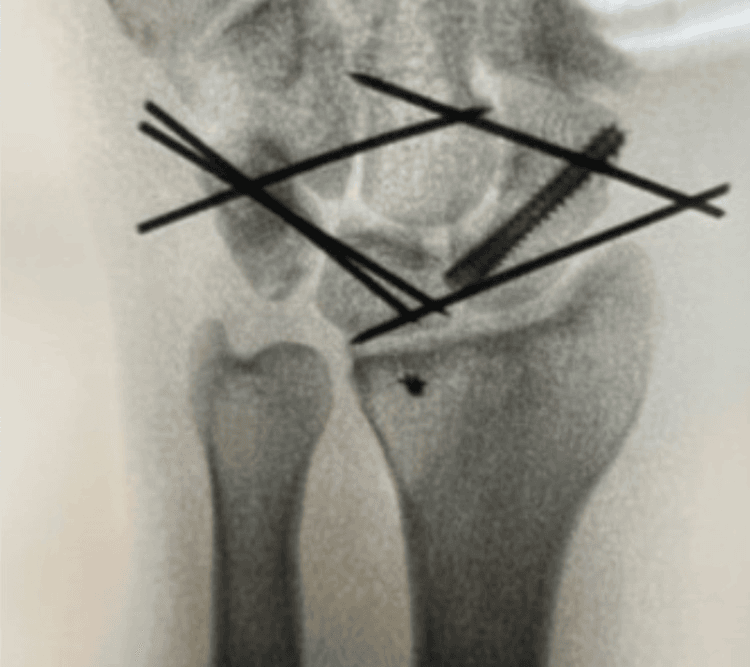
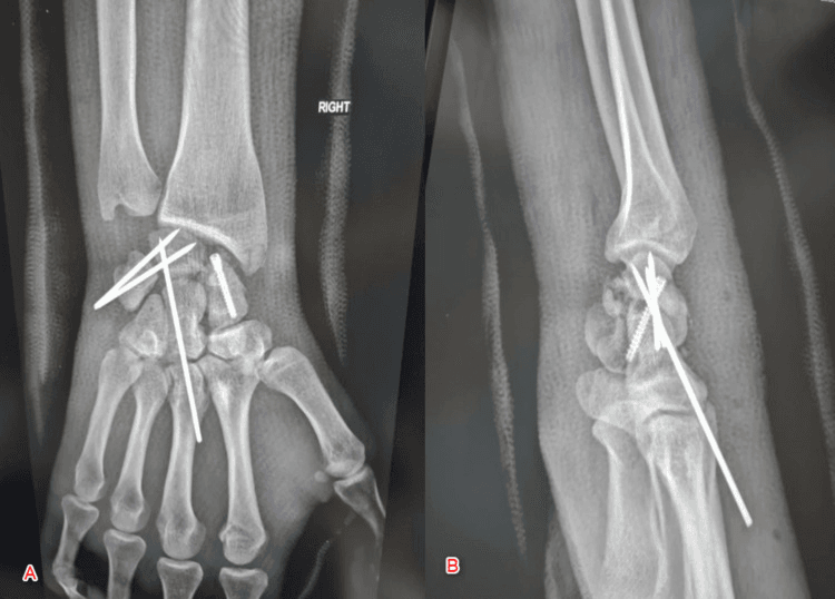
Positioning. Supine, arm on a hand table, upper-arm tourniquet, and exsanguination by elevation rather than an Esmarch bandage where there is concern that a bandage would displace a fracture. The volar approach is performed first and the dorsal second.
Complications
Median nerve injury. Acute compression by the displaced lunate in Stage IV, iatrogenic injury during reduction, or carpal tunnel syndrome postoperatively. It usually improves with reduction and carpal tunnel release.
Infection and failed reduction. Infection carries the standard surgical risk, higher after an open injury. A failure of reduction means interposed tissue, capsule or tendon, or inadequate surgical technique, and may require repeat surgery.
Arthritis and instability. Post-traumatic arthritis is the most common long-term complication and is covered under Outcomes. Carpal instability, recurrent scapholunate or lunotriquetral dissociation, progresses to collapse patterns and may require salvage procedures.
Scaphoid nonunion and lunate AVN. Nonunion complicates trans-scaphoid perilunate injuries, with delay, inadequate fixation and smoking as the risk factors, and requires revision surgery with bone grafting. Avascular necrosis of the lunate is rare but devastating: its blood supply is vulnerable during the injury, it may lead to a Kienböck's pattern, and the salvage options are limited.
Stiffness and CRPS. Some stiffness is expected, and therapy is critical to limit it. Complex regional pain syndrome is rare but serious; it needs early recognition and treatment and a multidisciplinary approach.
Postoperative Care
Strict elevation, multimodal analgesia, and neurovascular checks every 2 hours initially. Acute median neuropathy is common in this injury, so a nerve that was decompressed still needs watching and one that was not needs watching harder. Look for compartment syndrome: rare here but catastrophic. Finger range of motion starts immediately; the wrist is immobilised, the digits are not.
Volar splint or short arm cast with strict wrist immobilisation, continuing finger ROM throughout. First dressing change, wound check and suture removal at 2 weeks.
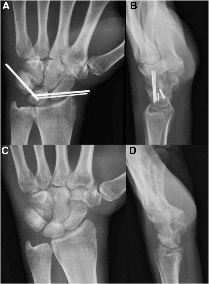
Continue immobilisation; a removable splint is reasonable for hygiene but there is no active wrist motion yet. K-wires stay in situ. Serial radiographs every 2 weeks: this is the window in which secondary loss of reduction declares itself, and it is the commonest complication of the whole injury (10.1% overall, and 24.2% after a pure dislocation versus 7.0% after a fracture-dislocation, Liechti). The pure ligamentous injury is the one to watch hardest, which is counterintuitive.
Radiographs to confirm maintained reduction, K-wires out in clinic under local anaesthesia, and gentle active ROM begins with hand therapy. Avoid forceful grip or loading.
Therapist-supervised progressive ROM and gentle strengthening; heat before exercise is reasonable. Still protect from forceful loading.
Unrestricted ROM, progressive strengthening with weights, putty and grip work, and functional activity.
Return to activity follows the timescales under Outcomes: light duty may be earlier, heavy labour last, and sport by individual progress. Set the expectation of the final range of motion and grip early rather than at this visit. A wrist that has had its perilunate ligament ring fail does not return to normal, and saying so at the first consultation is easier than saying it here.
Radiographs at 6 months and 1 year, then monitor. Counsel as set out under Outcomes: the X-ray will very likely look arthritic, and that does not reliably predict how the wrist will feel.
Outcomes/Prognosis
Range of motion. Expect 50-70% of normal wrist motion in the long term, and the loss is not evenly spread:
- Extension is typically the most affected, with a loss of 30-40 degrees
- Flexion loses 20-30 degrees
- Radial and ulnar deviation are relatively preserved
- Forearm rotation is usually normal
Strength and pain. Grip strength is typically 60-80% of the contralateral side; it improves with therapy and time and plateaus around 12-18 months. Most patients have some residual pain, usually activity-related, which improves with time if no arthritis develops and may worsen if it does.
Return to activities.
- Light work: 3-4 months
- Heavy labour: 6 months minimum
- Contact sports: 6-9 months
- Full recovery plateau: 12-18 months
Prognostic factors. The factors that predict a good or a poor result:
Good prognosis:
- Early treatment (within 7-14 days)
- Anatomic reduction achieved
- Secure ligament repair
- Greater arc injury (paradoxically, because of better bone healing)
- Young patient with good bone quality
- Compliance with therapy
Poor prognosis:
- Delayed diagnosis (over 4 weeks)
- Incomplete or malreduced carpus
- Failed ligament repair
- Lesser arc injury (poorer ligament healing)
- Associated nerve injury
- Multiple carpal fractures
- High-energy mechanism
Post-traumatic arthritis. It develops in 50-70% of patients by 5-10 years: Herzberg found 56% even in wrists treated early, and Liechti confirmed that it increases with follow-up duration. Radiographic arthritis is often asymptomatic initially, and the reassuring half is well evidenced: radiological osteoarthritis did not correlate with reduced wrist function or with patient dissatisfaction, and salvage surgery was needed in only 2.8%. So the counselling line is that the X-ray will very likely look arthritic and that this does not reliably predict how the wrist will feel.
The risk factors for its development:
- Residual carpal malalignment
- Articular cartilage damage at the time of injury
- Chronic instability
- Delayed treatment
Where it appears. The radiocarpal joint is most commonly affected and the midcarpal (capitolunate) joint is also common; the pattern may progress to a scaphoid nonunion advanced collapse (SNAC) or scapholunate advanced collapse (SLAC) equivalent.
Managing symptomatic arthritis. Conservative treatment first, NSAIDs, activity modification and splinting, with corticosteroid injection for temporary relief. The salvage procedures listed under chronic management are for symptomatic arthritis, not radiographic arthritis.
Guidelines, Registries & Global Practice
Global Epidemiology
Perilunate injuries are rare but consistently under-recognised. They constitute roughly 7% of all carpal traumas in a pooled systematic review (van der Oest 2022), and pure carpal-bone dislocations are far less common than the much more frequent scaphoid fracture in population series: in a Singapore cohort of 149 patients with 162 carpal fractures the scaphoid accounted for 99, and only 10 patients had multiple carpal fractures, of which 4 were perilunate fracture-dislocations (Hey 2011, PMID 21276891). Across the literature the demographic is strikingly uniform worldwide: young men injured by high-energy mechanisms (falls from height, road trauma, sport), reflecting the force required to fail the perilunate ligamentous ring. Hey's cohort matches that picture - 132 of 149 were male and 116 were under 40 - and found high-energy mechanism the only parameter associated with multiple carpal fractures (p=0.009).
- Reported Figure
- ~7% of all carpal traumas
- Evidence Source (PubMed)
- van der Oest 2022 (systematic review)
- Reported Figure
- 25% (41 of 166 injuries)
- Evidence Source (PubMed)
- Herzberg 1993 (7-centre multicentre)
- Reported Figure
- Trans-scaphoid perilunate fracture-dislocation (61%)
- Evidence Source (PubMed)
- Herzberg 1993
- Reported Figure
- ~47% across perilunate injuries
- Evidence Source (PubMed)
- Wickramasinghe 2015 (2 trauma centres)
- Reported Figure
- 15.0% overall; secondary loss of reduction 10.1%
- Evidence Source (PubMed)
- Liechti 2023 (550 patients, meta-analysis)
Guideline & Consensus Landscape
No major society publishes a disease-specific clinical practice guideline dedicated to perilunate/lunate dislocations; management rests on body-of-evidence consensus rather than formal AAOS/NICE guidance. The points of broad international agreement are summarised below.
- Position
- Universally recommended; decompress median nerve early
- Basis / Evidence Level
- Consensus; supported by Wickramasinghe 2015 (evidence level III)
- Position
- Operative (open reduction, ligament repair/fixation) for essentially all injuries; closed treatment alone inadequate
- Basis / Evidence Level
- Consensus; Herzberg 1993, Liechti 2023 (level IV)
- Position
- Surgery within ~7 days gives best outcomes; delay beyond 6 weeks worst
- Basis / Evidence Level
- van der Oest 2022 (systematic review, level IV; 16 included studies, qualitative comparison)
- Position
- Combined dorsal-volar widely taught; dorsal-only and arthroscopic-assisted have lower pooled complication rates in selected cases
- Basis / Evidence Level
- Practice variation; Liechti 2023 (level IV)
- Position
- Radiological osteoarthritis is frequent but often well tolerated; salvage rarely needed (2.8%)
- Basis / Evidence Level
- Liechti 2023; Herzberg 1993 (arthritis 56%)
Registries
There is no dedicated international perilunate-injury registry, and arthroplasty registries (AOANJRR, the UK NJR, the American AJRR) do not capture these soft-tissue/carpal injuries because they are not arthroplasty procedures. The best population-level data therefore come from trauma databases and pooled systematic reviews (Liechti 2023; van der Oest 2022) rather than from a formal registry.
Practice Variation & Service Delivery
Internationally, the dominant modifiable factor is timely recognition and referral rather than choice of implant. Practice variation centres on approach (combined dorsal-volar versus dorsal-only versus arthroscopic-assisted reduction and fixation) and on routine versus selective carpal tunnel release; pooled data favour less invasive techniques where the injury pattern allows (Liechti 2023). High-income trauma systems with 24/7 hand-surgery cover and ready CT access achieve earlier definitive fixation, whereas in many settings inter-hospital transfer from regional centres is the rate-limiting step to surgery within the optimal window. Beyond operative fixation, structured hand-therapy rehabilitation drives final outcome, yet its availability is uneven, with rural and remote regions most affected by limited access.
Viva Scenarios
Clinical Decision Scenarios
Practise clinical reasoning and management decisions out loud
“A 35-year-old motorcyclist presents 2 weeks after a fall with persistent wrist pain and weakness. Initial X-rays at another hospital were reported as 'normal'. Current examination shows median nerve paresthesias. You review the original X-rays.”
“A 28-year-old man presents to ED 4 hours after a high-speed motorcycle accident. He has severe wrist pain and reports numbness in his thumb, index, and middle fingers that is worsening. X-rays show a Stage IV lunate dislocation with the lunate tilted into the carpal tunnel.”
“A 32-year-old construction worker fell from scaffolding. X-rays show a perilunate dislocation with a scaphoid waist fracture. The carpus is displaced dorsally with the lunate remaining in the lunate fossa. No median nerve symptoms.”
MCQ Practice Points
Q: In Mayfield Stage III perilunate instability, where is the lunate located?
A: In Stage III (perilunate dislocation), the lunate remains in the lunate fossa of the radius. The capitate and rest of the carpus dislocate dorsally around it. In Stage IV (lunate dislocation), the lunate tips volarly out of the fossa.
Q: What is the "spilled teacup" sign?
A: The spilled teacup sign is seen on lateral X-ray in Stage IV lunate dislocation. Normally, the lunate looks like a cup holding the capitate. When the lunate dislocates volarly and rotates, it appears to have "spilled" out of its normal position - pathognomonic for lunate dislocation.
Q: What is the difference between greater and lesser arc injuries in perilunate dislocations?
A: Lesser arc injuries are pure ligamentous - the injury arc passes through the SL ligament, around the lunate, and through the LT ligament. Greater arc injuries involve fractures - the arc passes through bones (most commonly trans-scaphoid). Greater arc injuries have fractures that need fixation in addition to ligament repair.
Q: What is the urgency of treatment for a Stage IV lunate dislocation with median nerve symptoms?
A: This is a surgical emergency. The volarly dislocated lunate compresses the median nerve in the carpal tunnel. Urgent closed reduction should be attempted immediately (within hours), and definitive surgical treatment with carpal tunnel release is required. Permanent median nerve damage can occur with compression beyond 6-8 hours.
Q: What percentage of perilunate/lunate dislocations are missed on initial presentation?
A: 25% (one quarter) of these injuries are missed initially. This emphasizes the importance of careful examination of the lateral X-ray and maintaining high clinical suspicion. The lunate should articulate with both the radius and the capitate on a normal lateral view.
Mayfield Stages
- Stage I: SL dissociation
- Stage II: Capitolunate dissociation
- Stage III: Perilunate dislocation (lunate in fossa, carpus dorsal)
- Stage IV: Lunate dislocation (lunate volar = spilled teacup)
Key X-ray Findings
- Lateral: Lunate should articulate with radius AND capitate
- PA: Disrupted Gilula's arcs, triangular lunate
- Spilled teacup = Stage IV lunate dislocation
- 25% missed on initial X-ray
Greater vs Lesser Arc
- Lesser arc: Pure ligamentous (SL, LT)
- Greater arc: Fractures through bones (trans-scaphoid most common)
- Greater arc needs fracture fixation + ligament repair
Emergency Management
- Assess median nerve - acute CTS is emergency
- Closed reduction: TRAP (Traction, Recreate, Apply pressure, Push)
- Closed reduction is temporizing only
- All require definitive surgical treatment
Surgical Principles
- Combined volar + dorsal approach preferred
- CTR mandatory (even without acute CTS)
- K-wire fixation: SL, SC, LT
- Greater arc: Screw fixation of fractures + ligament repair
Evidence Base
Carpal Dislocations: Pathomechanics & Progressive Perilunar Instability (Landmark)
- Cadaveric study loading 32 wrists to failure produced 13 perilunate and 2 lunate dislocations via a reproducible mechanism of extension, ulnar deviation and intercarpal supination.
- Established the four sequential stages of perilunar instability (PLI), from scapholunate diastasis (Stage I) to complete lunate dislocation (Stage IV), with progressively greater carpal instability.
- Reduction was achieved by reversing the mechanism (intercarpal pronation, radial deviation, palmar flexion).
Perilunate Dislocations & Fracture-Dislocations: Multicentre Study (Landmark)
- 166 injuries from 7 centres, retrospectively studied; the diagnosis was MISSED INITIALLY IN 41 CASES (25%).
- Displacement was dorsal in 161 (97%) and palmar in only 5 (3%); fracture-dislocations outnumbered pure dislocations 2:1, and dorsal trans-scaphoid perilunate fracture-dislocation was 96% of dorsal fracture-dislocations and 61% of the whole series.
- Outcome analysed in 115 cases at minimum 1 year and mean 6 years 3 months: open injury and delay of treatment adversely affected clinical results, whereas anatomical type mattered less.
- Even in cases treated EARLY the clinical results were satisfactory but post-traumatic arthritis reached 56%.
- Fixing the scaphoid ALONE was not always sufficient - it left residual scapholunate dissociation, lunotriquetral dissociation, ulnar carpal translation or other collapse patterns, so appraise the ligamentous injury as well as the fracture.
Outcomes of Acute Perilunate Injuries: Systematic Review & Meta-Analysis
- Twenty-six studies pooling 550 patients with 553 operatively treated acute injuries (106 pure dislocations, 447 fracture-dislocations) - the largest pooled surgical dataset.
- Overall complication rate 15.0%, driven by secondary loss of reduction (10.1%); reoperation 10.4% and salvage procedures only 2.8%.
- Secondary loss of reduction was HIGHER for pure dislocations than fracture-dislocations (24.2% vs 7.0%, RR 3.5, 95% CI 1.6-7.5) - the pure ligamentous injury is the less stable one after fixation.
- APPROACH AND INVASIVENESS MATTERED: the combined dorsopalmar approach carried more complications than an isolated dorsal approach (17.4% vs 8.4%, RR 0.5, 95% CI 0.2-1.0, NNT 11.2), and open surgery more than arthroscopic (17.4% vs 4.8%, RR 0.3, 95% CI 0.1-0.9, NNT 8.0).
- Radiological osteoarthritis was common and increased significantly with follow-up duration, but did NOT correlate with reduced wrist function or patient dissatisfaction, and salvage surgery was rare.
Perilunate Injury Timing & Treatment Options: Systematic Review
- Perilunate injuries represent approximately 7% of all carpal traumas and are frequently missed; only 16 of 2,056 screened articles met inclusion.
- Timing was stratified as acute (under 7 days), delayed (7-45 days) and chronic (over 45 days). Outcomes are good when surgery is within 7 days, inferior with any delay, and worst beyond 6 weeks.
- Chronic PURE dislocations fare worse than chronic fracture-dislocations - the same direction as Liechti's reduction-loss finding.
- For fracture-dislocations the review suggests CLOSED reduction and internal fixation may give slightly better outcomes than open reduction and internal fixation - a direction that agrees with the modern preference for less invasive fixation.
Acute Median Neuropathy & Carpal Tunnel Release in Perilunate Injuries
- Across 71 patients with perilunate dislocation or fracture-dislocation over 10 years at two trauma centres, acute median neuropathy was diagnosed in 33 (47%).
- NO demographic or injury factor was associated with developing median nerve symptoms - the only significant difference between the dislocation and fracture-dislocation groups was younger age with fracture-dislocation.
- Carpal tunnel release at the initial operation was related to the presence of median nerve symptoms AND to which trauma centre treated the patient - so institutional habit, not just the neurology, determined who was decompressed.