The Infiltrative Sarcoma of the Elderly
- Myxofibrosarcoma (MFS, formerly 'myxoid malignant fibrous histiocytoma') is one of the COMMONEST soft-tissue sarcomas of the ELDERLY (typically 60s-80s), arising in the subcutaneous or deep soft tissues of the EXTREMITIES (lower limb more than upper) and trunk; histologically it has a MYXOID stroma with curvilinear blood vessels and pleomorphic spindle cells.
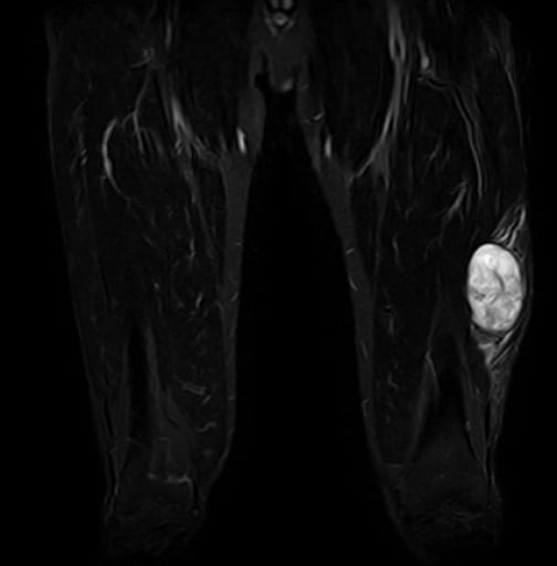
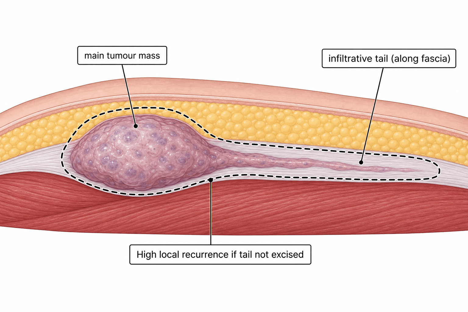
- Its defining feature is an INFILTRATIVE GROWTH PATTERN that extends along FASCIAL planes far beyond the clinically/radiologically obvious mass - seen on MRI as the 'TAIL SIGN' (a fascial 'tail' of tumour) - which makes the true extent easy to UNDERESTIMATE and underlies its VERY HIGH LOCAL RECURRENCE rate.
- On MRI the lesion is typically T2/STIR HYPERINTENSE (reflecting the myxoid content); the presence and extent of an infiltrative tail and of perilesional oedema are prognostically important, and DIFFUSE perilesional oedema is associated with WORSE survival - so detailed pre-operative MRI is essential to plan an adequate resection.
- MFS spans a GRADE spectrum from low to high; LOCAL RECURRENCE is high across grades, while DISTANT METASTASIS (especially to the LUNG) increases with grade, and low-grade tumours can recur at a HIGHER grade over time.
- TREATMENT is WIDE LOCAL EXCISION with GENEROUS margins planned on pre-operative MRI to encompass the infiltrative tail - inadequate margins frequently cause recurrence - usually combined with RADIOTHERAPY (neoadjuvant or adjuvant) for high-grade or large/deep tumours; chemotherapy has a limited role.
- Because of the infiltrative tail and high recurrence, these patients need MULTIDISCIPLINARY (sarcoma MDT) care, meticulous margin assessment, and LONG-TERM follow-up with surveillance imaging for both local recurrence and pulmonary metastases.
- “Myxofibrosarcoma = common soft-tissue sarcoma of the ELDERLY (extremities); myxoid stroma + curvilinear vessels.
- “Infiltrative growth along fascia -> MRI 'TAIL SIGN' (T2/STIR-bright myxoid mass) -> margins underestimated -> very high LOCAL recurrence.
- “Treat with WIDE excision (MRI-planned generous margins) + radiotherapy; metastatic risk (lung) rises with grade; long-term follow-up.
MFS spreads with an infiltrative tail along fascia beyond the visible mass (the MRI 'tail sign'), so margins are underestimated -> very high local recurrence.
Pre-operative MRI to map the tail, wide excision with generous margins to encompass it, plus radiotherapy for high-grade/large tumours.
Presentation, Imaging & Behaviour
Myxofibrosarcoma typically presents as a slowly enlarging soft-tissue mass in the extremity (often subcutaneous) of an elderly patient. Histologically it has a myxoid stroma with characteristic curvilinear vessels and pleomorphic spindle cells, and it spans a spectrum from low to high grade. Its hallmark is the infiltrative growth along fascial planes that produces the MRI 'tail sign' - a fascial tail of tumour extending beyond the main mass - so the true extent is easily underestimated. On MRI the lesion is typically T2/STIR hyperintense (myxoid content), and diffuse perilesional oedema is associated with a worse prognosis. The infiltrative behaviour drives a very high local recurrence rate, while distant metastasis (especially lung) increases with grade.
Management
- Pre-operative MRI to define the mass AND the infiltrative tail and perilesional oedema, and to plan the resection.
- Wide local excision with generous margins that encompass the infiltrative tail - because the extent is underestimated, inadequate margins are the main cause of recurrence.
- Radiotherapy (neoadjuvant or adjuvant) for high-grade, large or deep tumours and for close/positive margins (see our Radiotherapy in MSK Tumours topic for the neoadjuvant-vs-adjuvant trade-off).
- Chemotherapy has a limited role (selected high-grade/advanced disease).
- Multidisciplinary sarcoma care and long-term follow-up with surveillance for local recurrence and pulmonary metastases.
The recurring error with myxofibrosarcoma is to excise to the apparent edge of the mass and miss the infiltrative fascial tail, which leads to repeated local recurrence. Always assess the full extent on pre-operative MRI, plan a wide excision that includes the tail, and obtain clear histological margins; treat high-grade or large tumours within a sarcoma multidisciplinary team with radiotherapy, and follow patients long-term for both local recurrence and lung metastases. Remember that a low-grade lesion can recur at a higher grade, so recurrence should be re-staged and re-graded rather than assumed to be the same disease.
Evidence & Key Studies
Preoperative MRI evaluation of myxofibrosarcoma: prognostic value of imaging features
- Myxofibrosarcoma is characterised by an infiltrative growth pattern; the 'tail sign' was present in fewer than half of cases on MRI.
- Diffuse perilesional oedema on MRI was significantly associated with poorer overall survival (vs circumscribed oedema).
- Detailed pre-operative MRI with planning of the resection is a logical approach to achieve negative margins and recurrence-free survival.
Giant myxofibrosarcoma: infiltrative growth, wide margins and local recurrence
- Myxofibrosarcoma commonly arises in the extremities and has a high risk of local recurrence due to its infiltrative growth pattern; MRI showed the 'tail sign'.
- Wide-margin resection is essential, and insufficient margins frequently result in recurrence (this high-grade case recurred and was fatal).
- Highlights the importance of wide surgical margins given extensive infiltration, and the need for effective adjuvant therapy.
According to PubMed, the infiltrative growth/tail sign, the prognostic value of perilesional oedema and the role of pre-operative MRI in planning margins come from the cited Muhlhofer study, and the high local recurrence with infiltrative growth and the need for wide margins from the cited Zhong case/review. The elderly/ extremity demographic, the myxoid histology, the grade-dependent metastatic risk and adjuvant radiotherapy are standard, well-established soft-tissue-sarcoma teaching. (See also our Soft-Tissue Sarcoma Referral, Radiotherapy in MSK Tumours and Liposarcoma topics.)
Clinical Decision Scenarios
Practise clinical reasoning and management decisions out loud
“What is myxofibrosarcoma, what is the 'tail sign', and why does it have a high local recurrence rate?”
“How would you manage a high-grade myxofibrosarcoma of the thigh?”
Mnemonics & Memory Aids
TAIL
Hook:Myxofibrosarcoma = the TAIL sarcoma of the elderly.
MYXOID
Hook:MYXOID: bright on MRI, elderly, eXtensive tail, oedema = poor prognosis.
Demographics & histology
- One of the commonest soft-tissue sarcomas of the ELDERLY (60s-80s)
- Extremities (lower more than upper) and trunk; subcutaneous/deep
- Myxoid stroma + curvilinear vessels + pleomorphic spindle cells; low-to-high grade
Imaging
- MRI: T2/STIR hyperintense (myxoid)
- 'Tail sign' = infiltrative fascial spread beyond the mass
- Diffuse perilesional oedema = worse prognosis
Behaviour
- Very high LOCAL recurrence (infiltrative tail underestimates extent)
- Distant metastasis (lung) increases with grade
- Low grade can recur at higher grade
Treatment
- Pre-operative MRI planning -> wide excision with generous margins (encompass tail)
- Radiotherapy (neo/adjuvant) for high-grade/large; chemo limited
- Sarcoma MDT; long-term surveillance (local + lung)