Antibiotic Protocol | Wound Assessment | Soft Tissue Coverage | Fix-or-Flap
- Gustilo-Anderson is assessed INTRAOPERATIVELY - not in ED (wounds deceive)
- Antibiotics within 1 hour - reduces infection more than timing of surgery
- 6-hour rule is MYTH - early antibiotics matter, surgery timing less critical
- IIIB requires flap coverage - involve plastics within 72 hours (fix-or-flap)
- IIIC = vascular injury - revascularisation priority, consider amputation if over 6h warm ischaemia
- Farm injuries = add penicillin - Clostridium coverage for gas gangrene
- “Final classification is AFTER debridement in theatre - initial wound size often misleading
- “Antibiotics beat timing - can wait for ideal theatre conditions if antibiotics given early
- “4 Cs of muscle viability: Colour, Contractility, Consistency, Capacity to bleed
- “NPWT is bridge therapy - definitive coverage still needed by 72 hours
Overview and Epidemiology
The tibial diaphysis is the most common open long-bone fracture, and the reason is anatomical: its anteromedial surface is subcutaneous, so there is very little between the bone and the skin.
How common. Open fractures are roughly 3% of all fractures in a large epidemiological series (Court-Brown et al, 2386 open fractures over 15 years). Most are not the high-energy injuries the textbooks picture: only 22.3% are caused by road-traffic accidents or falls from height, and the rest are predominantly low-energy injuries, especially in older patients. The pattern is bimodal, with high-energy open fractures clustering in younger males and low-energy open fractures in older females.
Mechanism. Motor-vehicle and motorcycle crashes, pedestrian-versus-vehicle injuries and falls from height produce the larger, more contaminated Type III injuries. Simple falls and twisting injuries can still breach the skin over the subcutaneous tibia, and these are often Type I. Agricultural, marine and combat injuries carry distinct organism profiles and demand broader antibiotic cover.
Distribution by grade.
- Type I - 15-20%
- Type II - 35-40%
- Type IIIA - 25-30%
- Type IIIB - 10-15%
- Type IIIC - 5-10%
The grade also predicts infection, union and amputation; those figures are set out together under Outcomes.
Anatomy and Pathophysiology
Why the tibia is vulnerable. Three things. The anteromedial surface is subcutaneous, with minimal soft tissue over the medial tibia. The distal third is a watershed zone with a poor blood supply. And the tibia is the single weight-bearing bone of the leg, so the mechanical demands on it are high.
Blood supply. The nutrient artery enters the posterior cortex and runs proximally, supplying the inner two-thirds of the cortex; periosteal vessels supply the outer third. Intramedullary nailing disrupts the endosteal supply, but healing is usually adequate.
The compartments. The leg has four, and all of them are at risk in an open tibial fracture:
- Anterior - tibialis anterior, EDL, EHL, deep peroneal nerve
- Lateral - peroneus longus and brevis, superficial peroneal nerve
- Superficial posterior - gastrocnemius, soleus, plantaris
- Deep posterior - tibialis posterior, FDL, FHL, posterior tibial nerve and vessels
The zone of injury. It extends beyond the visible wound margins, and high-energy mechanisms create larger zones of devitalised tissue. Periosteal stripping compromises the blood supply to the bone.
Contamination and infection. Contamination is the presence of bacteria, and it is inevitable. Infection is bacterial multiplication with tissue invasion. The whole of open fracture care is aimed at stopping the first becoming the second, and the clock starts at the injury:
- 0-6 hours - contamination phase: bacteria present but not established
- 6-12 hours - bacterial multiplication begins
- Over 12 hours - biofilm formation, infection establishing
Early antibiotics prevent bacterial establishment regardless of when the surgery happens.
Classification Systems
Gustilo-Anderson. The universal language, and it is assigned in theatre. The grade is a judgement made after debridement and thorough wound inspection, because wounds are deceptive: a small external wound can mask severe deep injury, and the extent of the deep injury is only apparent at surgery. Never definitively classify from the emergency department; document the final classification in the operation note.
- Wound
- Under 1cm, usually a puncture, inside-out pattern
- Energy
- Low
- Soft tissue
- Minimal muscle damage, clean
- Contamination
- Minimal
- Wound
- 1-10cm
- Energy
- Moderate
- Soft tissue
- Moderate damage, no extensive periosteal stripping; bone coverage usually adequate
- Contamination
- Some
- Wound
- Over 10cm, or a segmental fracture
- Energy
- High
- Soft tissue
- Extensive, but muscle can still cover the bone
- Contamination
- May be significant
- Wound
- Over 10cm
- Energy
- High
- Soft tissue
- Extensive periosteal stripping; bone or hardware exposed; requires a flap
- Contamination
- Often severe
- Wound
- Variable
- Energy
- Very high
- Soft tissue
- Variable; arterial injury requiring repair
- Contamination
- -
Type III Subclassification - Defining the IIIA/B/C Split
- 87 Type III open fractures. The original single Type III grouping was too broad, so it was split into IIIA (adequate soft-tissue cover), IIIB (extensive soft-tissue loss with periosteal stripping/bone exposure) and IIIC (arterial injury requiring repair). Wound-sepsis rates were 4%, 52% and 42% and amputation rates 0%, 16% and 42% respectively.
IIIA or IIIB. This is the distinction that decides whether a flap is needed, and both grades start from the same wound: over 10cm or segmental, high energy. The question is whether the bone can be covered. In IIIA the periosteum may be stripped but muscle is present, closure is primary if the wound is clean and otherwise by delayed primary closure or a local flap, and plastics are involved case by case. In IIIB the stripping is significant, bone or hardware is exposed with nothing to cover it, the wound is often severely contaminated, and a flap is required, so early plastics referral is mandatory.
Type IIIC. Defined by an arterial injury requiring repair, regardless of the size of the wound, and limb-threatening for that reason. Suspect it when:
- Distal pulses are absent or diminished
- The foot is cool and pale
- Capillary refill is prolonged over 3 seconds
- The ankle-brachial index is under 0.9, if it can be measured
Warm ischaemia time over 6 hours leads to a high amputation rate. Revascularisation comes before or concurrent with skeletal stabilisation, and if there is a delay to definitive repair a shunt is considered for temporary perfusion.
Gustilo-Anderson is the universal language, but a strong answer also states its major limitation and the modern alternative.
- Poor interobserver reliability: agreement on the Gustilo-Anderson grade is only moderate at best (kappa often around 0.5-0.6), and it lumps several distinct injury features into a single grade. It is also a post-debridement judgement, so it cannot be applied reliably in the ED.
- The OTA Open Fracture Classification (OTA-OFC) was created to address this. Instead of one grade it scores each injury component separately - skin, muscle, arterial injury, contamination, and bone loss - each graded (e.g. mild/moderate/severe). This captures, for example, that a small skin wound can accompany severe muscle devitalisation, and it has shown better reliability and prognostic value for outcomes such as amputation, infection and the need for flap coverage.
- How to use both: still classify Gustilo-Anderson (everyone speaks it and it drives antibiotic choice), but recognise it is imperfect and that component-based grading (OTA-OFC) better describes the injury and predicts outcome.
Clinical Assessment
Open fractures are limb-threatening injuries but ATLS principles still apply. Complete primary and secondary survey before focusing on the open fracture. Associated injuries are common in high-energy trauma.
Primary survey. Polytrauma is common with high-energy mechanisms, so the ATLS assessment comes first. Control active haemorrhage, and assess for compartment syndrome (pain out of proportion, pain on passive stretch) from the outset.
The limb. Six steps, in this order:
- Neurovascular examination - documented before and after any manipulation
- Photograph the wound - a single high-quality photograph with a ruler for scale, then cover it
- Cover with saline-soaked gauze under an impervious dressing - do not probe or explore in the emergency department
- Align and splint - reduces bleeding, pain and further contamination
- Check tetanus status - immunise if not up to date
- Antibiotics within 1 hour of presentation
What not to do. Every repeat inspection of the wound increases contamination and probing introduces bacteria, so the wound is photographed once and left covered. Iodine-soaked gauze is controversial and packing with betadine has debated tissue toxicity, so plain saline is used. Nothing delays the antibiotics.
What else it might be. The diagnosis of an open fracture is usually obvious, but the critical reasoning is distinguishing what you are actually dealing with, because each scenario changes management.
- Discriminating Features
- Skin intact when cleaned; no communication with fracture
- Why It Matters
- Not an open fracture - antibiotic/theatre urgency differs
- Discriminating Features
- Wound communicates with fracture; fat globules/bone visible
- Why It Matters
- Needs early antibiotics, debridement, coverage
- Discriminating Features
- Pain out of proportion, pain on passive stretch, tense compartments - can coexist with open fracture
- Why It Matters
- Open wound does NOT decompress all compartments; needs fasciotomy
- Discriminating Features
- Absent pulses, cool/pale foot, ABI under 0.9 after realignment
- Why It Matters
- Limb-threatening - revascularisation priority
- Discriminating Features
- Non-reconstructable bone/soft tissue, prolonged warm ischaemia, unstable polytrauma
- Why It Matters
- Salvage may be worse than amputation - shared decision
- Discriminating Features
- Crepitus, rapidly spreading erythema, systemic toxicity, pain out of proportion
- Why It Matters
- Surgical emergency - radical debridement, not routine cover
Investigations and Imaging
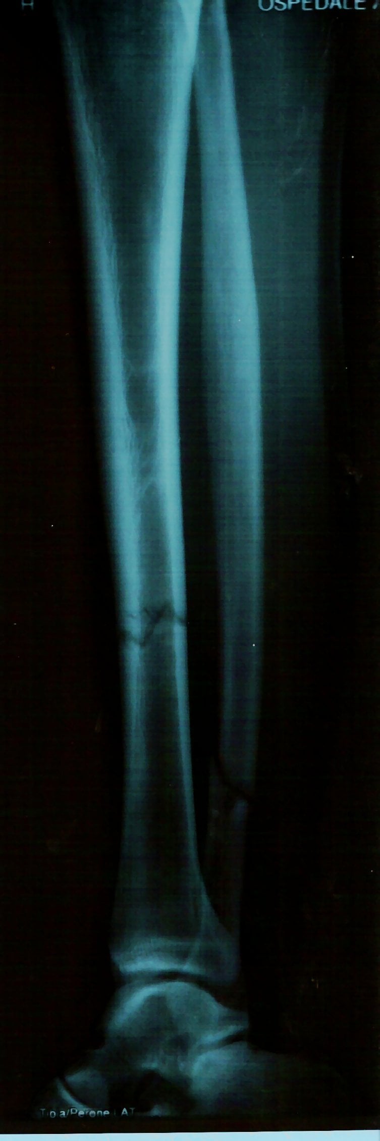
Radiographs. AP and lateral of the tibia, including the knee and ankle joints. CT is added if intra-articular extension is suspected. For a suspected IIIC the choice is angiography or going straight to theatre.
Management
In the Emergency Department
Early antibiotics are the single most important intervention to prevent infection. Studies consistently show antibiotics within 1 hour reduce infection more than timing of surgical debridement. Start in ED - do not wait for theatre.
The regimen. Cefazolin covers the gram-positives, above all Staphylococcus aureus, and every open fracture gets it. Gentamicin adds gram-negative cover for the severe Type III wound. Penicillin is added for farm injuries, for the Clostridia of tetanus and gas gangrene, and water contamination is the prompt to consider a fluoroquinolone for gram-negatives.
- First-Line Regimen
- Cefazolin 2g IV 8-hourly
- Duration
- 24-48h post-closure
- Additional Coverage
- -
- First-Line Regimen
- Cefazolin 2g IV 8-hourly
- Duration
- 24-48h post-closure
- Additional Coverage
- -
- First-Line Regimen
- Cefazolin 2g + Gentamicin 5mg/kg
- Duration
- 72h or until closure
- Additional Coverage
- -
- First-Line Regimen
- Above + Penicillin G 4MU IV 4-6 hourly
- Duration
- As above; penicillin for 7 days
- Additional Coverage
- Clostridium
- First-Line Regimen
- Consider fluoroquinolone
- Duration
- As above
- Additional Coverage
- Gram negatives
If the patient is penicillin-allergic:
- Type I-II: clindamycin 900mg IV 8-hourly
- Type III: clindamycin plus an aminoglycoside or aztreonam
Tetanus. The wounds at high risk are those contaminated with soil, faeces or saliva, puncture wounds, wounds with devitalised tissue present, and wounds over 6 hours old. Prophylaxis depends on what the patient has already had:
- Clean, Minor Wound
- Tdap
- Other Wounds
- Tdap + TIG
- Clean, Minor Wound
- Tdap
- Other Wounds
- Tdap
- Clean, Minor Wound
- None
- Other Wounds
- Tdap
- Clean, Minor Wound
- None
- Other Wounds
- None
TIG is tetanus immune globulin, 250 IU IM.
Booking theatre. Type I-II go urgently, within 24 hours; Type III the same day; Type IIIB with plastics warned as soon as it is suspected and consulted on day one; Type IIIC immediately, with vascular. Have an external fixator available and make sure of adequate lighting and irrigation. There is no six-hour rule: once antibiotics are in early the timing of surgery matters less, and the operation can wait for appropriate conditions and senior input rather than be rushed overnight.
The Principles of the Operation
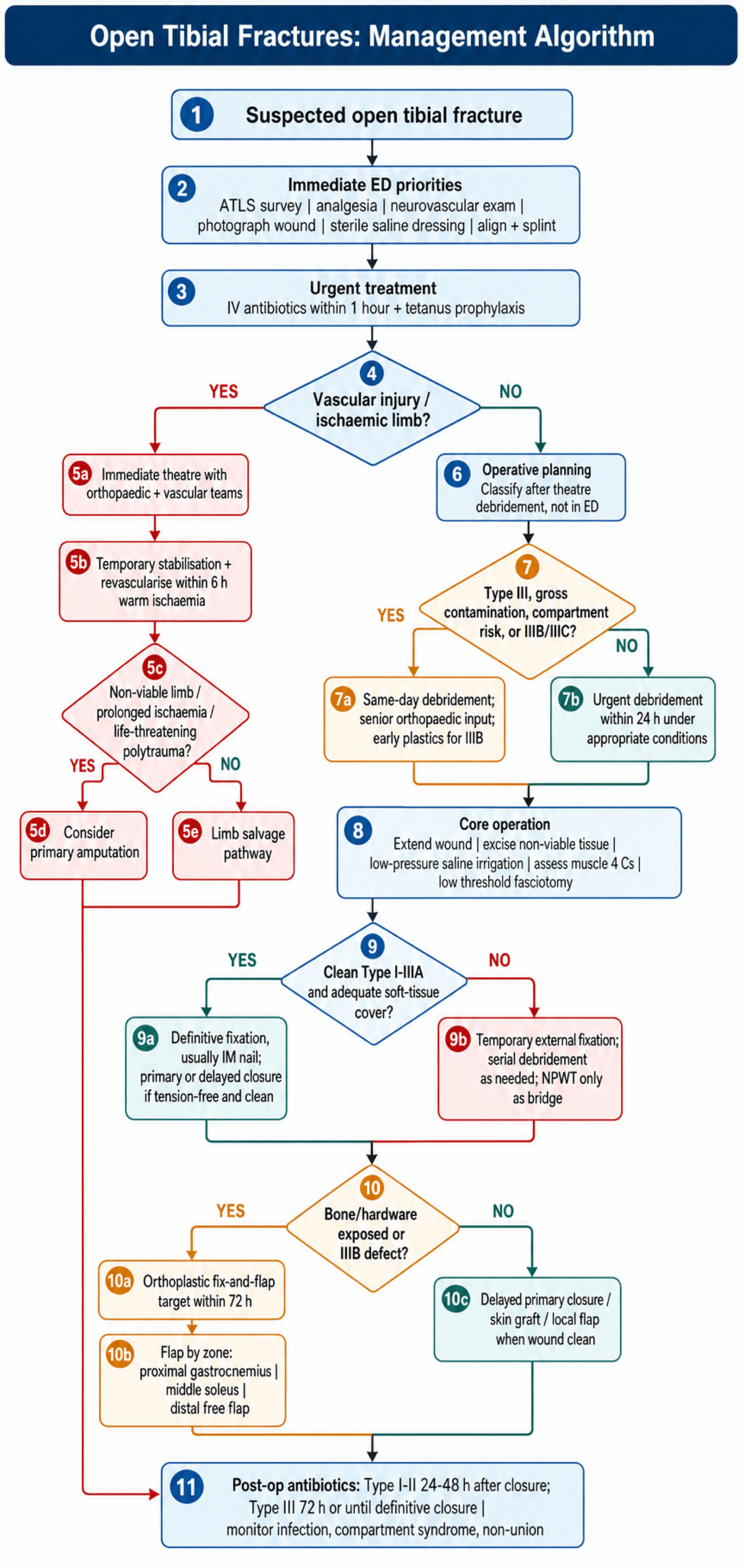
Conservative management is not appropriate for an open tibial fracture. Every one requires surgical debridement, skeletal stabilisation and definitive soft tissue coverage; the decisions are about how and when.
- Primary or staged fixation, which depends on contamination
- Primary or delayed wound closure
- Whether a flap is needed, which for IIIB is always
Wound Closure
By grade. The closure follows the grade assigned in theatre:
- Type I - primary closure is often possible; delayed primary closure at 3-5 days if there is any contamination
- Type II - delayed primary closure preferred, at 48-72 hours if the wound is clean; split-thickness skin graft if there is tension
- Type IIIA - may close primarily if clean; more commonly delayed primary closure or a local flap
- Type IIIB - cannot be closed, because bone is exposed; a muscle flap with or without a skin graft, targeted by 72 hours
Negative pressure wound therapy. NPWT is applied after debridement as a bridge to definitive coverage, and it is not definitive treatment. It reduces oedema, removes exudate, decreases the bacterial load and promotes granulation. Run it at continuous -75 to -125 mmHg and change it every 48-72 hours with the wound inspected at each change. It must not be left over exposed bone indefinitely; definitive coverage is still needed by 72 hours ideally.
Serial debridement. A planned second look at 24-48 hours is booked for:
- Severe contamination
- Questionable tissue viability
- Type IIIB and IIIC injuries
- A large zone of injury
Re-debride as needed and continue antibiotics until the wound is closed. Type IIIB injuries need on average 2-3 debridements.
Surgical Technique
Debridement
Extend the wound. The zone of injury is larger than the hole in the skin, so the wound is extended until healthy tissue is seen in every direction. Use longitudinal incisions, which respect the fasciocutaneous perforators, and avoid transverse incisions across the tibia.
Skin and subcutaneous tissue. Excise the non-viable skin edges, but keep the excision limited: more can always be taken later. Remove contaminated subcutaneous tissue with sharp debridement.
Fasciotomy. Keep a low threshold for prophylactic fasciotomy in Type III injuries and release all four compartments if there is any concern about compartment syndrome; it is better done and not needed than missed. The two-incision technique uses anterolateral and posteromedial incisions.
Muscle. Viability is judged on the four Cs, and all four must be present for muscle to be kept. Err on the side of debridement, because necrotic muscle causes infection; serial debridement is often needed.
4 CsMuscle Viability Assessment
Hook:All 4 Cs must be present - debride any muscle that fails ANY criterion!
Bone. Completely loose, devitalised fragments without soft tissue attachment are removed. Fragments that keep any periosteal blood supply are preserved, and large structural fragments may need to stay despite questionable viability. Strip as little periosteum from viable bone as possible.
Irrigation. High volume at low pressure, from a bulb syringe or gravity 30cm above the wound, in normal saline. Soap additives give no benefit (FLOW), and pulsatile lavage may drive bacteria deeper, so it is avoided. Volume scales with the grade:
- Type I - 3L
- Type II - 6L
- Type III - 9L or more
Fixation
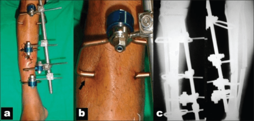
The fixation follows the wound. A clean Type I-IIIA fracture after adequate debridement is nailed in one stage; a IIIB or IIIC injury, a heavily contaminated wound or a damage-control situation is spanned with an external fixator and converted later. Plates have a very limited role.
Who. Type I-IIIA fractures with adequate debridement, a clean wound bed after irrigation and no gross contamination. The wound bed must be clean before the implant goes in.
Technique. Suprapatellar or infrapatellar approach, with blocking screws for short segments or metaphyseal extension. Reamed versus unreamed is debated: reamed gives better stability, and in open fractures SPRINT found no difference in reoperation between the two, so the open injury itself should not decide it.
Why it is the default. Definitive fixation in one stage, a load-sharing construct that allows early weight-bearing, minimal additional soft tissue trauma, and an infection rate lower than the historical data suggest. The infection risk in a contaminated wound is mitigated by adequate debridement; knee pain is the other complication, and the suprapatellar approach reduces it.
Soft Tissue Coverage
Fix and flap. The evidence for early coverage is Godina's, and it set the 72-hour target.
Godina Principle - Fix and Flap Within 72 Hours
- 532 extremity reconstructions stratified by timing. Free-flap failure 0.75% when transferred within 72 hours vs 12% at 72h-3 months. Post-operative infection 1.5% vs 17.5%. Bone-healing 6.8 vs 12.3 months and hospital stay 27 vs 130 days favouring early coverage.
Timing. Coverage within 72 hours is the optimal window, with the lowest infection and flap failure. Between 72 hours and 7 days the risk is intermediate and outcomes are still reasonable. Beyond 7 days infection, flap failure and non-union all rise.
Which flap. The choice is decided by the third of the tibia that needs covering. The distal third is the watershed zone with poor local options, which is why it is the free-flap zone.
- Primary Flap
- Gastrocnemius rotation
- Alternative
- Free flap
- Notes
- Medial head most common
- Primary Flap
- Soleus muscle flap
- Alternative
- Gastrocnemius if reach
- Notes
- Longer arc of rotation
- Primary Flap
- Free tissue transfer
- Alternative
- Propeller flaps if small
- Notes
- Watershed zone - poor local options
Reach. Proximal tibial coverage, and it can reach to the mid-tibia in some cases.
Technique. The medial head is used most often because it is larger. It is based on the sural artery from the popliteal, harvested through a medial incision, rotated into the wound and skin-grafted over the muscle.
Outcome. A reliable blood supply and minimal donor site morbidity, with some ankle plantarflexion weakness.
Complications
Infection. The risk factors:
- Type IIIB or IIIC classification
- Antibiotics delayed beyond 1 hour
- Inadequate debridement
- Soft tissue coverage delayed beyond 72 hours
- Smoking, diabetes, peripheral vascular disease
- Farm or water contamination
Prevention is key: early antibiotics and adequate debridement. Once infection is established, antibiotics become culture-directed, repeat debridement may be needed, antibiotic beads or cement spacers are considered, and the hardware is removed if infection persists.
Non-union. Type III injury, bone loss, infection, smoking and inadequate fixation all predispose. The options are:
- Exchange nailing
- Plate augmentation
- Bone grafting (autograft, RIA)
- Masquelet technique for segmental defects
- Free vascularised fibula for massive defects
Open fractures can still develop compartment syndrome! The wound does not adequately decompress all 4 compartments. Maintain high index of suspicion and low threshold for fasciotomy in Type III injuries.
Amputation. Primary amputation is considered for a crush injury with a non-viable limb (MESS over 7), prolonged warm ischaemia over 6 hours, severe polytrauma where limb salvage compromises life, or a mangled extremity with no prospect of function. The Mangled Extremity Severity Score adds up skeletal and soft tissue injury, limb ischaemia, shock and patient age; a score over 7 predicts amputation, but it is an imperfect predictor, which is the point LEAP made.
- Function is similar either way: at 2 and 7 years, patients who underwent limb salvage and those who had amputation had broadly equivalent functional outcomes (Sickness Impact Profile scores), with high complication and re-operation rates in both groups - salvage is not automatically the "better" choice.
- Severity scores poorly predict and should not dictate the decision: LEAP found that lower-extremity injury-severity scores (including the MESS) had poor sensitivity/specificity for who ultimately needs amputation - a high MESS supports but must not mandate amputation. The earlier "MESS over 7 = amputate" teaching is too rigid.
- What actually drives outcome: patient and psychosocial factors - return to work, self-efficacy, social support, education, smoking, and the presence of an insensate plantar foot (and notably, initial plantar insensation does NOT reliably predict a poor long-term sole) - influenced outcome more than the limb decision itself.
- Implication: the salvage-versus-amputation decision is a shared, multidisciplinary, patient-centred decision (orthoplastic + vascular + patient), informed by but not dictated by scores; counsel that a salvaged limb often means multiple operations and a long rehabilitation for function comparable to a well-fitted prosthesis.
Exam point: quote LEAP - salvage and amputation give similar long-term function, injury-severity scores (MESS) predict poorly and should guide not dictate, and psychosocial factors dominate outcome - so the decision is a shared one, not a number.
Postoperative Care
Antibiotics. Type I-II antibiotics can be discontinued once the wound is closed and clean. Type III cover may be extended if there are ongoing wound issues, but there is no benefit beyond 72 hours in a closed wound. Farm injuries continue penicillin for 7 days, and carry a higher risk of late clostridial complications.
The first 48 hours. Watch for compartment syndrome, which is still possible despite the open wound. Neurovascular checks are 4-hourly, and fever, increasing pain or discharge are the signs of infection to watch for; if there is concern, consider a second look.
Dressings. NPWT is changed every 48-72 hours with the wound inspected and its progression toward closure documented at each change, and the definitive coverage is planned within 72 hours.
Weight-bearing. It depends on the fixation and on whether a flap is healing:
- External fixator - touch weight-bearing initially, progressing as tolerated once callus is visible; usually 6-8 weeks to partial weight-bearing
- IM nail - touch to partial weight-bearing initially, full weight-bearing once callus bridges 3 cortices; usually 8-12 weeks for Type I-II and 12-16 weeks for Type III
- After a flap - non-weight-bearing for 2 weeks while the flap heals, then as the fracture stability allows
Rehabilitation. Early ankle and knee range of motion prevents stiffness, quadriceps strengthening starts on day 1, and gait training uses assistive devices. Return to work and sport expectations are addressed early.
Outcomes/Prognosis
By grade. Every outcome tracks the grade.
- Union Rate
- Over 95%
- Time to Union
- 16-20 weeks
- Infection Rate
- 0-2%
- Amputation
- Rare
- Secondary Procedures
- 5-10%
- Union Rate
- 90-95%
- Time to Union
- 20-24 weeks
- Infection Rate
- 2-5%
- Amputation
- Under 1%
- Secondary Procedures
- 10-20%
- Union Rate
- 85-90%
- Time to Union
- 24-32 weeks
- Infection Rate
- 5-10%
- Amputation
- 2-5%
- Secondary Procedures
- 20-30%
- Union Rate
- 70-80%
- Time to Union
- 32-40 weeks
- Infection Rate
- 10-25%
- Amputation
- 5-15%
- Secondary Procedures
- 40-60%
- Union Rate
- 50-70%
- Time to Union
- 40+ weeks
- Infection Rate
- 25-50%
- Amputation
- 25-50%
- Secondary Procedures
- 60-80%
Infection, early and late. Early infection, under 2 weeks, usually responds to debridement and antibiotics, may require hardware removal if persistent, and union can still be achieved. Late infection, over 6 weeks, is more difficult to eradicate: it often requires hardware removal, may need chronic suppressive antibiotics, and significantly increases the risk of non-union.
Function. After a Type I-II injury most patients return to their pre-injury function, 80-90% are satisfied, and return to work averages 3-6 months. After IIIA, 70-80% have good function, some with ankle or subtalar stiffness, and return to work takes 6-9 months. After IIIB or IIIC only 50-60% achieve good function, there is a high rate of chronic pain, return to work takes 9-18 months and many return with restrictions; an amputation may give better function than a poor salvage.
Prognostic factors. The favourable ones are age under 50, not smoking, no medical comorbidities, early soft tissue coverage and an adequate initial debridement. The unfavourable ones:
- Type IIIB or IIIC classification
- Smoking
- Diabetes, peripheral vascular disease
- Coverage delayed beyond 7 days
- Bone loss over 2cm
- Infection
In the long term. Post-traumatic arthritis develops in 20-30%, especially with intra-articular extension; chronic pain in 30-40% of Type III; leg length discrepancy is possible with bone loss; and 10-20% eventually need hardware removal.
Guidelines, Registries & Global Practice
Global Epidemiology
- Figure
- ~3%
- Source
- Court-Brown et al, 2386 open fractures over 15 years
- Figure
- Tibial diaphysis
- Source
- Subcutaneous anteromedial surface
- Figure
- 22.3% (most are LOW energy)
- Source
- Court-Brown et al
- Figure
- High-energy in younger males; low-energy in older females
- Source
- Court-Brown et al
Open fractures are a global problem with the heaviest burden in low- and middle-income countries (road-traffic trauma), where access to early antibiotics, theatre and microsurgical coverage is the principal driver of outcome variation rather than any difference in classification or technique.
Major Guidelines Side by Side
- Antibiotics
- Within 1 hour; cephalosporin for all, add gentamicin for high-grade; orthoplastic input
- Debridement timing
- No mandatory 6-hour rule; debride on a planned list with senior ortho + plastics
- Soft-tissue coverage
- Definitive cover within 72 hours where possible; single-stage fix-and-flap encouraged
- Antibiotics
- Early antibiotics emphasised; gram-positive cover for all, add gram-negative for Type III; aminoglycoside/penicillin for gross contamination
- Debridement timing
- Urgent but not strictly within 6 hours; emergent for vascular injury or gross contamination
- Soft-tissue coverage
- Early coverage; supports staged management for severe wounds
- Antibiotics
- Early antibiotic prophylaxis; debridement, stabilisation, coverage as core principles
- Debridement timing
- Adequate debridement prioritised over rigid clock
- Soft-tissue coverage
- Reconstructive ladder; ex-fix to definitive once soft tissues allow
- Antibiotics
- Co-amoxiclav or cefuroxime within 1 hour; reassess in theatre
- Debridement timing
- No fixed 6-hour cut-off; combined ortho-plastic decision
- Soft-tissue coverage
- Skeletal + soft-tissue reconstruction on the same list when feasible
Where guidance genuinely differs it is in emphasis, not direction: all converge on early antibiotics, thorough debridement and early specialist soft-tissue cover. The once-dogmatic 6-hour debridement rule is not endorsed by any current major guideline (Crowley et al; Pollak/LEAP).
Practice Variation
- High-resource systems run combined orthoplastic units allowing single-stage fix-and-flap, lowering deep infection and flap failure (Godina principle).
- Limited-resource settings more often rely on staged external fixation, serial debridement and delayed or local-flap coverage; transfer delay to a definitive centre is a key predictor of infection (Pollak/LEAP).
- Antibiotic regimens vary by local antimicrobial-resistance patterns; the principle (early gram-positive cover for all, broaden for high-grade and contaminated wounds) is universal even where specific agents differ.
Registry and System Evidence
There is no implant registry specific to open tibial fractures equivalent to the arthroplasty joint registries. System-level evidence instead comes from major trauma networks and trial collaboratives (FLOW, SPRINT, LEAP), which underpin the move away from high-pressure/soap irrigation and the rigid 6-hour rule toward early antibiotics and orthoplastic coverage.
Special Considerations
Farm injuries. Soil and manure bring organic contamination and higher bacterial loads, and Clostridium species are common in soil. The debridement is more aggressive, penicillin is added, and hyperbaric oxygen is considered if gas gangrene develops.
Farm injuries carry significant risk of Clostridium perfringens (gas gangrene) and tetani. ADD high-dose penicillin to standard regimen: Penicillin G 4 million units IV q4-6h.
Children. The classification is the same as in adults, but the healing potential is better and infection rates are lower, and a child with a Type I injury is more likely to tolerate non-operative management, the one qualification to the rule that every open tibial fracture goes to theatre. Growth plate injuries may affect management.
Segmental injuries. A segmental fracture is automatically at least Type IIIA. Non-union risk is higher, specialised fixation is often needed (an intramedullary nail with blocking screws), and bone loss may need bone transport.
Viva Scenarios
Clinical Decision Scenarios
Practise clinical reasoning and management decisions out loud
“32-year-old motorcyclist, MVA 2 hours ago. 15cm wound over tibial shaft, exposed bone, contaminated with gravel. Foot is warm with palpable pulses.”
“22-year-old, fall from height. Severely angulated tibial fracture with 8cm wound. Foot is cool and pale, no palpable pulses, capillary refill 5 seconds.”
“45-year-old farmer, tractor rollover 4 hours ago. Type II open tibial fracture (6cm wound), heavily contaminated with soil and manure. Neurovascularly intact.”
MCQ Practice Points
Q: When is Gustilo-Anderson classification definitively assessed?
A: Intraoperatively, after thorough debridement and wound inspection. Never in ED - wounds are deceptive and deep injury extent is only apparent in theatre.
Q: What antibiotics are indicated for a Type IIIB open tibial fracture on a farm?
A: Cefazolin 2g IV + Gentamicin 5mg/kg IV + Penicillin G 4MU IV. Penicillin is added for Clostridium coverage (tetanus/gas gangrene) in farm injuries with organic contamination.
Q: What flap is used for soft tissue coverage of the proximal third of the tibia?
A: Gastrocnemius rotation flap (medial head most commonly). Middle third = soleus. Distal third = free flap (latissimus dorsi, ALT).
Q: What defines a Type IIIC open fracture?
A: Arterial injury requiring repair, regardless of wound size or soft tissue injury. This is a limb-threatening emergency requiring urgent revascularization within 6 hours of warm ischemia.
Q: Is the 6-hour rule for debridement of open fractures evidence-based?
A: No - this is a myth. Studies show early antibiotics (within 1 hour) are more important than timing of surgical debridement. Surgery can be delayed for appropriate OR conditions if antibiotics given early.
Classification (in theatre)
- I: Under 1cm, clean, low energy
- II: 1-10cm, moderate
- IIIA: Over 10cm but adequate coverage
- IIIB: Bone exposed - needs flap
- IIIC: Vascular injury - emergency
Antibiotics (within 1 hour)
- All: Cefazolin 2g IV
- Type III: Add Gentamicin 5mg/kg
- Farm: Add Penicillin 4MU
- Duration: 24-72h until closure
- 6-hour surgery rule is MYTH
Debridement
- Extend wound for visualization
- 4 Cs for muscle viability
- High-volume, low-pressure irrigation
- Saline only (no soap benefit)
- Serial debridement for Type III
Flap Coverage (Fix-or-Flap 72h)
- Proximal: Gastrocnemius
- Middle: Soleus
- Distal: Free flap (lat dorsi, ALT)
- NPWT is bridge, not definitive
- Early plastics involvement
Type IIIC Emergencies
- Realign limb immediately
- Warm ischemia under 6 hours
- Ex-fix then vascular repair
- Consider shunting if delay
- Fasciotomy for reperfusion
Evidence Base
6-Hour Rule Has No Robust Evidence Base
- Systematic review of the timing of debridement and closure for lower-limb open fractures found no convincing evidence that debridement within 6 hours reduces infection. The historical 'six-hour rule' is not supported by the available data.
FLOW Trial - Irrigation Pressure and Solution
- 2447 patients, 2-by-3 factorial RCT. Reoperation rates were similar across high, low and very-low irrigation pressure (about 13%), so very-low pressure is an acceptable low-cost option. Castile soap had a HIGHER reoperation rate than normal saline (14.8% vs 11.6%, hazard ratio 1.32, P=0.01).
Immediate Antibiotics Minimise Type III Infection
- 137 Type III open tibia fractures. On multivariate analysis, antibiotics beyond 66 minutes (odds ratio 3.78) and wound coverage beyond 5 days (odds ratio 7.39) independently predicted deep infection. Time to debridement was NOT associated with infection. Infection was 2.8% when both antibiotics and coverage were early vs 40.5% when both were delayed.
Early Antibiotics - The Patzakis Landmark
- Analysis of 1104 open fracture wounds. The single most important factor in reducing infection was early administration of antibiotics with gram-positive and gram-negative activity, more influential than any other studied variable.
SPRINT Trial - Reamed vs Unreamed Nailing
- 1319 tibial shaft fractures randomised to reamed vs unreamed nailing. In OPEN fractures there was no difference in reoperation (relative risk 1.27, 95% CI 0.91-1.78, P=0.16). A benefit of reamed nailing was seen only in CLOSED fractures (relative risk 0.67, P=0.03).
LEAP - Time to Debridement vs Infection
- 315 patients with severe high-energy lower-limb injuries across 8 Level-I centres. Time from injury to debridement was NOT an independent predictor of infection. Time from injury to admission at a definitive trauma centre WAS an independent predictor, supporting early transfer to specialist care.
Gustilo-Anderson Classification (Original)
- Series of 1025 open long-bone fractures. Defined the Type I-III classification and showed Type III infection fell from 44% (retrospective) to 9% (prospective) with debridement, copious irrigation, delayed closure of Type III wounds and prophylactic cephalosporin.