& Sacroiliac Joint Infection in Children
- Pelvic osteomyelitis and septic sacroiliitis in children have a VAGUE, non-localising presentation - a limp or refusal to weight-bear, fever, and pain referred to the HIP, BUTTOCK, GROIN, THIGH or LOW BACK - so they are frequently MISDIAGNOSED or diagnosed LATE, mistaken for a septic hip, transient synovitis, discitis, a spinal problem or even an abdominal cause such as appendicitis.
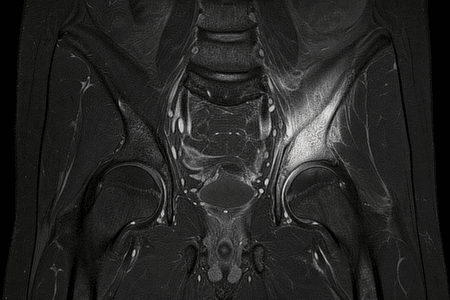
- Haematogenous osteomyelitis seeds 'METAPHYSEAL EQUIVALENTS' - portions of flat or irregular bone adjacent to cartilage with a similar sluggish vascular anatomy to a long-bone metaphysis; the pelvis has several, and the SACROILIAC JOINT region is the most frequently involved, with the ilium, pubis, ischium and the triradiate cartilage also affected - which is why pelvic infection often straddles bone and the adjacent SI joint (septic sacroiliitis).
- MRI is the KEY diagnostic test: it is sensitive early (showing bone-marrow oedema and surrounding soft-tissue change before plain radiographs become abnormal), it defines any abscess that would need drainage, and it distinguishes the diagnosis from the mimics; plain radiographs are often NORMAL in the early phase, and inflammatory markers (CRP/ESR) and blood cultures support the diagnosis.
- STAPHYLOCOCCUS AUREUS is the predominant organism (consider Kingella kingae in younger children and Salmonella in sickle-cell disease); a tissue or blood diagnosis should be sought, and a subacute focus can present as a BRODIE'S ABSCESS in pelvic bone, which mimics infection, tuberculosis and benign or malignant bone tumours - so biopsy/curettage is sometimes needed to confirm the diagnosis and exclude tumour.
- MANAGEMENT is ANTIBIOTIC-led: appropriate empirical antibiotics (covering S. aureus, guided by local protocols and culture) started after attempting to obtain a microbiological diagnosis, with most children responding to antibiotics alone; SURGICAL DRAINAGE is reserved for a drainable ABSCESS, for septic arthritis of the SI joint not responding, or for failure to improve.
- Because the diagnosis is so easily missed, the practical rule is to keep pelvic/SI-joint infection in the differential of the LIMPING, FEBRILE CHILD - especially when the hip examination is relatively unremarkable or pain localises to the buttock/back - and to use MRI early; timely treatment usually gives an excellent outcome, whereas delay risks abscess, chronicity and growth disturbance.
- “Pelvic osteomyelitis/septic sacroiliitis = the GREAT MIMIC of the limping febrile child (hip/buttock/back pain) - frequently missed/delayed.
- “Metaphyseal-equivalent concept: SI joint region most common pelvic site (also ilium/pubis/ischium/triradiate). MRI is the key test; plain films often normal early.
- “S. aureus predominates; ANTIBIOTIC-led management, drainage only for abscess/non-response; a subacute focus = Brodie's abscess (exclude tumour/TB).
Limp/refusal to weight-bear + fever with pain in hip, buttock, groin, thigh or low back - and a relatively unremarkable hip exam. A great mimic, often diagnosed late.
Get an MRI early (plain films often normal); cover S. aureus; drain only an abscess.
Presentation, Sites & Diagnosis
Pelvic osteomyelitis and septic sacroiliitis present vaguely: a child with a limp or refusal to weight-bear, fever, and pain that may localise to the hip, buttock, groin, thigh or low back - so they are frequently mistaken for a septic hip, transient synovitis, discitis or even appendicitis, and diagnosis is often delayed. The reason the pelvis is involved is the metaphyseal-equivalent concept: flat/irregular bones have regions adjacent to cartilage with metaphysis-like vascularity that seed haematogenous infection, and the sacroiliac joint region is the most frequent pelvic site (with ilium, pubis, ischium and triradiate cartilage). MRI is the key test - sensitive early and able to define an abscess - while plain films are often normal early; Staphylococcus aureus predominates.
Organisms & Management
- Get a diagnosis where possible. Inflammatory markers (CRP/ESR), blood cultures, and - for a localised or atypical focus - image-guided aspiration/biopsy; a subacute pelvic focus may be a Brodie's abscess that mimics tuberculosis and bone tumours, so biopsy/curettage is sometimes needed to confirm and exclude tumour.
- Cover the likely organism. S. aureus predominates; consider Kingella kingae in younger children and Salmonella in sickle-cell disease. Start empirical antibiotics per local protocol after sampling.
- Antibiotics are the mainstay. Most children respond to appropriately chosen antibiotics alone, with the usual transition from intravenous to oral guided by clinical and biochemical response.
- Drain when needed. Surgical drainage is reserved for a drainable abscess, for SI-joint sepsis not responding, or for failure to improve on antibiotics.
The recurring lesson of pelvic osteomyelitis and septic sacroiliitis is that they are MISSED because the presentation is non-specific and the hip examination can be relatively unremarkable, so a limping, febrile child with buttock or back pain - or a 'septic hip' that does not quite fit - should keep pelvic/SI-joint infection in the differential and prompt an EARLY MRI rather than waiting for plain films to change (which they often do not early). Equally, do not assume a destructive pelvic lesion is infective: a subacute focus can mimic, and be mimicked by, tuberculosis and bone tumours, so obtain tissue when the picture is atypical. Early diagnosis and antibiotic-led treatment usually give an excellent outcome; delay risks abscess, chronic infection and growth disturbance.
Evidence & Key Studies
Osteomyelitis of a sacral neurocentral synchondrosis: another pelvic metaphyseal equivalent
- Pelvic osteomyelitis occurs in 'metaphyseal equivalents' - portions of flat or irregular bone adjacent to cartilage.
- The pelvic bone has several metaphyseal equivalents, of which the sacroiliac joint is the most frequent site of involvement.
- Recognising additional metaphyseal equivalents (here a sacral neurocentral synchondrosis), especially in infants and younger children, aids diagnosis on MRI.
Brodie's abscess of the posterior ilium presenting as paediatric low back pain (gluteal syndrome)
- A Brodie's abscess (subacute osteomyelitis) of pelvic bone is rare and, in the posterior ilium, can present as low back pain and be easily missed or misdiagnosed.
- It resembles other pelvic and sacroiliac joint pathologies and the differential includes infection, tuberculosis and benign bone tumours - so biopsy/curettage may be needed; Staphylococcus aureus was cultured.
- Appropriate antibiotics (here six weeks after curettage) gave resolution without recurrence.
According to PubMed, the metaphyseal-equivalent concept and the sacroiliac joint being the most frequent pelvic site come from the cited Miyazaki report; the vague, easily-missed presentation of a subacute pelvic focus (Brodie's abscess) with its differential of infection, tuberculosis and bone tumour, the role of biopsy, the S. aureus aetiology and antibiotic resolution from the cited Behera report. The general 'limping febrile child' presentation, the central role of early MRI, the Kingella/Salmonella considerations and the antibiotic-led (drainage-for-abscess) management are standard, well-established teaching. (See also our Acute Haematogenous Osteomyelitis and Septic Arthritis of the Hip topics.)
Clinical Decision Scenarios
Practise clinical reasoning and management decisions out loud
“A child presents with a limp, fever and buttock pain, but the hip examination is relatively unremarkable. What are you worried about and how do you investigate?”
“How would you manage confirmed pelvic osteomyelitis or septic sacroiliitis in a child?”
Mnemonics & Memory Aids
PELVIS
Hook:PELVIS: referred Pain, Easily missed, Limp+fever, View on MRI, Infection (S. aureus), SI joint + antibiotics.
Recognition
- Limp/refusal to weight-bear + fever; pain in hip/buttock/groin/thigh/low back
- Relatively unremarkable hip exam; a great mimic, often diagnosed late
- Differential: septic hip, transient synovitis, discitis, appendicitis
Sites & concept
- Metaphyseal equivalents (flat/irregular bone adjacent to cartilage) seed infection
- SI joint region most frequent; also ilium, pubis, ischium, triradiate cartilage
- Septic sacroiliitis straddles bone and joint
Diagnosis
- MRI is the key test (sensitive early; defines abscess)
- Plain films often normal early; CRP/ESR and blood cultures support
- S. aureus predominates (Kingella younger; Salmonella sickle cell)
Management
- Antibiotic-led after sampling; most respond to antibiotics alone
- Drainage for abscess / non-responding SI sepsis / failure to improve
- Biopsy an atypical/destructive focus (Brodie's abscess mimics TB/tumour)