Os Trigonum | Dancers | Plantarflexion
- Os trigonum = accessory ossicle (unfused lateral talar process)
- Ballet dancers and footballers classically affected
- Pain with forced plantarflexion (en pointe, kicking downward)
- FHL tendon may be involved (adjacent, in groove)
- Endoscopic/arthroscopic excision is treatment of choice
- “Os trigonum present in around 9% of feet (meta-analysis pooled prevalence)
- “Posterior impingement test reproduces pain
- “Lateral X-ray shows os trigonum or large Stieda process
- “FHL tenosynovitis is common associated finding
Os trigonum = unfused secondary ossification center of lateral talar process. Stieda process = elongated lateral talar tubercle. Both impinge posteriorly with plantarflexion.
Ballet dancers (en pointe) and soccer players (kicking). Pain at posterior ankle with forced plantarflexion. May have triggering from FHL involvement.
Flexor hallucis longus runs in groove between lateral and medial talar tubercles. Can become inflamed (FHL tenosynovitis) causing great toe triggering ("dancer's tendinitis").
Endoscopic excision is preferred (less morbidity). Access through posterolateral and posteromedial portals. Excise os trigonum, release FHL. Excellent outcomes.
FHLSurgical Danger Structures (Posteromedial)
Hook:Stay LATERAL to the FHL — the neurovascular bundle is medial to it!
Overview and Anatomy
Posterior ankle impingement occurs when structures at the posterior ankle are pinched during plantarflexion. The most common cause is an os trigonum or prominent Stieda process.
Anatomy
The lateral talar process (posterior process of talus, lateral tubercle) is at the posterior talus. In a proportion of people this develops as a separate ossicle called the os trigonum (unfused secondary ossification center). A 2024 meta-analysis of 36,612 feet found a pooled prevalence of approximately 9% (wide reported range 1.7-32.5%, highest in East Asian populations and on cross-sectional imaging), bilateral in about a third of cases. The os trigonum appears in childhood (mean age 9-11 years) and fuses in roughly 70% of children by skeletal maturity, leaving the unfused remnant in adults. The Stieda process is an elongated lateral tubercle that is continuous with the talus (failure of separation of the same ossification center).
The flexor hallucis longus tendon runs in a groove between the medial and lateral talar tubercles. It can become involved in posterior impingement syndrome.
Pathophysiology
Forced or repetitive plantarflexion (as in ballet dancing en pointe or downward kicking) compresses the os trigonum or Stieda process between the posterior tibial plafond and the calcaneus ("nutcracker" mechanism). Repetitive loading causes synovitis, capsular thickening, bone marrow edema in the ossicle and, in chronic cases, disruption of the synchondrosis between the os trigonum and talus.
The adjacent flexor hallucis longus (FHL) runs in its fibro-osseous tunnel between the medial and lateral talar tubercles, immediately medial to the os trigonum. Repetitive plantarflexion and hallux motion (the FHL is maximally loaded with the ankle plantarflexed and the great toe pushing off) produce stenosing tenosynovitis — thickening of the tendon and sheath that can trigger or lock the hallux ("dancer's tendinitis"). PAIS and FHL pathology frequently coexist but behave as distinct entities and may occur in isolation.
OSSPosterior Impingement Causes
Hook:OSS = Os trigonum, Stieda, Soft tissue cause posterior impingement!
Clinical Presentation
History
Ballet dancers present with posterior ankle pain, particularly with en pointe work. Soccer players may have pain with downward kicking. Swimmers may have pain with push-off. Pain is worse with plantarflexion and may be associated with triggering of the great toe if FHL is involved.
Examination
Posterior Impingement Test: Passive forced plantarflexion of the ankle reproduces posterior pain. Highly specific.
Palpation: Tenderness at the posterolateral ankle, between the Achilles and peroneal tendons.
FHL Assessment: Passive flexion/extension of the great toe with the ankle in plantarflexion may reproduce symptoms or show triggering.
Exclude Achilles pathology: Tenderness at insertion, Thompson test.
DANCEClinical Features
Hook:DANCErs get posterior ankle impingement!
Investigations
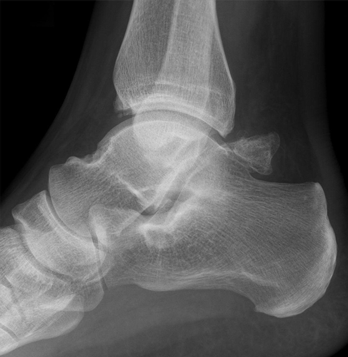
Lateral Radiograph: Shows os trigonum (separate ossicle) or prominent Stieda process (elongated tubercle). Best assessed on true lateral view.
CT Scan: Better defines anatomy, particularly for surgical planning. Shows relationship of ossicle to talus.
MRI: Shows bone edema in os trigonum or lateral tubercle. Shows FHL tenosynovitis. Shows associated soft tissue inflammation.
Ultrasound: Can assess FHL tendon dynamically.
Diagnostic Injection: Image-guided local anesthetic injection to the posterior ankle that relieves symptoms confirms the pain source. Greater than 70% relief of the posterior impingement test supports the diagnosis, particularly when imaging is equivocal.
Differential Diagnosis
- Pain pattern / trigger
- Deep posterior pain on forced plantarflexion (en pointe, kicking)
- Key examination
- Positive forced plantarflexion test, posterolateral tenderness
- Discriminating investigation
- Lateral X-ray ossicle/process; MRI bone edema; relief on diagnostic injection
- Pain pattern / trigger
- Posteromedial pain, may trigger/lock great toe
- Key examination
- Pain on resisted hallux flexion and passive hallux extension in plantarflexion
- Discriminating investigation
- MRI/US thickened FHL (over 6mm), fluid in sheath; dynamic US shows stenosis
- Pain pattern / trigger
- Superficial posterior pain, worse on push-off and dorsiflexion stretch
- Key examination
- Tender Achilles insertion or midportion, NOT deep on plantarflexion
- Discriminating investigation
- US/MRI tendon thickening; impingement test negative
- Pain pattern / trigger
- Posterior heel pain at bursa, shoe pressure
- Key examination
- Tender retrocalcaneal space, posterosuperior calcaneal prominence
- Discriminating investigation
- Lateral X-ray Haglund deformity; MRI bursal fluid
- Pain pattern / trigger
- Hindfoot pain on uneven ground, stiffness
- Key examination
- Restricted/painful subtalar motion
- Discriminating investigation
- CT (coalition); weightbearing X-ray/MRI
- Pain pattern / trigger
- Acute post-traumatic posterior pain
- Key examination
- Acute tenderness, history of injury (Shepherd fracture)
- Discriminating investigation
- CT distinguishes acute fracture from chronic os trigonum (smooth corticated margins)
Management

Activity Modification: Avoid provocative plantarflexion activities.
Physiotherapy: Ankle conditioning, avoid excessive plantarflexion.
NSAIDs/Analgesia: Symptomatic relief.
Injection: Corticosteroid to the posterior ankle may provide relief. Both diagnostic and therapeutic.
Conservative treatment may be successful in mild cases but dancers and athletes often require surgery to return to activity.
Complications
Complications of the Condition (untreated)
- Persistent posterior ankle pain limiting plantarflexion-dependent activity (dance, kicking sports).
- Progressive FHL tenosynovitis / stenosis with hallux triggering or locking if untreated.
- Synchondrosis disruption / chronic os trigonum fragmentation with ongoing mechanical symptoms.
- Career-limiting loss of performance in elite dancers and athletes.
Complications of Surgery
- Nerve injury (most important). Portal-related neurological complication ~1.9% overall in large ankle arthroscopy series — sural nerve laterally, tibial nerve / medial calcaneal and plantar branches medially; most are transient (resolve within ~6 months). Posterior tibial neurovascular bundle lies just medial to the FHL, the key danger structure on the posteromedial side.
- Incomplete excision — residual os trigonum/Stieda fragment is the commonest cause of persistent symptoms and surgical failure (overall failure ~5-15%).
- Untreated coexisting FHL pathology causing persistent symptoms despite osseous excision.
- Posterior tibial vessel injury / haematoma, wound problems, infection (rare).
- Stiffness / scarring of the posterior recess; FHL weakness if excessive tendon is debrided (critical to avoid in dancers).
- Persistent neuritis / scar dysaesthesia at portal sites (reported in up to ~15% transiently in some series).
The Posterior Impingement Test
The posterior impingement test is named as the topic's key clinical sign ("highly specific," "reproduces posterior pain") and is used in every scenario, but the body never describes how to perform or interpret it.
- How to perform it (van Dijk's test). With the patient seated or prone and the knee flexed to relax the gastrocnemius, the examiner grasps the foot and performs a quick, repetitive passive forced hyperplantarflexion of the ankle; a recognisable reproduction of the patient's deep posterior pain is positive. A small rotatory ("grinding") movement at end-range increases posterior compression and heightens sensitivity, and the manoeuvre is often repeated in slight external and internal rotation.
- What a positive and a negative result mean. The test has high sensitivity, so a negative (pain-free) forced-plantarflexion test essentially rules out bony posterior impingement. Its specificity is lower, however - forced plantarflexion also loads the FHL, Achilles and posterior capsule - so a positive test is not by itself diagnostic and should be correlated with imaging and, when equivocal, a diagnostic injection (over 70% relief of the reproduced pain confirms the source).
- Separating coexisting FHL involvement. Because the FHL is stretched and loaded in plantarflexion too, add passive hallux dorsiflexion and resisted hallux flexion with the ankle plantarflexed: pain or triggering there points to coexisting FHL tenosynovitis rather than pure osseous impingement.
Q: How is the posterior impingement test performed and interpreted? A: With the knee flexed, the examiner performs a quick passive forced hyperplantarflexion of the ankle (adding a small rotatory grind at end-range); reproduction of the deep posterior pain is positive. It is highly sensitive, so a negative test essentially excludes bony posterior impingement, but its specificity is lower (forced plantarflexion also loads the FHL/Achilles/capsule), so a positive test needs imaging and often a confirmatory diagnostic injection (over 70% relief). Adding hallux dorsiflexion and resisted hallux flexion helps separate coexisting FHL tenosynovitis.
Rehabilitation and Return to Sport
The topic quotes return-to-sport rates (around 89-94%) and times, and every viva leans on a recovery timeline, but the body never sets out the actual rehabilitation protocol after excision.
- Early phase (0-2 weeks). A short period in a removable boot or backslab with protected weight-bearing; endoscopic patients often weight-bear as comfort allows within days. The key principle is to begin early active ankle plantar/dorsiflexion and hallux motion as soon as the wounds allow - this prevents re-scarring and adhesion of the posterior recess, a recognised cause of recurrent symptoms and stiffness.
- Intermediate phase (2-6 weeks). Progress to full weight-bearing out of the boot, restore the full range (including the plantarflexion arc), and add calf and FHL strengthening and proprioception. If an FHL release/tenosynovectomy was performed, protect against forced hallux dorsiflexion early but keep the toe gliding to avoid adhesion.
- Return to sport (6 weeks onward). Introduce sport-specific plantarflexion loading in a graded manner - relevé/pointe work for dancers, kicking for footballers, turn push-off for swimmers. Typical return is around 6-12 weeks for general sport and around 12-16 weeks (or longer) for elite dance and forced-plantarflexion athletes; isolated FHL tendinopathy recovers more slowly (about 16 weeks) than osseous impingement (Rietveld), and endoscopic excision returns athletes faster than open (about 7 versus 11-12 weeks to previous level in the one RCT).
- Why it matters. Setting this staged, early-motion protocol - and counselling the longer timeline when the FHL is involved - is what delivers the high return-to-sport figures the topic quotes and avoids the posterior-recess stiffness the complications section warns about.
Q: Outline the rehabilitation and return-to-sport timeline after posterior ankle impingement excision. A: A short period of protected weight-bearing (boot), then early active ankle and hallux motion to prevent posterior-recess re-scarring, progressing to full weight-bearing, range and calf/FHL strengthening by about 6 weeks, then graded sport-specific plantarflexion loading. Typical return is around 6-12 weeks for general sport and around 12-16 weeks for dance/elite plantarflexion athletes; isolated FHL tendinopathy takes longer (about 16 weeks), and endoscopic excision returns athletes faster than open (about 7 versus 11-12 weeks in the RCT).
Guidelines, Registries & Global Practice
Global Epidemiology
Posterior ankle impingement is predominantly an overuse condition of athletes who repetitively plantarflex — classically ballet dancers (en pointe / relevé) and football/soccer players (instep kicking), and also gymnasts, divers, swimmers (turn push-off) and downhill runners. An os trigonum is present in roughly 9% of feet worldwide (highest in East Asian populations), but is asymptomatic in most. There is no dedicated implant registry for this soft-tissue/osseous procedure; outcome evidence comes from case series, one RCT and systematic reviews rather than national arthroplasty registries.
Society Guidance — Side by Side
There are no disease-specific clinical practice guidelines from the major bodies for posterior ankle impingement; recommendations are derived from expert/society positions on hindfoot endoscopy and from sports-medicine consensus:
- Position relevant to PAIS
- Endorse hindfoot arthroscopy as a recognized, effective option for posterior impingement; emphasize confirming the diagnosis (exam + imaging ± diagnostic injection) before surgery
- Position relevant to PAIS
- Support a trial of non-operative care first, then minimally invasive (endoscopic) or open excision; stress neurovascular safety with portal placement
- Position relevant to PAIS
- Two-portal hindfoot endoscopy (van Dijk) regarded as the standard minimally invasive technique; recognized in foot and ankle arthroscopy teaching
- Position relevant to PAIS
- In dancers, distinguish isolated PAIS, PAIS with FHL tendinopathy and isolated FHL injury; counsel longer return when FHL is involved
Registry & Outcome Notes
No joint registry captures this procedure. The strongest data point is the single RCT (endoscopic vs open, Level II) plus Level IV series and a complications study reporting an overall ankle-arthroscopy complication rate of ~3.5% with portal-related neurological injury ~1.9%.
High- vs Limited-Resource Practice Variation
- Well-resourced settings: MRI and image-guided diagnostic injection are standard, and endoscopic two-portal excision is widely available, allowing faster return to sport in elite athletes.
- Limited-resource settings: Diagnosis often rests on clinical examination and plain lateral radiographs; open excision through a posterolateral or posteromedial approach remains an entirely valid, effective treatment where endoscopic equipment or expertise is unavailable, with the trade-off of slower recovery and a higher (but still acceptable) complication rate.
Controversies & Areas of Uncertainty
- Endoscopic versus open excision. Only one randomized trial exists (Georgiannos & Bisbinas, Level II); it favors endoscopy for faster return and fewer complications, but most other evidence is Level IV/V. Open excision remains acceptable, especially where endoscopic expertise is limited or extensive FHL work is anticipated.
- Posteromedial versus posterolateral open approach. Posteromedial gives direct access to the FHL but risks the tibial nerve and posterior tibial vessels; posterolateral is simpler but FHL access is harder. No high-level comparative data; choice is surgeon-dependent.
- Routine FHL release. Whether to release/decompress the FHL when it looks normal intraoperatively is debated. Under-treating coexisting FHL pathology is a recognized cause of surgical failure, yet unnecessary release adds morbidity. Current practice is to address the FHL only when pre-operative or intraoperative pathology is demonstrated.
- Soft-tissue versus osseous impingement. A substantial minority of cases have no ossicle; the true proportion and the optimal treatment of pure soft-tissue impingement are not well defined by high-level evidence.
- Conservative management. Evidence for non-operative treatment is very weak (few patients, retrospective), so no evidence-based protocol exists; a trial of activity modification, physiotherapy and image-guided injection remains reasonable before surgery in non-elite patients.
- Incidental os trigonum. An os trigonum is a common asymptomatic anatomical variant; imaging findings must correlate with a positive impingement test and ideally a confirmatory diagnostic injection before attributing symptoms to it.
MCQ Practice Points
Q: What is an os trigonum? A: An unfused secondary ossification center of the lateral talar process (posterior process of talus). Pooled prevalence is around 9% of feet (meta-analysis). Causes posterior ankle impingement when symptomatic.
Q: What tendon pathology is commonly associated with posterior ankle impingement? A: Flexor hallucis longus tenosynovitis ("dancer's tendinitis"). The FHL runs in a groove between the medial and lateral talar tubercles and can be compressed.
Q: Which structure is the key landmark protecting the neurovascular bundle in two-portal hindfoot endoscopy? A: The FHL tendon. Working lateral to the FHL keeps you away from the posterior tibial neurovascular bundle (tibial nerve and posterior tibial artery), which lies medial to it. Portal-related nerve injury is the commonest complication (~1.9%).
Exam Viva Scenarios
Practise clinical reasoning and management decisions out loud
“A 22-year-old ballet dancer has posterior ankle pain worse when going en pointe. How do you assess and manage her?”
“You are seeing a 28-year-old competitive swimmer in your sports medicine clinic who has been experiencing progressive posterior right ankle pain for the past 8 months. She describes deep posterior ankle pain that is particularly worse during the push-off phase of her turns when she plantarflexes forcefully against the wall. The pain started insidiously without any specific injury and has progressively worsened to the point where it is affecting her training and competition times. She has tried 3 months of rest from competitive swimming, physiotherapy focusing on ankle strengthening and flexibility, and NSAIDs without significant improvement. She is frustrated and concerned as she has national championships in 4 months. On examination, she has deep tenderness to palpation in the posterior ankle between the Achilles tendon and peroneal tendons. You perform a posterior impingement test (passive forced plantarflexion) which clearly reproduces her deep posterior ankle pain. Her Achilles tendon is non-tender and normal on palpation. The retrocalcaneal bursa is non-tender. When you assess her flexor hallucis longus (FHL) by asking her to flex and extend her great toe with the ankle in different positions, she has pain with passive hallux extension when the ankle is in plantarflexion (stretches the FHL), but there is NO triggering or clicking of the great toe. Active and passive ankle range of motion is full and symmetrical, but extremes of plantarflexion reproduce her posterior pain. She has brought radiographs from her sports physician. The lateral ankle X-ray report states: 'Normal bony alignment. No fracture. No os trigonum identified. The lateral talar process appears normal in size and morphology (no prominent Stieda process). Achilles tendon insertion appears normal.' The radiologist specifically notes: 'No osseous cause for posterior impingement identified.' The patient is confused and asks: (1) The X-ray shows no bone problem - so what is causing my posterior impingement? (2) How do we diagnose what's causing my pain if there's no os trigonum? (3) Do I still need surgery even though there's no bone to remove? (4) If I do need surgery, what would you actually do if there's nothing to take out?”
“You are seeing a 26-year-old professional contemporary dancer in your complex foot and ankle clinic for a second opinion. She underwent endoscopic excision of a symptomatic right os trigonum 9 months ago performed by another surgeon at a different institution. The initial presentation was classic posterior ankle impingement - deep posterior ankle pain worse with plantarflexion during dance, positive posterior impingement test, lateral X-ray showing os trigonum, MRI confirming bone marrow edema in the ossicle. She had failed 6 months of conservative management (rest, physiotherapy, two corticosteroid injections) prior to surgery. The operative report from the previous surgery describes: 'Endoscopic excision of os trigonum via two-portal posterior ankle approach. Posterolateral and posteromedial portals established. Os trigonum identified and excised arthroscopically. FHL visualized and appeared normal. No release performed as no evidence of stenosis. Wounds closed. No complications.' Post-operatively, she was initially better for about 6-8 weeks. However, her symptoms have gradually returned and she now describes similar posterior ankle pain to pre-operative levels. She is extremely frustrated and concerned that the surgery 'didn't work'. She reports: (1) Deep aching posterior ankle pain, particularly with plantarflexion during dance, (2) The pain is in a similar location to before surgery, perhaps slightly more medial, (3) She now also experiences occasional clicking or snapping sensation in the posterior ankle when she moves her great toe, which she didn't have before surgery, (4) The pain is affecting her ability to perform and she is considering whether she should retire from professional dancing. On examination, she has well-healed posterolateral and posteromedial arthroscopic portal scars with no signs of infection or wound complications. There is deep tenderness to palpation in the posterior ankle, slightly more prominent medially than laterally. Passive forced plantarflexion reproduces her posterior ankle pain (positive posterior impingement test - similar to pre-op). When you assess her flexor hallucis longus by asking her to actively flex and extend her great toe, you can palpate and hear a distinct SNAP or CLICK in the posterior ankle, and she reports this is the clicking she has been experiencing. This was NOT documented in her pre-operative assessment. Resisted plantarflexion of the hallux is painful. Passive forceful extension of the hallux with the ankle in plantarflexion reproduces deep posterior pain. Her ankle range of motion is full but extremes of plantarflexion reproduce her posterior pain. You review the post-operative radiographs she brought (taken at 3 months post-op by the previous surgeon): Lateral ankle X-ray report states: 'Post-surgical changes. Partial excision of os trigonum with small residual ossicle fragment noted posterior to talus (approximately 5mm x 3mm). Alignment normal.' You also order a NEW MRI which reports: 'Post-surgical changes in posterior ankle. Small residual os trigonum fragment present (5mm) with surrounding bone marrow edema. Flexor hallucis longus tendon is markedly thickened (8mm diameter, normal less than 6mm) with high T2 signal consistent with tendinopathy. Moderate fluid around FHL tendon sheath (tenosynovitis). The FHL appears stenosed in the fibro-osseous tunnel at the level of the posterior talus. Post-surgical scarring noted in the posterior ankle recess. Findings suggestive of: (1) Incomplete os trigonum excision with residual symptomatic fragment, (2) FHL tendinopathy and stenosing tenosynovitis (possibly missed at initial surgery or developed post-operatively).' The patient has multiple questions: (1) Why didn't the surgery work - did the surgeon not remove the whole bone? (2) The report mentions my FHL tendon - could this be the problem now rather than the bone? Was this missed initially? (3) Do I need another operation? If so, what would be different this time? (4) Is there a risk that revision surgery could make things worse? I'm worried about ending my dance career. (5) Could there be something else causing my pain that everyone has missed?”
Causes (OSS)
- Os trigonum (unfused ossicle)
- Stieda process (elongated tubercle)
- Soft tissue (FHL, capsule)
Clinical
- Dancers, footballers
- Pain with plantarflexion
- Posterior impingement test positive
- FHL triggering may be present
Diagnosis
- Lateral X-ray shows ossicle/process
- MRI shows bone edema, FHL pathology
- Injection confirms diagnosis
Treatment
- Conservative first if possible
- Endoscopic excision preferred
- Two-portal posterior approach
- ~89-94% return to sport
Evidence Base
- Randomized controlled trial, 52 athletes (26 endoscopic vs 26 open), 5-year follow-up
- Return to training 4.6 vs 9.6 weeks and to previous sports level 7.1 vs 11.5 weeks, both favoring endoscopic (p less than 0.001)
- Complication rate 3.8% (1/26) endoscopic vs 23% (6/26) open
- AOFAS hindfoot scores higher with endoscopic; VAS-FA similar between groups
- Describes the prone two-portal (posterolateral + posteromedial) hindfoot endoscopy approach
- Single platform to address os trigonum, FHL release, posterior tendon/joint pathology
- Lower morbidity, reduced postoperative pain and day-case treatment versus open surgery
- Companion ICL series reported no major complications in 240 consecutive procedures
- 1305 ankle arthroscopies (anterior and posterior two-portal hindfoot) over 19 years
- Overall complication rate 3.5%; neurological complications 1.9%, related to portal placement
- Most complications transient and resolved within 6 months with no functional limitation
- Posterior two-portal hindfoot approach compared favourably with anterior arthroscopy