Failed Fusion | Nonunion After Arthrodesis | Symptomatic vs Asymptomatic
- Pseudarthrosis = failure to achieve solid bony fusion by 1 year post-surgery
- Smoking is the single biggest modifiable risk factor (~2x nonunion risk; 26.5% vs 14.2% in Glassman 2000)
- CT scan is gold standard - look for bridging trabecular bone
- Symptomatic pseudarthrosis causes persistent axial pain at fusion level
- Revision includes debridement, fresh autograft, rigid fixation, interbody support
- “Single-level posterolateral fusion: 5-10% pseudarthrosis rate
- “Multi-level fusion plus smoking compounds nonunion risk
- “Not all radiographic pseudarthrosis is symptomatic
- “Hardware breakage suggests pseudarthrosis with instability
- “BMP can augment fusion but smoking still impairs outcomes
Failure to achieve solid bony fusion by 1 year post-surgery. Fibrous or cartilaginous tissue instead of bridging bone at intended fusion site. May be asymptomatic (incidental finding) or symptomatic (persistent pain).
Smoking roughly doubles nonunion risk (meta-analysis RR 2.2; Glassman 2000 nonunion 26.5% in continuing smokers vs 14.2% non-smokers). Nicotine directly inhibits osteoblast function, reduces blood flow to bone, and creates a hypoxic environment. Smoking cessation for 6 weeks minimum before surgery is essential. Even with revision, smoking impairs outcomes.
CT scan with thin cuts and sagittal/coronal reconstructions is gold standard. Look for continuous bridging trabecular bone through graft. Lucency at graft-host interface indicates nonunion. Plain X-rays less sensitive. Dynamic films show motion but less reliable than CT.
Revision strategy (GRIFT): Remove fibrous tissue, fresh autograft (iliac crest), revise or extend instrumentation, add interbody support (PLIF/TLIF/ALIF), address risk factors (smoking cessation mandatory). Success rate 70-80% if risk factors controlled.
Overview and Epidemiology
Spinal pseudarthrosis (also called spinal nonunion) refers to the failure to achieve solid bony fusion following an intended arthrodesis procedure. It represents a recognized complication of spinal fusion surgery and can be either symptomatic or asymptomatic.
Definition
Pseudarthrosis is defined as the absence of bridging trabecular bone across the intended fusion site by 1 year post-operatively. Instead of solid bone, fibrous tissue or fibrocartilage fills the fusion gap. This fibrous union lacks the mechanical strength of bony fusion.
Epidemiology
Incidence varies significantly based on multiple factors:
- Single-level posterolateral fusion (PLF): 5-10% pseudarthrosis rate
- Multi-level PLF: 15-25% nonunion rate
- Combined anterior-posterior fusion: 2-5% (lower than PLF alone)
- Interbody fusion (PLIF/TLIF/ALIF): 3-8% nonunion rate
- Smokers vs non-smokers: roughly two-fold increase in nonunion (meta-analysis RR 2.2; Glassman 26.5% vs 14.2%)
- Multi-level fusion in smokers: risk factors compound, with reported nonunion rates well above single-level baselines
Clinical Significance
Not all pseudarthrosis requires treatment. Asymptomatic pseudarthrosis discovered incidentally on imaging in a pain-free patient can be observed. Symptomatic pseudarthrosis causes persistent or recurrent axial back pain at the fusion site and typically requires revision surgery.
The key distinction is clinical correlation. A patient with CT-confirmed pseudarthrosis who is completely pain-free and functional does not need revision surgery. Conversely, persistent pain with imaging-confirmed nonunion is an indication for revision.
Historical Context
The term "pseudarthrosis" comes from Greek: pseudo (false) + arthrosis (joint). Early spinal fusion techniques had high nonunion rates. Recognition of risk factors (especially smoking) and development of improved techniques (instrumentation, interbody cages, BMP) have reduced but not eliminated pseudarthrosis.
Risk Factors
Smoking
The single most important modifiable risk factor.
- Roughly doubles pseudarthrosis risk (meta-analysis RR 2.2; Glassman 26.5% vs 14.2%)
- Even second-hand smoke exposure increases risk
- Nicotine patches and replacement therapy also impair fusion (nicotine is the problem)
- Smoking cessation 6 weeks minimum before surgery is essential
- Risk reduction requires complete abstinence, not just reduction
Diabetes Mellitus
Impairs bone healing through multiple mechanisms:
- Microangiopathy reduces blood supply to fusion bed
- Advanced glycation end-products (AGEs) impair bone quality
- Increased infection risk further compromises healing
- HbA1c should be optimized to less than 7% before elective fusion
Obesity
BMI over 30 associated with increased pseudarthrosis:
- Increased mechanical stress on fusion construct
- Metabolic syndrome (insulin resistance, inflammation)
- Greater soft tissue dissection increases infection risk
- Longer operative times
Nutritional Deficiency
Poor nutrition impairs bone formation:
- Vitamin D deficiency: Impairs calcium absorption and bone formation
- Protein malnutrition: Insufficient substrate for collagen synthesis
- Calcium deficiency: Inadequate mineral for bone matrix
- Preoperative nutritional optimization recommended
Age
Older age associated with higher nonunion:
- Decreased osteoblast activity
- Reduced bone healing capacity
- More medical comorbidities
- Osteoporosis common in elderly
Osteoporosis
Low bone density impairs fusion substrate:
- Poor quality bone for graft incorporation
- Hardware loosening more common
- Consider bone anabolic therapy (teriparatide) in high-risk cases
This completes the patient factors overview.
Pathophysiology of Nonunion
Normal Fusion Biology
Successful spinal fusion requires:
- Osteoconduction: Scaffold for bone growth (provided by graft)
- Osteoinduction: Signals for bone formation (BMPs, growth factors)
- Osteogenesis: Viable cells that form bone (osteoblasts from graft or host)
- Mechanical stability: Immobilization allows bone formation
- Vascular supply: Blood flow delivers nutrients and cells
Pseudarthrosis Pathway
When fusion fails, the process follows this sequence:
Week 0-6: Initial graft placement. If stability inadequate or biology impaired, granulation tissue forms instead of organized bone.
Week 6-12: Fibrous tissue fills the fusion gap. Without rigid fixation, micromotion prevents bone formation.
Month 3-6: Fibrocartilage may develop at areas of compression. Sclerotic bone forms at graft-host interface without bridging.
Month 6-12: Established fibrous nonunion. If hardware is present, cyclic loading may cause fatigue failure and breakage.
Why Smoking Impairs Fusion
Nicotine effects on bone healing:
- Direct osteoblast toxicity: Inhibits alkaline phosphatase and collagen synthesis
- Vasoconstriction: Reduces blood flow to fusion bed (hypoxic environment)
- Reduced growth factors: Decreased local BMP-2 and TGF-beta
- Immune dysfunction: Impaired macrophage and fibroblast function
- Increased fibrous tissue: Promotes fibrous nonunion over bone
For both primary fusion and revision surgery, smoking cessation for minimum 6 weeks before and 12 weeks after surgery is essential. Glassman (2000) showed nonunion fell from 26.5% in continuing smokers towards 17.1% in those who quit for more than 6 months, approaching the 14.2% non-smoker rate, and meta-analysis confirms smokers carry roughly twice the nonunion risk.
Biomechanical Factors
Excessive motion at the fusion site prevents bone formation:
- Posterolateral fusion alone: Relies on tension band effect, higher motion
- Interbody fusion: Anterior column support, compressive loading, more stable
- Combined 360-degree fusion: Most stable, lowest pseudarthrosis risk
- Multi-level fusion: Greater lever arms, increased stress at each segment
Clinical Presentation
History
Symptomatic pseudarthrosis typically presents with:
Persistent axial back pain:
- Localized to fusion level
- Worsened by activity, standing, walking
- Improved with rest and lying down
- May be constant or intermittent
Pattern of pain:
- Never improved: Pain present from surgery, never resolved
- Recurrent pain: Initial improvement (3-6 months), then pain returns
- Progressive pain: Gradual worsening over months
Associated symptoms:
- Pseudarthrosis itself causes axial pain, not radiculopathy
- If radicular symptoms present, consider adjacent segment disease or hardware-related nerve compression
- Hardware breakage may cause sudden increase in pain
Examination
Inspection:
- Observe posture and gait
- Assess spinal alignment (scoliosis, kyphosis)
Palpation:
- Tenderness over fusion site
- Palpable hardware prominence
Range of Motion:
- Painful motion at fusion level suggests pseudarthrosis
- Patient may report feeling "motion" or "giving way"
- Flexion-extension may reproduce pain
Neurological Examination:
- Typically normal in isolated pseudarthrosis
- Document motor, sensory, reflexes to establish baseline
- If deficits present, consider alternative diagnoses
Persistent axial back pain more than 6-12 months post-fusion, especially if there was initial improvement followed by recurrence, should raise suspicion for pseudarthrosis. Pain localized to the fusion level, worsened by activity.
Differential Diagnosis
Other causes of persistent pain after fusion:
- Adjacent segment disease: Pain at level above or below fusion
- Hardware prominence: Localized pain over screws/rods
- Infection: Fever, wound drainage, elevated inflammatory markers
- Facet arthropathy: At unfused levels
- Sacroiliac joint pain: If lumbosacral fusion performed
- Persistent stenosis: Radicular symptoms, inadequate decompression
- Psychosocial factors: Depression, secondary gain, disability
- Typical pain pattern
- Axial pain at fusion level, activity-related; classically improved then recurred
- Key discriminator
- Hardware breakage / motion at the fused segment
- Best test
- Thin-cut CT (correlate with dynamic films)
- Typical pain pattern
- Axial +/- radicular pain at level above or below fusion
- Key discriminator
- New degeneration/stenosis at adjacent, not fused, level
- Best test
- MRI / standing radiographs
- Typical pain pattern
- Constant rest pain, systemic features
- Key discriminator
- Fever, wound issues, raised CRP/ESR
- Best test
- CRP/ESR, MRI, image-guided biopsy/cultures
- Typical pain pattern
- Focal pain over implants, posture-related
- Key discriminator
- Tenderness directly over screws/rods, often thin patient
- Best test
- Radiographs / CT; diagnostic local block
- Typical pain pattern
- Buttock/groin pain below L5, worse rising/sitting
- Key discriminator
- Provocation tests positive; common after lumbosacral fusion
- Best test
- Image-guided SI joint block
- Typical pain pattern
- Neurogenic claudication, radicular leg pain
- Key discriminator
- Walking-limited leg symptoms, not axial
- Best test
- MRI (or CT myelogram if hardware artefact)
Diagnostic Workup
CT Scan - Gold Standard
CT with thin cuts (1-2mm slices) and multiplanar reconstructions is the gold standard for assessing spinal fusion.
Technique:
- Thin-cut axial images through fusion levels
- Sagittal and coronal reconstructions
- Bone windows for optimal visualization
Signs of Solid Fusion:
- Continuous bridging trabecular bone through graft on all planes
- Graft incorporation: Remodeling and trabecular integration with host bone
- No lucency: Absence of radiolucent line at graft-host interface
- Bilateral bridging (for posterolateral fusion, need bone bridge on both sides)
Signs of Pseudarthrosis:
- Lucency at graft-host junction: Radiolucent line indicates fibrous tissue
- Discontinuous bone: Gap in expected bone bridge
- Sclerotic margins: Dense bone at edges without bridging suggests nonunion
- Resorption: Graft material absorbed without new bone formation
- Hardware loosening: Lucency around screws (5mm halo sign)
- Hardware breakage: Rod or screw fracture indicates motion and nonunion
For posterolateral fusion, assess both sides. Unilateral bridging may be insufficient. Need continuous bone bridge bilaterally for solid fusion.
Sensitivity and Specificity:
- CT sensitivity for pseudarthrosis: 80-90%
- CT specificity: 90-95%
- Superior to plain radiographs (sensitivity 50-60%)
This completes the CT imaging section.
- Sensitivity
- 80-90%
- Specificity
- 90-95%
- Advantages
- Gold standard, visualizes bone detail
- Disadvantages
- Radiation, cost
- Sensitivity
- 50-60%
- Specificity
- 70-80%
- Advantages
- Low cost, widely available
- Disadvantages
- Overlapping structures, less sensitive
- Sensitivity
- 85-95%
- Specificity
- 85-90%
- Advantages
- Shows metabolic activity
- Disadvantages
- High radiation, cost, availability
- Sensitivity
- Variable
- Specificity
- Variable
- Advantages
- Soft tissue detail, no radiation
- Disadvantages
- Metal artifact, not for fusion assessment
The topic says to "look for bridging trabecular bone", but examiners expect the named grading systems used to report fusion - and an honest statement of what the true gold standard actually is:
- Bridwell interbody fusion grades (for PLIF/TLIF/ALIF): I = fused, remodelled with trabeculae crossing the disc space; II = graft intact, not fully remodelled, no lucency; III = graft intact but with a lucent line above or below; IV = graft resorbed/collapsed (clear pseudarthrosis). Grades III-IV are nonunion.
- Lenke posterolateral fusion grades (for PLF on the AP film): A = bilateral solid trabeculated fusion masses; B = unilateral solid mass with a contralateral thin mass; C = thin masses bilaterally; D = bilateral resorption/pseudarthrosis. Remember the topic's own rule that PLF needs a bilateral bridge - a unilateral mass (Lenke B/C) is not a solid fusion.
- The reference-standard caveat: thin-cut CT is the best non-invasive test but over-calls fusion (low specificity - Carreon), so surgical exploration remains the true gold standard. Correlate an equivocal CT with dynamic flexion-extension films (motion = nonunion) and hardware integrity (a broken rod/screw means nonunion regardless of the CT read).
Exam point: report interbody fusion by the Bridwell grade (I-IV) and posterolateral fusion by the Lenke grade (A-D, needing a bilateral mass), and remember CT over-reads fusion so surgical exploration is the definitive standard - corroborate with dynamic films and hardware status.
Management Algorithm
Observation for Asymptomatic Pseudarthrosis
Definition: Radiographic evidence of pseudarthrosis on CT but patient is pain-free and functional.
Management Approach:
- No surgery indicated
- Clinical observation
- Serial imaging not necessary if patient asymptomatic
- Educate patient about diagnosis but reassure no treatment needed
Rationale:
- Not all pseudarthrosis is symptomatic
- Revision surgery has risks (infection, neurological injury, dural tear)
- If pain-free, fibrous union may provide adequate stability
- Surgery should be for symptoms, not radiographic finding
Follow-up:
- Return if symptoms develop
- No routine imaging required
A patient with CT-confirmed pseudarthrosis who is completely pain-free and functional does not need revision surgery. This is an important exam point - avoid overtreatment.
This completes the asymptomatic management section.
Complications
Complications of Pseudarthrosis Itself
Persistent Pain:
- Chronic axial back pain at fusion level
- Impaired quality of life and function
- Disability and inability to work
- Psychological impact (depression, anxiety)
Hardware Failure:
- Rod fracture: Cyclic loading causes metal fatigue
- Screw breakage: Loosening and fracture
- Screw pullout: Loss of fixation
- Requires revision surgery with hardware removal
Progressive Deformity:
- Kyphosis or scoliosis progression
- Loss of sagittal balance
- Adjacent segment degeneration
- Neurological compromise
Instability:
- Motion at intended fusion site
- Mechanical back pain
- Risk of neurological injury
Complications of Revision Surgery
Intraoperative Complications:
Dural Tear (10-15%, higher than primary):
- Epidural scarring makes dissection difficult
- CSF leak requiring repair
- Risk of headache, infection, meningitis
Nerve Root Injury (2-5%):
- Scarring obscures anatomy
- Manipulation during hardware removal
- May cause radiculopathy or motor deficit
Vascular Injury (rare, under 1%):
- Great vessels at risk in anterior approach
- Segmental vessels during pedicle screw placement
- Can be catastrophic
Excessive Blood Loss:
- Revision surgery more vascular due to scarring
- May require transfusion
- Cell saver use recommended
Postoperative Complications:
Infection (5-10%, higher than primary):
- Superficial wound infection
- Deep infection requiring irrigation and debridement
- May require hardware removal
- Chronic infection with biofilm
Persistent Pseudarthrosis (20-30% for revision):
- Failure to achieve fusion again
- Each revision decreases success
- May require multiple revision attempts
Adjacent Segment Disease (10-15% at 5 years):
- Degeneration of level above or below fusion
- May require extension of fusion
Hardware Complications:
- Malposition requiring revision
- Prominence causing pain
- Loosening or breakage
Medical Complications:
- Deep vein thrombosis and pulmonary embolism
- Pneumonia
- Urinary tract infection
- Cardiovascular events in elderly
The risk of dural tear is 2-3 times higher in revision surgery (10-15%) compared to primary fusion (3-5%) due to epidural scarring and adhesions. Careful dissection and liberal use of magnification recommended.
Long-term Complications
Chronic Pain (20-30%):
- Persistent pain despite solid fusion
- May be from adjacent segments or other sources
- Requires multidisciplinary pain management
Functional Impairment:
- Reduced mobility and activity tolerance
- Inability to return to work
- Disability claims
Psychological Impact:
- Depression from chronic pain
- Anxiety about future surgeries
- Impact on quality of life
Prevention Strategies
Preoperative Optimization
Smoking Cessation:
- Mandatory minimum 6 weeks before surgery
- Consider 12 weeks for best outcomes
- Verify with urine cotinine levels (nicotine metabolite)
- Provide smoking cessation resources and support
Diabetes Control:
- Optimize HbA1c to less than 7% before elective fusion
- Tighter perioperative glucose control (less than 180 mg/dL)
Nutritional Optimization:
- Correct vitamin D deficiency (target 25-OH vitamin D over 30 ng/mL)
- Adequate protein intake (1-1.5 g/kg/day)
- Calcium supplementation if deficient
Osteoporosis Treatment:
- DEXA scan if risk factors present
- Consider bone anabolic therapy (teriparatide) in severe osteoporosis
- Optimize bisphosphonate therapy timing (controversial if affects fusion)
Weight Optimization:
- Weight loss if BMI over 35 before elective fusion
- Bariatric surgery consideration in morbid obesity
Intraoperative Techniques
Adequate Fusion Bed Preparation:
- Thorough decortication: Remove cartilage down to bleeding cancellous bone
- Achieve punctate bleeding (paprika sign)
- Larger surface area for fusion
Optimal Graft Selection:
- Autograft preferred: Iliac crest gold standard
- Sufficient volume (critical mass of bone)
- Minimize cautery near graft (heat kills osteoblasts)
- Consider BMP augmentation in high-risk cases
Rigid Fixation:
- Pedicle screw instrumentation for stability
- Compression across fusion site (interbody cages)
- Appropriate rod diameter and material
Interbody Support:
- Consider interbody fusion (PLIF/TLIF/ALIF) in high-risk cases
- Provides anterior column support
- Lower pseudarthrosis risk than PLF alone
Biologics:
- BMP (rhBMP-2): Osteoinductive, improves fusion rates
- Controversial in anterior cervical (swelling risk)
- Expensive but effective in high-risk lumbar fusions
- Bone marrow aspirate concentrate (BMAC) as adjunct
Postoperative Measures
Activity Restrictions:
- No bending, lifting, twisting (BLT) for 12 weeks
- Gradual return to activity after 3 months
- Compliance critical in first 3 months (fusion consolidation)
Bracing (controversial):
- Some surgeons use TLSO brace for 3 months
- Evidence mixed on whether bracing improves fusion
- May improve patient compliance with restrictions
Avoid NSAIDs:
- Many surgeons avoid NSAIDs for 3-6 months post-fusion
- Use acetaminophen, opioids for pain control instead
Smoking Cessation Continuation:
- Continue abstinence for minimum 12 weeks post-surgery
- Ideally permanent cessation
Monitoring:
- Serial plain radiographs at 6 weeks, 3 months, 6 months, 1 year
- CT scan at 1 year to confirm fusion
The topic mentions teriparatide once, only "for severe osteoporosis", but it is increasingly used as a fusion-enhancing biologic in its own right - a distinct concept from BMP worth holding:
- What it is: teriparatide (recombinant PTH 1-34) is an anabolic agent that, given intermittently (daily subcutaneous), stimulates osteoblasts and net bone formation - unlike the antiresorptive bisphosphonates.
- Why it helps fusion: it improves bone quality and accelerates graft incorporation. In postmenopausal/osteoporotic patients it has been shown to raise spinal fusion rates and speed union (and to improve pedicle-screw purchase/reduce loosening), making it the rational adjunct precisely in the low-bone-density population at high pseudarthrosis risk.
- How it differs from BMP: BMP (rhBMP-2) is a local osteoinductive implant placed at the fusion bed; teriparatide is a systemic anabolic agent given perioperatively for weeks to months. They target different parts of the fusion biology and are not interchangeable.
- Caveats: it does not overcome ongoing smoking; historical osteosarcoma signal in rodents prompted duration limits (now relaxed) and it is avoided in patients with prior skeletal radiation, bone metastases or Paget disease.
Exam point: in the osteoporotic/postmenopausal high-risk fusion, add teriparatide (anabolic PTH 1-34, daily subcutaneous) to raise fusion rates and screw purchase - a systemic anabolic adjunct distinct from local osteoinductive BMP, but not a substitute for smoking cessation.
Guidelines, Registries & Global Practice
Pseudarthrosis is a complication of an extremely common operation: lumbar fusion volumes have risen markedly across high-income healthcare systems over the last two decades, so even a single-digit percentage nonunion rate generates a large absolute burden of symptomatic patients and revision surgery. The evidence base is dominated by North American and European cohorts, randomised trials and systematic reviews rather than by dedicated spinal-fusion registries, which remain less mature than the arthroplasty registries.
Global Epidemiology and Evidence Base
- High-level evidence (40-study meta-analysis, 7516 procedures) shows smokers carry roughly 2.2x the nonunion risk across fracture, fusion and arthrodesis (Pearson 2016, BMJ Open).
- Reported nonunion rates vary widely by technique: posterolateral fusion alone has the highest rate, falling progressively with instrumentation, interbody support and circumferential (360-degree) fusion.
- Instrumentation roughly doubled radiographic fusion (82% vs 45%) in a landmark RCT, although short-term clinical outcome was similar (Fischgrund 1997, Spine).
- Symptomatic nonunion matters at long-term follow-up: excellent/good outcomes 86% with solid fusion vs 56% with pseudarthrosis at mean 7.7 years (Kornblum 2004, Spine).
Guideline and Society Guidance (Side-by-Side)
- Position relevant to pseudarthrosis
- Evidence-based guidelines on fusion for degenerative conditions; smoking cessation and risk-factor optimisation emphasised before fusion
- Evidence basis
- Systematic-review-derived recommendations
- Position relevant to pseudarthrosis
- Education and classification frameworks; promotes interbody/circumferential support and rigid fixation to reduce nonunion in high-risk constructs
- Evidence basis
- Expert consensus + cohort data
- Position relevant to pseudarthrosis
- Cautious stance on lumbar fusion for non-specific low back pain; restricts elective fusion, indirectly limiting fusion-related pseudarthrosis burden
- Evidence basis
- HTA / guideline appraisal
- Position relevant to pseudarthrosis
- rhBMP-2 approved for stand-alone ALIF with specific cages; off-label posterolateral/cervical use carries recognised adverse-effect warnings
- Evidence basis
- IDE trials (Burkus) + post-marketing safety
Registry and Adjunct Evidence
National spine-fusion registries are less established than joint-replacement registries; the strongest pooled data are from systematic reviews. Postoperative electrical stimulation increased the odds of fusion 2.5-fold across 7 RCTs (Akhter 2020, Sci Rep), and the NSAID effect on fusion is dose-dependent — high-dose ketorolac raised nonunion (RR 2.87) while short-course normal-dose NSAIDs did not (Li 2011, Spine).
Practice Variation
Spinal fusion is performed worldwide for degenerative disease, deformity and trauma, with thin-cut CT the standard modality for assessing fusion at major centres. Across high-income settings, many units operate mandatory preoperative smoking-cessation pathways (abstinence for a minimum of 6 weeks before elective fusion, sometimes verified with urine cotinine) alongside HbA1c optimisation (commonly targeting less than 7%) before elective surgery. Recombinant human BMP-2 is regulator-approved for stand-alone ALIF, but reimbursement and access vary internationally, so in practice its use is largely confined to high-risk and revision cases; iliac crest autograft remains the biological gold standard, typically harvested posteriorly to limit donor-site morbidity. Practice variation internationally is driven mainly by indication thresholds (notably the restrictive NICE position on fusion for non-specific back pain), choice of interbody versus posterolateral technique, and access to biologics, rather than by differing definitions of nonunion.
MCQ Practice Points
Q: What is the gold standard imaging modality for diagnosing spinal pseudarthrosis?
A: CT scan with thin cuts (1-2mm) and multiplanar reconstructions. Look for continuous bridging trabecular bone (fusion) or lucency at graft-host interface (pseudarthrosis). CT has 80-90% sensitivity and 90-95% specificity, superior to plain radiographs (50-60% sensitivity).
Q: What is the single most important modifiable risk factor for spinal pseudarthrosis?
A: Smoking. Smokers carry roughly twice the nonunion risk (meta-analysis RR 2.2; Glassman 26.5% in continuing smokers vs 14.2% in non-smokers). Nicotine directly inhibits osteoblasts, reduces blood flow, and creates a hypoxic environment. Smoking cessation minimum 6 weeks before surgery is essential.
Q: What is the definition of spinal pseudarthrosis?
A: Failure to achieve solid bony fusion by 1 year post-surgery. Fibrous or fibrocartilaginous tissue instead of bridging bone at the intended fusion site. May be symptomatic (persistent axial pain) or asymptomatic (incidental finding).
Q: A patient has rod fracture on 1-year follow-up X-ray after spinal fusion. What does this indicate?
A: Hardware breakage is highly specific for pseudarthrosis. Cyclic loading and motion at the fusion site causes metal fatigue and fracture. This only occurs with nonunion. CT scan needed to confirm extent of pseudarthrosis and plan revision.
Q: What is the expected fusion rate for revision surgery for pseudarthrosis?
A: 70-80% for first revision if risk factors controlled (especially smoking cessation). Second revision decreases to 50-60%. BMP augmentation improves rates by 10-15%. Continued smoking dramatically reduces success.
Q: How do you manage a patient with CT-confirmed pseudarthrosis who is completely pain-free?
A: Observation only. Asymptomatic pseudarthrosis does not require surgery. Fibrous union may provide adequate stability. Revision surgery risks not justified when patient pain-free. Advise return if symptoms develop. Treat the patient, not the radiograph.
At a Glance
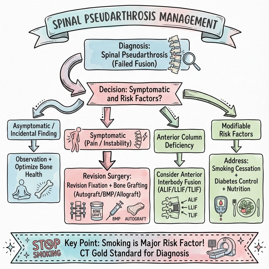
Spinal pseudarthrosis is failure to achieve solid bony fusion by 1 year post-surgery, with fibrous or cartilaginous tissue instead of bridging bone. Smoking is the single biggest modifiable risk factor (meta-analysis RR ~2.2; Glassman reported 26.5% nonunion in continuing smokers vs 14.2% in non-smokers)—nicotine directly inhibits osteoblasts and creates hypoxia. Other risk factors include multi-level fusion, diabetes, obesity, NSAIDs, and inadequate surgical technique. CT scan with thin cuts is the gold standard—look for continuous bridging trabecular bone; lucency at graft-host interface indicates nonunion. Hardware breakage suggests instability at the pseudarthrosis site. Revision surgery requires debridement of fibrous tissue, fresh autograft, rigid fixation, and interbody support (PLIF/TLIF/ALIF), with 70-80% success if risk factors are controlled. Smoking cessation for ≥6 weeks is mandatory before revision.
SMOKINGPseudarthrosis Risk Factors
Hook:SMOKING reminds you that smoking is the number one risk factor for spinal pseudarthrosis
BRIDGECT Findings of Fusion vs Pseudarthrosis
Hook:Look for BRIDGE on CT - bridging bone means successful fusion
GRIFTRevision Surgery Principles
Hook:Use GRIFT to remember revision pseudarthrosis strategy
PAINSIndications for Revision Surgery
Hook:Revision for PAINS - not all pseudarthrosis needs surgery
Exam Viva Scenarios
Practise clinical reasoning and management decisions out loud
“A 55-year-old male smoker presents with persistent axial back pain 18 months after L4-L5 posterolateral fusion with pedicle screw instrumentation. He had partial improvement for 6 months but pain has returned. How do you assess and manage?”
“A 62-year-old woman with diabetes presents for routine 1-year follow-up after L3-L5 posterolateral fusion. X-ray shows rod fracture at L4 level. She has mild back pain but is otherwise functional. What does this finding indicate and how do you proceed?”
“A 48-year-old woman had L5-S1 ALIF with posterior instrumentation 2 years ago for degenerative spondylolisthesis. She is completely pain-free and functional. CT scan obtained for unrelated reason shows lucency at graft-host interface suggesting pseudarthrosis. What is your management?”
Definition
- Failure to achieve solid bony fusion by 1 year
- Fibrous tissue instead of bridging bone
- May be symptomatic (pain) or asymptomatic
- Single-level PLF: 5-10%, multi-level: 15-25%
Risk Factors (SMOKING)
- Smoking: ~2x nonunion risk; 26.5% vs 14.2% (biggest modifiable factor)
- Multi-level fusion (each level adds risk)
- Obesity (mechanical and metabolic)
- Key nutrients lacking (Vit D, Ca, protein)
- Inadequate graft/technique
- NSAIDs and steroids
- Glucose intolerance (diabetes)
Clinical Presentation
- Persistent axial back pain at fusion level
- Pain worsened by activity, better with rest
- Pattern: never improved OR recurrent after initial improvement
- Painful motion on examination
- No radicular symptoms (unless other pathology)
Diagnosis - CT Gold Standard
- CT: thin cuts with multiplanar reconstructions
- Fusion: continuous bridging trabecular bone
- Pseudarthrosis: lucency at graft-host junction
- Hardware breakage highly specific for nonunion
- SPECT/CT if equivocal
Prevention
- Smoking cessation 6 weeks minimum before surgery
- Optimize diabetes (HbA1c less than 7%)
- Nutritional optimization (Vit D, Ca, protein)
- Adequate decortication (bleeding bone)
- Sufficient graft volume (autograft preferred)
- Rigid fixation with instrumentation
- Consider interbody fusion in high-risk cases
- BMP augmentation in revision/high-risk
Management
- Asymptomatic: observation only (no surgery)
- Symptomatic: trial conservative 3-6 months
- Revision indications: persistent pain + CT confirmed + failed conservative
- Smoking cessation MANDATORY before revision
- Revision success: 70-80% first revision, 50-60% second
Revision Principles (GRIFT)
- Graft: fresh autograft (iliac crest)
- Remove: fibrous tissue, sclerotic bone
- Instrumentation: revise/extend for rigid fixation
- Fix: risk factors (smoking cessation essential)
- Thorough: decorticate to bleeding bone
- Add interbody support (PLIF/TLIF/ALIF)
- Consider BMP augmentation
Key Exam Points
- CT scan is gold standard (80-90% sensitive)
- Smoking roughly doubles nonunion risk
- Hardware breakage = pseudarthrosis
- Asymptomatic pseudarthrosis = observation
- Treat patient not X-ray
- Revision needs smoking cessation
Evidence Base
- Retrospective review of 357 instrumented L4-L5/L4-S1 fusions
- Nonunion 14.2% in non-smokers vs 26.5% in patients who continued to smoke postoperatively (P less than 0.05)
- Patients who quit for more than 6 months had a 17.1% nonunion rate (intermediate)
- Return-to-work: 75% in quitters vs 53% in continuing smokers
- Landmark study validating postoperative smoking cessation
- Meta-analysis of 40 studies (7516 procedures) of fracture, fusion, osteotomy and arthrodesis
- Smokers had 2.2x (95% CI 1.9-2.6) the risk of delayed and/or non-union
- Increased risk was at least 1.6x in every subgroup analysed
- Time to union prolonged by a mean of 27.7 days in smokers
- 49 patients / 69 levels with surgical exploration as reference standard
- Fine-cut CT sensitivity 70-97% but specificity only 28-85% for fusion
- Raters consistently over-read fusion (low specificity)
- Posterior sentinel sign accuracy 74% vs sentinel sign 61%
- FDA IDE cohort, 277 enrolled; 6-year clinical/radiographic follow-up
- 98% (128/130) fused at 6 years with stand-alone cages plus rhBMP-2
- Worst-case fusion rate 91% (128/141) including reoperations for pseudarthrosis
- Durable improvement in ODI, SF-36 and pain scores to 6 years
- 76 instrumented posterolateral fusions with autograft plus autologous growth factor (AGF) vs matched controls
- Nonunion 25% with AGF vs 17% with autograft alone (P = 0.18, not significant)
- Platelet concentrate did not enhance fusion despite high platelet concentration
- Authors recommend against platelet gel as an autograft supplement
- RCT of 76 patients, instrumented vs non-instrumented posterolateral fusion
- Successful arthrodesis 82% instrumented vs 45% non-instrumented (P = 0.0015)
- Pedicle screws substantially raise fusion rate
- Clinical outcome was not significantly different between groups at 2 years
- Prospective RCT cohort, 47 patients with degenerative spondylolisthesis, mean 7.7-year follow-up
- Excellent/good outcome 86% with solid fusion vs 56% with pseudarthrosis (P = 0.01)
- Solid fusion superior for back/leg pain and symptom-severity scores
- Demonstrates that symptomatic nonunion does worsen long-term results
- Meta-analysis of 5 comparative studies (1403 patients)
- High-dose ketorolac increased nonunion risk (RR 2.87, 95% CI 1.53-5.38)
- Normal-dose short-course NSAIDs (less than 14 days) did not increase nonunion (RR 1.39, 95% CI 0.74-2.61)
- Effect on fusion appears dose-dependent
- Meta-analysis of 7 RCTs (941 patients); moderate-quality evidence
- Electrical stimulation increased odds of successful fusion 2.5-fold (OR 2.53, 95% CI 1.86-3.43)
- Benefit across PEMF, direct current and capacitive coupling
- No significant subgroup interaction by smoking or number of levels