Neuraxial and Peripheral Blocks | Ultrasound-Guided | LAST Prevention and Management
- Ultrasound guidance reduces complications and improves success rates
- LAST (Local Anaesthetic Systemic Toxicity) - recognise CNS then cardiac signs
- Interscalene block for shoulder - expect phrenic nerve palsy
- Adductor canal block preserves quadriceps strength vs femoral block
- Lipid emulsion 20% is first-line treatment for LAST
- “Regional anaesthesia reduces opioid use, improves pain control, and enables early mobilisation
- “Ultrasound has become standard of care for most peripheral nerve blocks
- “LAST presents with CNS symptoms first (perioral numbness, tinnitus) then cardiovascular collapse
- “Continuous catheter techniques provide prolonged analgesia (48-72 hours)
Overview and Role in Orthopaedics
Regional anaesthesia covers the neuraxial techniques (spinal and epidural) and the peripheral nerve blocks that give targeted anaesthesia and analgesia for orthopaedic procedures. It has become integral to Enhanced Recovery After Surgery (ERAS) protocols and to multimodal analgesia.
What it offers. Compared with systemic opioids it gives superior analgesia and reduces opioid consumption, and less opioid means less postoperative nausea and vomiting (PONV). Motor-sparing blocks allow earlier mobilisation, hospital stay is shorter, and patients are more satisfied.
How practice evolved. From the 1970s to the 1990s blocks were placed by anatomical landmarks and the nerve stimulator. Ultrasound guidance arrived in the 2000s and brought direct visualisation and real-time needle placement, and from the 2010s motor-sparing blocks (adductor canal, IPACK, PENG) followed. Continuous catheter techniques prolong analgesia after complex surgery.
The block follows the operation. In the upper limb the interscalene block serves the shoulder, the supraclavicular and infraclavicular blocks the elbow and forearm, and the axillary block the hand. In the lower limb the femoral, adductor canal, sciatic and popliteal blocks do the work.
Anatomy
Neuraxial anatomy
Where the cord ends. The spinal cord ends at L1-L2 in adults and at L3 in children, while the dural sac continues to S2. Total CSF volume is 120-150 mL.
The epidural space. In the lumbar region it measures 3-5 mm, and the ligamentum flavum is the key resistance met on the way in. It contains fat, which cushions; lymphatics and Batson's venous plexus; blood vessels, the source of the haematoma risk; and the nerve roots exiting at each level, with the dural cuffs where local anaesthetic spreads.
Brachial plexus
The plexus runs from the roots (C5-T1) to the terminal branches, and each level has its own block. Where the needle meets the plexus decides what is covered.
- Structure
- Exit interscalene groove
- Approach
- Interscalene block
- Clinical Relevance
- Covers shoulder surgery; spares ulnar (C8-T1)
- Structure
- Upper, middle, lower
- Approach
- Supraclavicular block
- Clinical Relevance
- Most complete arm block; pneumothorax risk
- Structure
- Anterior/posterior
- Approach
- N/A (not targeted)
- Clinical Relevance
- Transition zone behind clavicle
- Structure
- Lateral, posterior, medial
- Approach
- Infraclavicular block
- Clinical Relevance
- Elbow to hand (often spares shoulder); deeper access
- Structure
- Terminal nerves
- Approach
- Axillary block
- Clinical Relevance
- Mid-forearm to hand; multiple injections needed
- Origin
- Lateral cord
- Motor Function
- Elbow flexion (biceps, brachialis)
- Sensory Territory
- Lateral forearm
- Origin
- Lateral + medial cords
- Motor Function
- Forearm pronation, wrist/finger flexion
- Sensory Territory
- Palmar 3.5 digits, thenar
- Origin
- Medial cord
- Motor Function
- Intrinsics, finger flexion (4-5)
- Sensory Territory
- Ulnar 1.5 digits, hypothenar
- Origin
- Posterior cord
- Motor Function
- Elbow, wrist, finger extension
- Sensory Territory
- Posterior arm, dorsal hand
- Origin
- Posterior cord
- Motor Function
- Deltoid, teres minor
- Sensory Territory
- Regimental badge area
Lower limb
Lumbar plexus (L1-L4). It lies within psoas major and is blocked in the psoas compartment or beneath the fascia iliaca.
- Femoral nerve (L2-L4): anterior thigh and knee
- Lateral femoral cutaneous nerve (L2-L3): lateral thigh
- Obturator nerve (L2-L4): medial thigh and hip
Sacral plexus (L4-S3). The sciatic nerve divides in the popliteal fossa into the tibial and common peroneal nerves, and it is blocked at gluteal, subgluteal or popliteal level.
- Sciatic nerve (L4-S3): posterior thigh, leg and foot
- Posterior femoral cutaneous nerve (S1-S3): posterior thigh
- Pudendal nerve (S2-S4): perineum
Fascial planes
Fascial spread is the key to a successful block.
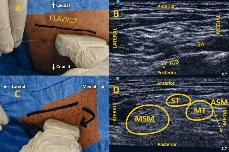
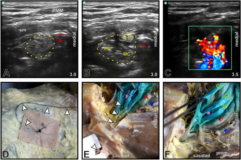
Upper limb. The interscalene groove lies between the anterior and middle scalenes. At the first rib the plexus forms a nerve cluster in the supraclavicular fossa, below the clavicle the infraclavicular space lies deep to the pectoralis muscles, and in the axilla the sheath is a continuation of the prevertebral fascia.
Lower limb. The fascia iliaca covers the femoral nerve and allows the block to spread. In the popliteal fossa the dividing sciatic nerve is surrounded by fat, and at the ankle the nerves are superficial and lie in several compartments. The IPACK block uses the interspace between the popliteal artery and the knee.
Classification
Regional techniques are grouped as neuraxial, peripheral or local.
- Technique
- Spinal (subarachnoid)
- Indications
- Lower limb, perineal surgery
- Duration
- 90-180 minutes (single shot)
- Technique
- Epidural
- Indications
- Labour, post-op analgesia, surgery
- Duration
- Continuous (catheter)
- Technique
- Combined spinal-epidural
- Indications
- Arthroplasty, prolonged procedures
- Duration
- Surgical + extended
- Technique
- Single-shot nerve block
- Indications
- Day surgery, post-op analgesia
- Duration
- 8-24 hours
- Technique
- Continuous catheter block
- Indications
- Prolonged analgesia
- Duration
- Days (catheter)
- Technique
- Local infiltration
- Indications
- Minor procedures, wound edges
- Duration
- 2-6 hours
- Technique
- Local infiltration analgesia (LIA)
- Indications
- Arthroplasty, multimodal
- Duration
- 12-24 hours
Upper limb blocks. Besides the four brachial plexus blocks, the suprascapular block covers the shoulder with limited motor block. At the wrist the median nerve block covers the palmar thumb, index and middle fingers, the ulnar block the palmar and dorsal ulnar hand, and the radial block the dorsal radial hand. Digital blocks anaesthetise individual fingers, and WALANT avoids a formal block altogether.
Lower limb blocks. The lumbar plexus (psoas) block serves THA and femur fractures, the fascia iliaca block hip fractures and anterior THA, the femoral nerve block knee surgery and femur fractures, and the PENG block the hip. The lateral femoral cutaneous nerve can be blocked for thigh graft harvest and the saphenous nerve for the medial leg and ankle. Below the knee, the sciatic block by its several approaches serves below-knee surgery, the popliteal sciatic block the foot and ankle, and the ankle block (five nerves) the foot.
Local Anaesthetic Pharmacology
How they work. Local anaesthetics bind to voltage-gated sodium channels, preventing depolarisation of the nerve fibre and so blocking propagation of the action potential. Of the agents below, only chloroprocaine is an ester; the rest are amides.
Differential blockade. Small fibres are blocked first, the C fibres carrying pain and the A-delta fibres carrying temperature. Large fibres, motor (A-alpha) and proprioceptive, are blocked last, which explains why sensory block comes before motor block.
- Class
- Amide
- Onset
- Fast (5-10 min)
- Duration
- 60-120 min
- Max Dose plain / with adrenaline (mg/kg)
- 4.5 / 7
- Use
- Short procedures
- Class
- Amide
- Onset
- Fast (5-10 min)
- Duration
- 2-4 hours
- Max Dose plain / with adrenaline (mg/kg)
- 4.5 / 7
- Use
- Intermediate duration
- Class
- Amide
- Onset
- Slow (15-30 min)
- Duration
- 240-480 min
- Max Dose plain / with adrenaline (mg/kg)
- 2 / 3
- Use
- Long procedures
- Class
- Amide
- Onset
- Slow (15-30 min)
- Duration
- 240-480 min
- Max Dose plain / with adrenaline (mg/kg)
- 3 / 4
- Use
- Motor-sparing, long duration
- Class
- Amide
- Onset
- Slow (15-30 min)
- Duration
- 240-480 min
- Max Dose plain / with adrenaline (mg/kg)
- 2 / 3
- Use
- Less cardiotoxic than bupivacaine
- Class
- Ester
- Onset
- Very fast
- Duration
- 30-60 min
- Max Dose plain / with adrenaline (mg/kg)
- 11 / 14
- Use
- -
Adjuncts that prolong the block.
- Adrenaline (1:200,000 to 1:400,000) vasoconstricts, prolonging the block by 30-50% and reducing systemic absorption, which lowers the LAST risk. It also marks an intravascular injection by causing tachycardia. Avoid it in the interscalene block, where it prolongs the phrenic palsy.
- Dexamethasone (4-8 mg) prolongs the block by 30-50% and, being anti-inflammatory, reduces postoperative pain; perineural and IV routes are both effective. Perineural use prolongs the block by 8-12 hours but is off-label and controversial, while IV use adds 4-8 hours and is the safer option.
- Clonidine (1-2 mcg/kg, or 75-150 mcg), an alpha-2 agonist, prolongs sensory and motor block with a modest effect, at the cost of hypotension and sedation. It is less commonly used with longer-acting agents.
- Dexmedetomidine (50-100 mcg) gives modest prolongation, with sedation.
Adjuncts that do not help. Perineural opioids give no significant benefit and add systemic side effects, ketamine has no proven perineural benefit, and the evidence for magnesium is limited. Sodium bicarbonate speeds onset but does not prolong the block, and hyaluronidase may speed onset but shorten duration.
Neuraxial Anaesthesia
Spinal anaesthesia (subarachnoid block)
Technique. With the patient sitting or in the lateral decubitus position, the needle enters the L3-L4 or L4-L5 interspace, below the conus medullaris at L1-L2. A 25G or 27G pencil-point needle (Whitacre, Sprotte) reduces post-dural puncture headache (PDPH), and CSF flow is the endpoint. The lower-limb dose is 10-15 mg of heavy 0.5% bupivacaine.
Onset and duration. Onset takes 5-10 minutes. The block lasts 90-150 minutes with plain solution and 120-180 minutes with heavy bupivacaine and an opioid.
Uses. Total hip and knee arthroplasty, lower-limb fracture fixation, and foot and ankle surgery.
Strengths and costs. The block is rapid in onset, dense in both motor and sensory fibres and predictable in duration, and the controlled hypotension reduces blood loss. Against that, the duration of a single shot is fixed, sympathetic blockade causes hypotension, urinary retention can follow, and a dural puncture with a large needle risks headache.
Epidural anaesthesia
Technique. The needle goes in at lumbar level (L2-L3, L3-L4) for the lower limb, or thoracic level for upper abdominal and thoracic surgery. A 16-18G Tuohy needle finds the epidural space by loss of resistance to saline or air, and a catheter threaded through it allows continuous infusion of bupivacaine 0.25-0.5% or ropivacaine 0.2-0.5%.
Onset and duration. Onset takes 15-30 minutes, and with a catheter the block is continuous.
Uses. Major lower-limb surgery (bilateral TKA, complex trauma), postoperative analgesia after spine surgery, and rib fractures (thoracic epidural).
Strengths and costs. The block is titratable, continuous through the catheter and usable for postoperative analgesia, and its gradual onset keeps the circulation stable. It is slower in onset than a spinal, needs more drug, carries the risk of epidural haematoma under anticoagulation, and can be patchy, with a 5-10% failure rate.
Combined spinal-epidural (CSE)
The spinal component gives rapid onset and the epidural catheter prolonged analgesia, placed needle-through-needle or through separate spaces. It suits surgery of long or unpredictable duration and operations after which postoperative analgesia is required, such as THA and TKA.
- Spinal
- Subarachnoid
- Epidural
- Epidural (potential) space
- CSE
- Both
- Spinal
- 5-10 minutes
- Epidural
- 15-30 minutes
- CSE
- 5-10 minutes
- Spinal
- 90-180 minutes
- Epidural
- Continuous
- CSE
- Continuous
- Spinal
- Less than 5%
- Epidural
- 5-10%
- CSE
- Less than 5%
- Spinal
- High
- Epidural
- Moderate
- CSE
- High initially
- Spinal
- Less than 1%
- Epidural
- 1-2%; rare unless dural puncture
- CSE
- Less than 1%
- Spinal
- Short procedures
- Epidural
- Long procedures
- CSE
- Long with rapid onset
Contraindications to neuraxial block are set out with the others under Clinical Assessment, and anticoagulant timing has its own section.
Upper Extremity Peripheral Nerve Blocks
The four brachial plexus blocks step down the plexus from the roots to the terminal branches, as laid out in the organisation table under Anatomy.
Interscalene block (ISB)
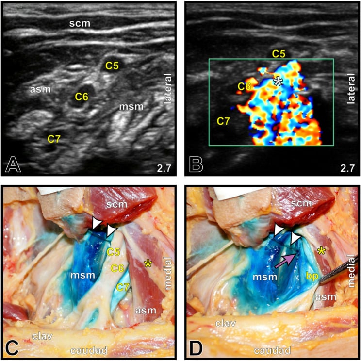
Target. The brachial plexus roots at the level of the cricoid cartilage, between the anterior and middle scalene muscles. It blocks C5, C6 and C7, primarily the superior trunk, and often misses C8 and T1, the ulnar territory of the hand.
Uses. Shoulder surgery (rotator cuff, arthroplasty, arthroscopy), proximal humeral fractures and clavicle fractures.
Technique. With the patient supine and the head turned away, a high-frequency linear probe is placed transversely on the lateral neck at the cricoid level, where the roots lie between the scalenes like "traffic lights". The needle goes in-plane or out-of-plane, and 15-20 mL of 0.5% ropivacaine or bupivacaine is injected.
What to expect. Onset takes 15-30 minutes, and the block lasts 12-18 hours as a single shot or 48-72 hours with a catheter. It covers the shoulder and proximal arm, with incomplete coverage of the hand.
Complications.
- Phrenic nerve palsy: temporary hemidiaphragm paralysis in 100%
- Horner syndrome (ptosis, miosis, anhidrosis): 25-75%
- Recurrent laryngeal nerve block (hoarseness): 5-10%
- Vertebral artery injection: rare but catastrophic
- Pneumothorax: under 1% with ultrasound
The phrenic nerve decides who can have it. Avoid bilateral ISB, which risks bilateral phrenic palsy and respiratory compromise, and take care in respiratory disease such as COPD and obstructive sleep apnoea. Low-volume techniques (5-10 mL) reduce phrenic nerve involvement.
Intravenous regional anaesthesia (IVRA, Bier block)
The Bier block is a simple, reliable technique for short distal-limb procedures (hand and forearm, and occasionally foot) that needs no nerve localisation.
Technique. An IV cannula is sited in the operative hand or foot, the limb is exsanguinated (elevation, then an Esmarch bandage), and a double (two-cuff) tourniquet is inflated above arterial pressure. Dilute lidocaine or prilocaine (for example 0.5% lidocaine, roughly 3 mg/kg) is then injected through the cannula; it diffuses from the vasculature into the nerves and produces anaesthesia distal to the cuff within about 5-10 minutes.
The two cuffs. The proximal cuff is inflated first. Once the block is established the distal cuff, now over anaesthetised skin, is inflated and the proximal cuff released to relieve tourniquet pain.
Deflation is the danger. Letting the cuff down releases the sequestered local anaesthetic into the systemic circulation as a bolus and risks LAST. The cuff must therefore stay inflated for a minimum of around 20-30 minutes however quickly surgery finishes, and at the end it is deflated in cycles (deflate, reinflate) while watching for toxicity. This hazard is why bupivacaine, which is cardiotoxic, must never be used for IVRA. The tourniquet-use topic covers cuffs and pressures.
Uses and limits. Fast, technically easy, with high success and rapid recovery, it is ideal for day-case carpal tunnel release, ganglion excision or fracture manipulation. Anaesthesia lasts only while the cuff is up, so there is no postoperative analgesia; tourniquet pain limits it to roughly an hour; and it is unsuitable when a bloodless field is needed beyond tourniquet tolerance.
WALANT (wide-awake local anaesthesia, no tourniquet)
WALANT is a hand, and increasingly foot, technique that uses local anaesthetic with adrenaline alone: no tourniquet, no sedation and no formal block, so the patient is fully awake and comfortable.
Technique. Lidocaine (e.g. 1%) with adrenaline (e.g. 1:100,000) is infiltrated subcutaneously along the planned operative field in a tumescent approach, usually buffered with bicarbonate to reduce the sting of injection. Waiting about 25-30 minutes for maximal adrenaline vasoconstriction gives a near-bloodless field without a tourniquet.
Adrenaline in the fingers. The old teaching that adrenaline is dangerous in the fingers has been refuted: large series show that lidocaine with adrenaline is safe in the digits, and phentolamine is available as a rescue reversal agent if vasoconstriction is excessive. That removed the rationale for the tourniquet and sedation in many hand procedures.
Why use it. The patient can actively move the hand during the operation, which is invaluable for flexor tendon repair (testing the repair for gliding and gapping), tenolysis and tendon transfers. There is no tourniquet pain and therefore no sedation, no general or neuraxial anaesthetic and no fasting, and it is efficient and low-cost, suited to minor-procedure and office settings.
Typical uses. Carpal tunnel release, trigger-finger release, flexor and extensor tendon repair, fracture K-wiring and Dupuytren surgery.
Lower Extremity Peripheral Nerve Blocks
Femoral nerve block (FNB)
Target. The femoral nerve lateral to the femoral artery and deep to the fascia iliaca, below the inguinal ligament in the femoral triangle (VAN: vein, artery, nerve). It supplies the anterior thigh, the knee joint and, through its saphenous branch, the medial leg.
Uses. Analgesia for femoral fractures, knee surgery combined with a sciatic block or local infiltration, and quadriceps tendon repair.
Technique. With the patient supine and the leg slightly abducted, a linear probe is placed at the inguinal crease. The needle goes in-plane from lateral, and 20-30 mL is injected.
What to expect. Onset takes 15-30 minutes and the block lasts 12-18 hours, covering the anterior thigh, knee joint and medial leg.
The price is the quadriceps. Quadriceps weakness occurs in 100% and is the major disadvantage: the patient cannot weight-bear, so falls are a risk. The femoral artery is adjacent, so vascular puncture is possible, and nerve injury is rare with ultrasound.
The fascia iliaca block. This more lateral approach beneath the fascia iliaca has an easier landmark and a lower risk of nerve injury. Its coverage is less consistent and it needs a higher volume, 40-50 mL (0.25-0.375% ropivacaine).
Ultrasound Guidance in Regional Anaesthesia
The evidence. Ultrasound has become the standard of care for most peripheral nerve blocks. In the Abrahams meta-analysis (BJA 2009), compared with nerve stimulation it reduced block failure (RR 0.41, 95% CI 0.26-0.66) and vascular puncture (RR 0.16, 95% CI 0.05-0.47), made onset 29% faster and the block 25% longer, and improved block density and coverage. The trials were not powered to prove a reduction in LAST.
Why it is safer. The operator sees the nerves and the vessels, pleura and peritoneum around them, tracks the needle in real time to prevent vascular puncture, and confirms that the spread is adequate. If the injection is intravascular or intraneural, it can be stopped.
Probes. High-frequency linear probes (8-15 MHz) image superficial structures under 4 cm and serve most blocks, while low-frequency curvilinear probes (2-5 MHz) image deep structures over 4 cm. Compound imaging improves tissue definition, Doppler identifies vessels to avoid, and 3D/4D imaging is emerging for complex anatomy.
- Description
- Needle parallel to probe
- Advantages
- Full needle shaft visualised
- Disadvantages
- Requires needle steering skill
- Description
- Needle perpendicular to probe
- Advantages
- Short skin-target distance
- Disadvantages
- Only see needle tip (cross-section)
- Description
- Angled between in/out plane
- Advantages
- Compromise approach
- Disadvantages
- Variable visualisation
Reading the image. Nerves appear as round or oval honeycomb structures, multiple hypoechoic fascicles within a bright hyperechoic rim. Arteries are pulsatile and non-compressible, veins compressible and non-pulsatile, fascia forms hyperechoic lines, and muscle is striated and contracts. Understanding this sonoanatomy is now essential.
A good injection. Local anaesthetic spreads circumferentially around the nerve, the "donut sign", and separates it from the adjacent structures. Slight distension of the nerve during injection can accompany a successful block.
Stop immediately if there is any sign of intraneural injection:
- High resistance to injection
- Severe paraesthesia or pain reported by the patient
- Nerve expansion without spread around the nerve
- No visible spread despite injection
If intraneural injection is suspected, stop, withdraw the needle slightly and reassess its position.
Injection pressure. Normal injection pressure is under 15 psi, and higher pressure (over 15-20 psi) suggests intraneural injection. It can be judged with a commercial opening-pressure device or by syringe feel, and the needle is repositioned before injection is attempted again.
The nerve stimulator. In modern practice ultrasound is primary and the stimulator an adjunct, not mandatory, used to confirm the identity of a nerve. Start at 0.5-1.0 mA and look for a motor twitch at 0.3-0.5 mA. No response at 0.2 mA indicates a good position, not intraneural, while a twitch below 0.2 mA may mean the needle is intraneural and should be repositioned.
Training. Basic skills take 20-30 blocks, competency 50-100 and expert level 200 or more. The recommended pathway runs from didactic learning (anatomy, physics) through simulation and phantom practice and supervised clinical practice to independent practice with backup.
Clinical Assessment
History. Ask about allergies (local anaesthetics, latex, antiseptics), anticoagulation (type, dose and timing of the last dose), previous blocks (success, complications, nerve injury), and cardiac, respiratory and neurological comorbidity. Note anything that affects consent, such as language, comprehension or anxiety.
Examination. Look for infection at the proposed site and for skin lesions, scarring or burns, and assess the landmarks, any deformity, body habitus and peripheral vascular disease. Examine and document any pre-existing neurological deficit before the block.
Contraindications
- Type
- Absolute
- Blocks Affected
- All
- Management
- Alternative anaesthesia
- Type
- Absolute
- Blocks Affected
- All
- Management
- Alternative site or GA
- Type
- Absolute
- Blocks Affected
- All LA blocks
- Management
- Amide/ester switch or GA
- Type
- Absolute
- Blocks Affected
- Deep/neuraxial blocks
- Management
- Peripheral or GA
- Type
- Absolute
- Blocks Affected
- Neuraxial, deep blocks
- Management
- Timing per guidelines
- Type
- Absolute
- Blocks Affected
- Neuraxial
- Management
- -
- Type
- Absolute
- Blocks Affected
- Neuraxial
- Management
- -
- Type
- Relative
- Blocks Affected
- Neuraxial
- Management
- Timing critical
- Type
- Relative
- Blocks Affected
- Block in affected territory
- Management
- Document, discuss with patient (medicolegal)
- Type
- Relative
- Blocks Affected
- Interscalene, high neuraxial
- Management
- Motor-sparing alternatives
- Type
- Relative
- Blocks Affected
- Neuraxial
- Management
- Bacteraemia risk of seeding
- Type
- Relative
- Blocks Affected
- Neuraxial
- Management
- -
- Type
- Relative
- Blocks Affected
- Neuraxial
- Management
- Difficult technique
- Type
- Relative
- Blocks Affected
- Neuraxial
- Management
- Altered anatomy
Consent. Describe the procedure and its expected benefits (pain relief, opioid sparing), the common risks (bruising, numbness, discomfort) and the serious ones (LAST, nerve injury, infection), the alternatives (GA, IV analgesia, other blocks) and when sensation will return. Record the risks discussed for that block, the pre-existing motor and sensory examination, the patient's understanding and any concerns, and have the consent signed before sedation.
Anticoagulation and Regional Blocks
Bleeding into a confined space. The risk is not just bleeding but bleeding in a confined space, as when an epidural haematoma compresses the cord. Superficial sites can be compressed and deep sites cannot, so the block type sets the level of risk.
- Compressible sites (lower risk): axillary (superficial), femoral (below the inguinal ligament), popliteal, ankle (multiple superficial sites), and wrist or digital blocks (very low risk)
- Non-compressible sites (higher risk): neuraxial (catastrophic if a haematoma forms), lumbar plexus (deep within psoas), infraclavicular (behind the clavicle, near vessels), supraclavicular (risk of subclavian puncture) and deep cervical plexus (near the vertebral artery)
- Time Before Block
- No restriction
- Time After Block
- No restriction
- Notes
- Safe for neuraxial; can continue
- Time Before Block
- No restriction
- Time After Block
- No restriction
- Notes
- Safe for neuraxial
- Time Before Block
- 5-7 days
- Time After Block
- Immediately
- Notes
- High-risk; alternatives preferred
- Time Before Block
- 4-6 hours
- Time After Block
- 1 hour
- Notes
- aPTT normal before block; check aPTT if over 4 days
- Time Before Block
- 4-6 hours + normal aPTT
- Time After Block
- 1 hour
- Notes
- Higher risk; check aPTT
- Time Before Block
- 12 hours
- Time After Block
- 4 hours
- Notes
- Daily dosing; twice-daily dosing = 24 hours
- Time Before Block
- 24 hours
- Time After Block
- 4 hours
- Notes
- Twice-daily dosing; consider anti-Xa if uncertain
- Time Before Block
- 5 days, INR under 1.4
- Time After Block
- After catheter removal
- Notes
- Check INR before block; assess with other agents
- Time Before Block
- 72 hours
- Time After Block
- 6 hours
- Notes
- NOACs require longer intervals; longer for renal impairment
- Time Before Block
- 72-120 hours
- Time After Block
- 6 hours
- Notes
- Highly renal dependent
Catheters. Remove the catheter before restarting anticoagulation, wait the same interval after removal as for block placement, and observe for 4 hours after removal for signs of haematoma.
Deciding at the bedside. Identify the block type (neuraxial, deep or superficial peripheral) and the anticoagulant (type, dose, last administration), then apply ASRA and local guidelines. Consider the alternatives, superficial blocks, GA or multimodal analgesia, and document the risk-benefit discussion.
Investigations
Test only what will change management. Routine screening is not required for healthy patients having peripheral blocks, and coagulation testing is needed only if the patient is anticoagulated or coagulopathy is suspected clinically.
- When Required
- Warfarin use, liver disease
- Target/Normal
- Under 1.4 for neuraxial
- Action if Abnormal
- Hold warfarin or use peripheral block
- When Required
- UFH use
- Target/Normal
- Normal (less than 40 sec)
- Action if Abnormal
- Wait 4-6 hours post-heparin
- When Required
- Suspected thrombocytopenia
- Target/Normal
- Greater than 80,000 for neuraxial
- Action if Abnormal
- Consider peripheral alternatives
- When Required
- DOAC use
- Target/Normal
- Calculate dose adjustments
- Action if Abnormal
- Extend hold times for renal impairment
- When Required
- Not routine
- Target/Normal
- Cardiac history only
- Action if Abnormal
- Optimise before elective surgery
- Measures
- Whole clot formation and lysis
- Time to Result
- 30-60 min (full)
- Limitations
- Requires training, not specific to LA agents
- Measures
- Similar to TEG
- Time to Result
- 30-60 min
- Limitations
- Expensive, operator dependent
- Measures
- Heparin effect (UFH)
- Time to Result
- 5 min
- Limitations
- Only measures heparin; not warfarin/DOACs
- Measures
- LMWH, rivaroxaban, apixaban
- Time to Result
- 1-2 hours
- Limitations
- Lab-based; not true POCT
- Measures
- Aspirin/clopidogrel effect
- Time to Result
- 10-15 min
- Limitations
- Not widely available
The ultrasound pre-scan. Now standard of care for most peripheral blocks, the pre-scan finds the nerve's location and depth, maps the vessels to avoid puncture, and detects pathology such as cysts, tumours or anomalies. It also plans the needle trajectory, and the patient's habitus guides the choice of probe and needle.
Other imaging. Fluoroscopy can guide lumbar plexus and neuraxial blocks but is not routine, CT guidance helps with complex anatomy or tumours, and MRI serves pre-procedural planning, not real-time guidance. Injection pressure monitoring is a safety adjunct.
Management Algorithm
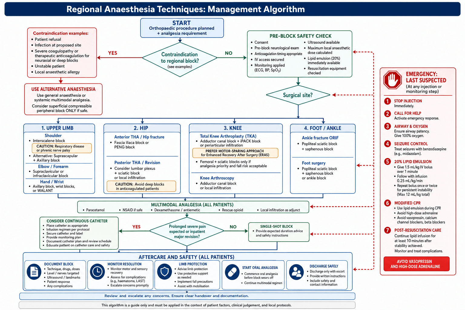
Planning a block. Work through six questions:
- What surgery? The site determines which nerves to block and the sensory and motor requirements.
- How long? The duration of surgery and the postoperative analgesic need decide between a single shot with a long-acting agent and a catheter.
- Ambulatory or inpatient? Motor-sparing blocks suit ambulation.
- Anticoagulated? This governs site selection and timing, and deep blocks are contraindicated.
- What comorbidities? In respiratory disease avoid a phrenic block and avoid bilateral interscalene blocks; consider cardiac disease too.
- What multimodal plan? Integrate the block with paracetamol, NSAIDs and gabapentinoids.
The best block is the one that provides adequate analgesia with minimal motor impairment for the specific surgery and patient.
Block selection by surgery
- First Choice
- Interscalene
- Alternatives
- Supraclavicular; suprascapular + axillary
- Coverage, Motor Effect and Catheter
- C5, C6, C7 (superior trunk); catheter for major open surgery
- First Choice
- Interscalene
- Alternatives
- Supraclavicular
- Coverage, Motor Effect and Catheter
- C5-C7, partial C8-T1
- First Choice
- Supraclavicular
- Alternatives
- Infraclavicular
- Coverage, Motor Effect and Catheter
- Complete C5-T1; catheter rarely
- First Choice
- Supraclavicular
- Alternatives
- Infraclavicular or axillary
- Coverage, Motor Effect and Catheter
- C6-T1; catheter rarely
- First Choice
- Axillary
- Alternatives
- Wrist blocks
- Coverage, Motor Effect and Catheter
- Median nerve only
- First Choice
- Axillary
- Alternatives
- Supraclavicular
- Coverage, Motor Effect and Catheter
- C7-T1
- First Choice
- Axillary or wrist blocks
- Alternatives
- WALANT
- Coverage, Motor Effect and Catheter
- No catheter
- First Choice
- Adductor canal + IPACK or periarticular infiltration
- Alternatives
- Femoral + sciatic; adductor canal + sciatic
- Coverage, Motor Effect and Catheter
- Nerves: femoral + sciatic (or saphenous + popliteal); quadriceps preserved with ACB; catheter for major surgery or poor pain control
- First Choice
- Fascia iliaca or PENG
- Alternatives
- LIA
- Coverage, Motor Effect and Catheter
- Femoral, LFCN, obturator; quadriceps preserved; catheter for major revision
- First Choice
- Lumbar plexus + sciatic
- Alternatives
- LIA
- Coverage, Motor Effect and Catheter
- Sciatic, lumbar plexus; catheter for major revision
- First Choice
- Femoral or fascia iliaca
- Alternatives
- -
- Coverage, Motor Effect and Catheter
- Quadriceps blocked (acceptable); superior analgesia
- First Choice
- Adductor canal or LIA
- Alternatives
- Femoral; intra-articular
- Coverage, Motor Effect and Catheter
- Motor sparing with adductor canal; no catheter
- First Choice
- Adductor canal
- Alternatives
- -
- Coverage, Motor Effect and Catheter
- Quadriceps preserved; allows quadriceps exercises
- First Choice
- Popliteal sciatic + saphenous
- Alternatives
- Ankle block
- Coverage, Motor Effect and Catheter
- Sciatic (tibial + common peroneal) + saphenous; foot drop overnight; catheter rarely
- First Choice
- Popliteal sciatic + saphenous, or ankle block (5 nerves)
- Alternatives
- -
- Coverage, Motor Effect and Catheter
- No motor block needed; ankle block avoids sciatic motor block; no catheter
Block timing
- Advantages
- Pre-emptive analgesia, awake positioning, reduced GA requirements
- Disadvantages
- Block room time, risk of incomplete block
- Best For
- Major surgery, day surgery (efficiency)
- Advantages
- Confirmed surgical success, neurological exam possible
- Disadvantages
- Delayed analgesia, patient in pain
- Best For
- When neurological exam critical, rescue analgesia
Multimodal analgesia
Before surgery.
- Paracetamol 1 g orally
- An NSAID if not contraindicated (e.g. celecoxib 200-400 mg)
- A gabapentinoid, pregabalin 75-150 mg, which reduces opioid requirement
- Dexamethasone 8 mg IV, which extends the block and is antiemetic
- Midazolam 1-2 mg for anxiolysis if needed
During and after surgery.
- The regional block as planned for the surgery, with LIA as an adjunct or alternative
- Regular paracetamol 1 g four times daily, and NSAIDs continued if tolerated
- Oxycodone 5-10 mg as needed for breakthrough pain
- Antiemetics: ondansetron, dexamethasone
Surgical Technique
Preparation. Apply monitoring (SpO2, ECG, blood pressure) before any sedation, and secure IV access, which is essential before any block. Have resuscitation equipment and lipid emulsion available, use sterile technique (skin preparation, sterile probe cover), and position the patient comfortably with access to the block site.
Equipment. A high-frequency linear ultrasound probe suits most blocks, and a nerve stimulator is an adjunct. Block needles are 50-100 mm, echogenic preferred, and the local anaesthetic is pre-drawn in labelled syringes, with extension tubing for aspiration and injection.
- Action
- Identify target nerve/structures
- Key Points
- Optimise image depth, gain, frequency
- Action
- Determine needle path
- Key Points
- In-plane preferred for visualisation
- Action
- Antiseptic, sterile field
- Key Points
- Allow to dry; probe in sterile sheath
- Action
- Subcutaneous LA at entry point
- Key Points
- Small volume, reduce patient discomfort
- Action
- Advance under real-time visualisation
- Key Points
- Keep needle tip in view at all times
- Action
- Small test injection (D5W or LA)
- Key Points
- Confirms tip position; opens tissue planes
- Action
- Check for blood before main injection
- Key Points
- Negative aspiration does not exclude IV placement
- Action
- 5mL aliquots with aspiration
- Key Points
- Watch for spread around nerve; reposition if needed
If in doubt, don't inject. Never inject when unsure where the needle tip is, and stop and reassess if the patient reports paraesthesia or severe pain.
Continuous catheters
When to use one. A catheter is for analgesia needed for more than 24 hours: major surgery (TKA, THA, shoulder reconstruction), trauma (rib fractures, complex fractures), chronic cancer pain, and sympathetic block to improve perfusion.
- Technique
- Assess on ultrasound
- Troubleshooting
- Deeper nerves need longer catheters
- Technique
- Tip at target, small injection
- Troubleshooting
- Confirm spread before threading
- Technique
- Advance 3-5cm past needle tip
- Troubleshooting
- Resistance = redirect needle slightly
- Technique
- Inject through catheter, watch spread
- Troubleshooting
- No spread = reposition before securing
- Technique
- Dressing, tape, loop at skin
- Troubleshooting
- Prevent traction, mark depth at skin
What goes wrong. Dislodgement needs repositioning in 5-10%, infection risk rises after 72-96 hours, local anaesthetic can leak around the site, and motor block may impair rehabilitation. The infusion runs from an elastomeric or electronic pump.
Local Anaesthetic Systemic Toxicity (LAST)
Mechanism. In the CNS, local anaesthetic inhibits the inhibitory neurons first, producing excitation, and then all neurons, producing depression. In the heart it blocks sodium and potassium channels and dysregulates calcium. The lipid sink theory holds that lipid emulsion sequesters local anaesthetic from the tissues.
Who is at risk.
- A high total dose, exceeding the maximum recommended
- Accidental intravascular injection, especially into an artery
- A highly vascular site, such as intercostal or paracervical blocks
- Extremes of age, cardiac disease, hepatic dysfunction and low-protein states
Which agent. Bupivacaine has the highest cardiotoxicity, ropivacaine is intermediate, and lidocaine is the least toxic.
Presentation
LAST progresses in stages, and the CNS signs come before the cardiac ones:
- CNS excitation: perioral numbness, metallic taste, tinnitus, visual disturbances, agitation and confusion, muscle twitching
- CNS depression: seizures, loss of consciousness, respiratory depression, coma
- Cardiovascular toxicity: bradycardia, hypotension, arrhythmias (ventricular tachycardia, fibrillation) and cardiac arrest, which is often resistant to standard ACLS
The first sign is often perioral numbness or tinnitus in an awake patient. If the patient reports these symptoms during injection, stop immediately and do not continue injecting.
Management
Immediate actions.
- Stop injecting local anaesthetic.
- Call for help: LAST is a team emergency.
- Manage the airway with 100% oxygen, ventilating or intubating if needed and avoiding hyperventilation.
- Suppress seizures with benzodiazepines, not propofol initially.
- Give 20% lipid emulsion (Intralipid or equivalent), the first-line treatment.
- Dose
- 1.5 mL/kg over 1 minute
- Notes
- Approximately 100 mL for a 70 kg adult
- Dose
- 0.25 mL/kg/min
- Notes
- Continue for at least 10 minutes after cardiovascular stability
- Dose
- 1.5 mL/kg
- Notes
- If cardiovascular instability persists after 5 minutes or collapse occurs; can repeat 1-2 times
- Dose
- 10 mL/kg over the first 30 minutes
- Notes
- Higher doses used in refractory cases
Cardiac arrest. Start CPR by the ACLS protocol and continue lipid emulsion throughout the resuscitation. Give adrenaline only in small doses (under 1 mcg/kg) and avoid vasopressin, which may worsen cardiac toxicity. Resuscitation may take over 60 minutes, so do not give up early.
After resuscitation. Observe for at least 4-6 hours (12-24 hours if severe), monitoring the ECG, cardiac enzymes and lipid levels, because toxicity can recur as local anaesthetic redistributes from the tissues.
Propofol is NOT a substitute for lipid emulsion. While propofol contains lipid, the concentration is too low (10% vs 20% in Intralipid), and propofol itself is a myocardial depressant. Using propofol for LAST can worsen cardiovascular collapse. Always use 20% lipid emulsion (Intralipid).
Prevention
- Use ultrasound guidance, which reduces accidental intravascular injection and the volume of local anaesthetic required
- Calculate the weight-based maximum dose before drawing up, and respect it
- Aspirate for blood before injecting
- Fractionate the injection into 3-5 mL aliquots with aspiration and pauses, which allows an intravascular injection to be detected
- Divide the total dose between blocks or sites when several are needed
- Add adrenaline 1:200,000 as a marker or test dose: a rising heart rate suggests intravascular injection
- Talk to the patient and ask about symptoms during injection
Complications
Sudden deterioration after a block
Acute collapse or distress shortly after local anaesthetic injection has a focused differential, and telling the causes apart quickly matters because their management differs.
- Typical timing/clues
- Seconds to minutes; perioral numbness, tinnitus, seizures then arrhythmia
- Key discriminator
- CNS signs precede cardiac; recent large/vascular LA dose
- Immediate action
- Stop injection, lipid emulsion 20%, ACLS
- Typical timing/clues
- Minutes; rising sensory level, bradycardia, hypotension, apnoea
- Key discriminator
- Ascending block after neuraxial or deep injection
- Immediate action
- Airway/ventilation, fluids, vasopressors, atropine
- Typical timing/clues
- Minutes; urticaria, bronchospasm, hypotension
- Key discriminator
- Skin/airway features; often to antiseptic, latex or antibiotic, not LA
- Immediate action
- Adrenaline, fluids, remove trigger
- Typical timing/clues
- During/just after block; bradycardia, pallor, nausea, rapid recovery supine
- Key discriminator
- Self-limiting, responds to position and atropine
- Immediate action
- Lie flat, reassure, atropine if bradycardic
- Typical timing/clues
- 15-30 min; dyspnoea, reduced ipsilateral air entry
- Key discriminator
- Isolated respiratory symptoms after interscalene block
- Immediate action
- Oxygen, upright posture, reassurance
- Typical timing/clues
- Delayed; pleuritic pain, dyspnoea, reduced breath sounds
- Key discriminator
- Supraclavicular/infraclavicular approach; confirm on imaging
- Immediate action
- Oxygen, imaging, drain if tension/large
- Typical timing/clues
- Often hours-days post-op; hypoxia, tachycardia, pleuritic pain
- Key discriminator
- Not temporally linked to injection; VTE risk factors
- Immediate action
- Oxygen, anticoagulation, CTPA
Neuraxial complications
- Incidence
- 1:150,000 to 1:220,000
- Risk Factors
- Anticoagulation, coagulopathy, difficult insertion
- Management
- Emergency MRI, surgical decompression within 8 hours
- Incidence
- 1:10,000 to 1:100,000
- Risk Factors
- Catheter duration, immunocompromise, diabetes
- Management
- MRI, IV antibiotics, surgery if neurological deficit
- Incidence
- 1 in 1,000
- Risk Factors
- High epidural injection
- Management
- -
- Incidence
- Rare
- Risk Factors
- High-dose hyperbaric LA, narrow canal
- Management
- Emergency imaging, decompression if structural cause
- Incidence
- 0.5-1% (small needle)
- Risk Factors
- Young, female, large needle, multiple attempts; lower with pencil-point needles
- Management
- Conservative (fluids, caffeine), then blood patch if persistent
- Incidence
- 2-7%
- Risk Factors
- Lidocaine spinal, lithotomy position
- Management
- Reassurance, NSAIDs; usually resolves in 3-5 days
- Incidence
- 20-40%
- Risk Factors
- Sympathetic blockade
- Management
- Fluids, vasopressors
- Incidence
- 10-30%
- Risk Factors
- -
- Management
- Resolves as block wears off
Peripheral nerve block complications
How often. Nerve injury occurs in under 1 in 1,000 blocks, fewer with ultrasound, and vascular injury in under 1 in 500, usually a self-limiting haematoma. The block fails to give complete anaesthesia in 5-10%. Block-specific complications, such as phrenic palsy and pneumothorax, are listed with each block; the lumbar plexus block adds epidural spread and renal injury.
Other problems. Infection is rare after a single shot and more likely with catheters, and a retained catheter may need imaging and extraction. True allergy is rare, and commoner with esters than amides.
- Mechanism
- Pressure, ischaemia, stretching
- Prognosis
- Complete recovery (weeks-months)
- Prevention
- Ultrasound guidance, low pressure injection
- Mechanism
- More severe pressure/trauma
- Prognosis
- Recovery possible (months)
- Prevention
- Avoid intraneural injection
- Mechanism
- Needle transection (rare)
- Prognosis
- Poor; may need surgery
- Prevention
- Keep needle tip visible; stop if paraesthesia
Managing a new deficit. Document it at once and compare it with the pre-block examination. Most resolve within 4-6 weeks; if symptoms persist, obtain EMG/NCS at 3-4 weeks and refer to a specialist if there is no improvement by 6 weeks, with neurology or hand surgery review for persistent or worsening deficits.
Minimising the risk. Use ultrasound guidance, avoid injecting against high resistance, stop if the patient reports severe pain, respect the anatomy without excessive force, and use atraumatic needles.
Postoperative Care
- Block Type
- Peripheral block
- Monitoring Requirements
- 30-60 min observation
- Discharge Criteria
- Stable vitals, protective sensation returning, escort home
- Block Type
- Spinal/epidural
- Monitoring Requirements
- Full motor recovery required
- Discharge Criteria
- Walking, voiding, stable BP
- Block Type
- Peripheral block
- Monitoring Requirements
- Routine ward observations
- Discharge Criteria
- Document block resolution on chart
- Block Type
- Continuous catheter
- Monitoring Requirements
- Daily catheter checks, motor/sensory assessment
- Discharge Criteria
- Remove if infection signs or no longer needed
Protecting the insensate limb. Position it to prevent nerve compression, protect it from heat, cold and sharp objects, and support weight-bearing with crutches if the leg is blocked; the patient receives written instructions. An arm block needs a sling, gait training with physiotherapy waits until the block resolves, and DVT prophylaxis continues despite immobility. Record the expected time of block resolution.
Discharge after a peripheral block. Protective sensation must be returning or the limb protected, and a responsible adult should be at home for the duration of the block. After a motor-blocking block such as a femoral block, provide crutches or gait aids and clear instructions about the fall risk, and consider an overnight stay for a high-risk patient.
Give verbal and written instructions covering:
- The expected time for sensation and movement to return
- How to keep the insensate limb safe from injury
- Oral analgesia, supplied and taken before the block wears off
- When to seek help: numbness beyond 24 hours, increasing weakness, signs of infection
- A contact number for concerns
Continuous catheter management
- Normal Finding
- Clean, dry, no erythema
- Action if Abnormal
- Remove if infected; culture tip
- Normal Finding
- Appropriate dermatomal coverage
- Action if Abnormal
- Adjust rate; check position
- Normal Finding
- Minimal impairment for rehab
- Action if Abnormal
- Reduce concentration if excessive
- Normal Finding
- VAS less than 4 at rest
- Action if Abnormal
- Bolus, increase rate, or add systemic analgesia
- Normal Finding
- Easy injection, no leakage
- Action if Abnormal
- Resite if dysfunctional; consider removal
Regimens. A background infusion of 5-10 mL/hour (0.1-0.2% ropivacaine) with patient-controlled demand boluses (5 mL every 30-60 minutes) is the commonest arrangement. Intermittent boluses of 10-15 mL every 4-6 hours use less local anaesthetic and potentially spread better, and lower concentrations (0.1-0.15%) spare motor function.
Troubleshooting. For inadequate analgesia, check the position with ultrasound and bolus through the catheter; for motor block, reduce the concentration or switch to intermittent boluses. Leakage needs the dressing resecured and may need a new catheter, and a disconnected catheter is not reconnected because of the contamination risk.
Infection. Inspect the site daily and teach the patient to report redness, discharge or fever. Consider removal at 72-96 hours, and remove the catheter immediately if it is infected, sending it for culture and giving antibiotics if there is cellulitis; chlorhexidine-impregnated dressings may reduce infection.
Transition to oral analgesia
- Action
- Give first dose of oral analgesia
- Rationale
- Prevents rebound pain gap
- Action
- Regular paracetamol + NSAID
- Rationale
- Steady state before block ends
- Action
- Have breakthrough opioid available
- Rationale
- For rescue if pain exceeds baseline analgesia
- Action
- Assess pain and adjust regimen
- Rationale
- May need continued opioids for 24-48 hours
Outcomes
- Surgery
- Shoulder surgery
- Pain Score Reduction
- VAS reduced by 3-4 points
- Duration of Effect
- 12-18 hours
- Surgery
- TKA
- Pain Score Reduction
- VAS reduced by 2-3 points
- Duration of Effect
- 12-24 hours
- Surgery
- Foot/ankle surgery
- Pain Score Reduction
- Excellent analgesia
- Duration of Effect
- 18-24 hours
- Surgery
- Hip fracture
- Pain Score Reduction
- Reduces opioid in ED and periop
- Duration of Effect
- 8-12 hours
- Surgery
- Major surgery
- Pain Score Reduction
- Sustained analgesia
- Duration of Effect
- Days (while catheter in situ)
- Evidence
- Lower 30-day mortality with neuraxial (0.10% vs 0.18%; Memtsoudis 2013)
- Level of Evidence
- Low (large observational cohort)
- Clinical Impact
- Supports use of regional in high-risk patients
- Evidence
- Neuraxial reduces DVT risk
- Level of Evidence
- Moderate
- Clinical Impact
- Part of multimodal thromboprophylaxis
- Evidence
- 30-50% reduction; 50-70% in RCT and ERAS data
- Level of Evidence
- High (RCTs)
- Clinical Impact
- Reduces opioid-related side effects
- Evidence
- Less opioid = less nausea
- Level of Evidence
- High
- Clinical Impact
- -
- Evidence
- Motor-sparing blocks preserve function
- Level of Evidence
- Moderate
- Clinical Impact
- -
- Evidence
- Reduced in ERAS pathways
- Level of Evidence
- Moderate
- Clinical Impact
- Regional is one component of multimodal approach
- Evidence
- Consistently higher with regional
- Level of Evidence
- High
- Clinical Impact
- -
- Evidence
- Possible reduction with regional
- Level of Evidence
- Low-Moderate
- Clinical Impact
- More research needed; biological plausibility
Motor-sparing trends. IPACK adds posterior knee coverage, PENG gives hip analgesia without motor block, suprascapular plus axillary block is an alternative to interscalene, and catheters use lower concentrations such as 0.1% ropivacaine.
- Finding
- 0.04-0.1% permanent injury
- Follow-up Period
- 1 year
- Finding
- Inconsistent evidence
- Follow-up Period
- 3-12 months
- Finding
- May be faster with motor-sparing blocks
- Follow-up Period
- 6 weeks - 6 months
- Finding
- Potentially faster with better early analgesia
- Follow-up Period
- Variable
What a service should track. A block failure rate under 5% is the target, and LAST should be rare with proper technique. Nerve injuries, most of them transient, falls after femoral and other motor blocks, and catheter infections are each documented and tracked.
Guidelines, Registries & Global Practice
OrthoVellum is a worldwide resource: regional anaesthesia is practised on every continent, and the major society guidance is largely convergent. The differences a candidate is most likely to be examined on relate to anticoagulation timing, LAST rescue protocols, and the local availability of ultrasound and lipid emulsion.
Epidemiology and burden
- Local anaesthetic systemic toxicity (LAST) is the most feared complication of regional anaesthesia. Contemporary registry and audit data place clinically apparent LAST in the low single digits per 10,000 peripheral nerve blocks, with severe cardiac events rarer still.
- Permanent peripheral nerve injury after blockade is rare (of the order of 0.02-0.04% at long-term follow-up), with most postoperative neurological deficits being transient and multifactorial (surgery, tourniquet, positioning).
- Adoption of ultrasound guidance has risen to near-universal in high-resource settings; nerve stimulation and landmark techniques remain important where ultrasound is unavailable.
Major guidelines, side by side
- Focus
- Neuraxial + anticoagulation
- Key recommendation
- Agent-specific hold/restart intervals (e.g. prophylactic LMWH 12 h before, therapeutic 24 h; DOACs typically 72 h)
- Evidence basis
- Consensus, evidence-based (4th ed, Horlocker 2018)
- Focus
- LAST
- Key recommendation
- Lipid emulsion 20% first-line; reduced adrenaline dosing; avoid vasopressin; displayed checklist
- Evidence basis
- Consensus advisory + checklist (2020)
- Focus
- Neuraxial + antithrombotics
- Key recommendation
- Broadly aligned intervals; some European thresholds differ for specific agents and renal impairment
- Evidence basis
- European consensus guidelines
- Focus
- LAST management; Stop Before You Block
- Key recommendation
- Quick reference LAST guideline; pre-block pause to prevent wrong-side block
- Evidence basis
- Consensus / safety initiative
- Focus
- Procedural safety
- Key recommendation
- Surgical Safety Checklist and team time-out before invasive procedures
- Evidence basis
- Global standard
The headline principles (ultrasound where available, lipid emulsion immediately available, respect maximum doses, document pre-block neurology) are universal. Anticoagulation intervals are the main area of divergence between ASRA and European bodies, particularly for DOACs and in renal impairment — always quote the local/most recent guideline and the principle that the risk is bleeding into a confined space for neuraxial and deep blocks.
LAST rescue: the global consensus
- 20% lipid emulsion is the agreed first-line therapy worldwide and should be stocked wherever local anaesthetic is injected in potentially toxic doses.
- Bolus 1.5 mL/kg then infusion 0.25 mL/kg/min, with repeat boluses for persistent instability and an approximate upper limit of 10-12 mL/kg.
- Reduce adrenaline dosing, avoid vasopressin, calcium channel blockers and beta-blockers, and anticipate prolonged resuscitation with consideration of cardiopulmonary bypass in refractory arrest.
Registry and practice variation
- Regional anaesthesia is not implant-based, so it is not tracked by the arthroplasty joint registries (NJR, AOANJRR, AJRR, SHAR); registry insight instead comes from national audit projects and adverse-event databases.
- High- vs limited-resource settings: where ultrasound machines, single-use block needles or lipid emulsion are scarce, nerve stimulation and landmark spinal anaesthesia remain mainstays, and drug selection favours cheaper agents (lidocaine, bupivacaine) over ropivacaine/levobupivacaine.
- ERAS integration is global: regional anaesthesia is a cornerstone of enhanced recovery for arthroplasty everywhere, with motor-sparing combinations (ACB + IPACK for TKA; PENG or fascia iliaca for hip; interscalene for shoulder) reducing opioid use and enabling early mobilisation.
MCQ Practice Points
Q: What is Local Anesthetic Systemic Toxicity (LAST) and how is it treated?
A: LAST occurs when local anesthetic reaches toxic plasma levels, affecting CNS (tinnitus, circumoral numbness, seizures, coma) and cardiovascular system (arrhythmias, cardiovascular collapse). Treatment: Stop injection, call for help, manage airway, give Intralipid 20% (1.5 mL/kg bolus then infusion). Avoid propofol (lipid-soluble) and vasopressin. Bupivacaine is most cardiotoxic; ropivacaine and levobupivacaine are safer alternatives.
Q: What are the maximum safe doses of commonly used local anesthetics?
A: Lidocaine: 4.5 mg/kg plain, 7 mg/kg with adrenaline. Bupivacaine: 2 mg/kg (150 mg max, regardless of adrenaline). Ropivacaine: 3 mg/kg (225 mg max). Prilocaine: 6 mg/kg (can cause methaemoglobinaemia). Levobupivacaine: 2 mg/kg. For regional blocks, total dose matters more than concentration. Always calculate dose before injection.
Q: What nerve block provides anesthesia for knee arthroscopy and TKA?
A: Adductor canal block (saphenous nerve) combined with iPACK (interspace between popliteal artery and capsule of knee) provides excellent analgesia while preserving quadriceps strength. Alternative: femoral nerve block gives good analgesia but causes quadriceps weakness (fall risk). Sciatic block adds posterior knee coverage. For TKA, multimodal including periarticular infiltration is standard.
Q: What blocks comprise the brachial plexus approaches and what are their indications?
A: Interscalene: Shoulder/proximal humerus (C5-6 predominant). Risks: phrenic nerve palsy (100%), Horner's, recurrent laryngeal. Supraclavicular: Arm/elbow ("spinal of the arm"). Risk: pneumothorax. Infraclavicular: Forearm/hand. Axillary: Hand/forearm - safest, no pneumothorax risk. Choose level based on surgical site and risk tolerance. Ultrasound guidance is now standard for all approaches.
Q: What are the contraindications to neuraxial anesthesia (spinal/epidural)?
A: Absolute: Patient refusal, coagulopathy/anticoagulation (ASRA guidelines for timing), infection at injection site, severe hypovolemia, increased ICP. Relative: Pre-existing neurological disease, severe spinal stenosis, previous spinal surgery (relative for epidural). For anticoagulation: stop warfarin 5 days (INR less than 1.4), LMWH 12-24 hours, heparin 4-6 hours, DOACs 3-5 days depending on agent and renal function.
Summary
Regional anesthesia is a cornerstone of modern orthopaedic perioperative care, offering superior analgesia, reduced opioid consumption, and facilitation of early mobilization. The evolution from landmark-based techniques to ultrasound-guided approaches has dramatically improved safety and efficacy.
Key exam points:
- LAST is a life-threatening complication treated with lipid emulsion 20%
- Ultrasound guidance is now standard of care for peripheral nerve blocks
- Motor-sparing blocks (ACB, IPACK, PENG) enable ERAS protocols and same-day mobilization
- Block selection must match surgical site and patient goals
- Anticoagulation timing is critical for neuraxial safety
Future directions include development of novel motor-sparing blocks, longer-acting local anesthetics, and integration of continuous catheter techniques with ambulatory surgery protocols.
Regional Anesthesia Viva Scenarios
Practise clinical reasoning and management decisions out loud
“You are performing an interscalene block for a shoulder arthroscopy. After injecting 15 mL of 0.5% ropivacaine, the patient suddenly reports perioral numbness and ringing in the ears. What is happening and how do you manage this?”
“You are planning anesthesia for a 68-year-old patient undergoing primary total knee arthroplasty as part of an ERAS protocol. The surgeon wants the patient ambulating on the day of surgery. What regional anesthesia options would you consider and why?”
“You have just performed an ultrasound-guided interscalene block for shoulder arthroscopy. Thirty minutes later, the patient reports difficulty breathing and feels short of breath. Oxygen saturation is 92% on room air. What are your differential diagnoses and management?”
LAST Management
- Early signs: perioral numbness, metallic taste, tinnitus
- Late signs: seizures, arrhythmias, cardiac arrest
- STOP injecting immediately
- Lipid emulsion 20%: 1.5 mL/kg bolus, then 0.25 mL/kg/min
- Prolonged CPR may be needed (over 60 minutes)
- Avoid vasopressin, propofol is NOT lipid therapy
Upper Extremity Blocks
- Interscalene: shoulder (C5-C7, 100% phrenic palsy)
- Supraclavicular: elbow/forearm (complete block, low pneumothorax risk with US)
- Infraclavicular: elbow/hand (cords, good for catheters)
- Axillary: hand/wrist (safe, no pneumothorax, multi-injection)
- 15-25 mL per block, 12-18 hour duration
Lower Extremity Blocks
- Femoral: anterior thigh/knee (50% quadriceps weakness)
- ACB: knee analgesia (motor-sparing, 8% quad weakness)
- IPACK: posterior knee capsule (no motor block)
- Sciatic: posterior thigh, leg/foot below knee
- Popliteal: ankle/foot (foot drop expected)
- PENG: hip joint (motor-sparing hip block)
Block Selection by Surgery
- Shoulder arthroscopy: Interscalene
- TKA: ACB + IPACK (motor-sparing for ERAS)
- THA: PENG or fascia iliaca (motor-sparing)
- Ankle ORIF: Popliteal sciatic + saphenous
- Hand surgery: Axillary or supraclavicular
- Femur fracture: Femoral or fascia iliaca
Ultrasound Benefits
- Reduced block failure (RR 0.41 vs nerve stimulation)
- Reduced vascular puncture (RR 0.16 vs nerve stimulation)
- Faster onset (29%) and longer block duration (25%)
- Direct visualization of nerves, needle, spread
- Real-time adjustment prevents intravascular injection
- Standard of care for peripheral nerve blocks
Anticoagulation Timing
- Aspirin/NSAIDs: no restriction for neuraxial
- Prophylactic LMWH: 12 hours before, 4 hours after
- Therapeutic LMWH: 24 hours before, 4 hours after
- Warfarin: 5 days before, INR under 1.4
- NOACs (rivaroxaban, apixaban): 72 hours before, 6 hours after
- Remove catheter before restarting anticoagulation
Evidence Base
Neuraxial vs General Anaesthesia for Hip/Knee Arthroplasty
- Large US cohort (382,236 primary hip/knee arthroplasties, 2006-2010)
- Neuraxial anaesthesia: lower 30-day mortality (0.10% vs 0.18% general)
- For TKA, general anaesthesia carried higher adjusted mortality (OR 1.83, 95% CI 1.08-3.1)
- Neuraxial associated with fewer in-hospital complications, shorter stay and lower cost
Adductor Canal Block vs Femoral Nerve Block and Quadriceps Strength
- Randomised, double-blind, placebo-controlled crossover study in healthy volunteers (n=11 analysed)
- Quadriceps strength fell only 8% from baseline with ACB vs 49% with FNB
- Ambulation test performance preserved with ACB compared with FNB
- Demonstrated the predominantly sensory nature of the adductor canal block
Ultrasound vs Nerve Stimulation for Peripheral Nerve Block
- Systematic review and meta-analysis of 13 randomised controlled trials
- Ultrasound reduced block failure (RR 0.41, 95% CI 0.26-0.66)
- Ultrasound reduced vascular puncture (RR 0.16, 95% CI 0.05-0.47)
- Faster onset (29% shorter) and longer block duration (25% longer) than nerve stimulation
Treatment of Local Anaesthetic Systemic Toxicity (LAST)
- Narrative review establishing the modern LAST treatment paradigm
- Airway, oxygenation and seizure suppression are the foundation of resuscitation
- Lipid emulsion should be considered early once LAST is suspected
- Recommends avoiding vasopressin and using only small doses of adrenaline
100% Incidence of Hemidiaphragmatic Paresis with Interscalene Block
- Ultrasonography showed ipsilateral hemidiaphragmatic paresis in all 13 patients after interscalene block
- Paresis developed within 5 minutes (most by 2 minutes) of injection
- Diaphragmatic motion returned to normal within 3-5 hours as the block resolved
- Established phrenic palsy as an essentially inevitable consequence of the classic interscalene block
ASRA Local Anaesthetic Systemic Toxicity Checklist (2020)
- Updated ASRA cognitive aid for managing LAST, revised from simulation studies and user feedback
- Reinforces lipid emulsion 20% as the cornerstone pharmacological therapy
- Recommends reduced adrenaline dosing and avoidance of vasopressin, calcium channel blockers and beta-blockers
- Endorses prolonged resuscitation and consideration of cardiopulmonary bypass in refractory arrest