Progressive Arthritis from SL Dissociation | Watson Classification | Radiolunate Preserved
- Most common degenerative wrist arthritis - from chronic SL ligament incompetence
- Radiolunate joint preserved until very late (Stage IV rare) - allows motion-preserving salvage
- Identical pattern to SNAC wrist - only aetiology differs (SL dissociation vs scaphoid nonunion)
- Watson classification guides treatment: Stages II-III are most common presentation
- Four-corner fusion vs PRC both preserve approximately 50% motion with comparable outcomes
- DISI pattern - dorsal intercalated segment instability from scaphoid flexion and lunate extension
- “SLAC = SNAC in pattern, different in cause
- “Radiolunate preserved = motion-preserving salvage possible
- “4CC fuses capitate-lunate-hamate-triquetrum after scaphoid excision
- “PRC removes scaphoid-lunate-triquetrum, capitate articulates with radius
Overview and Definition
SLAC (scapholunate advanced collapse) wrist is a pattern of progressive degenerative arthritis that follows chronic scapholunate ligament injury. Watson and Ballet coined the term in 1984 to describe the predictable sequence of arthritic change, and it is the most common pattern of degenerative wrist arthritis.
Causes. Every route runs through an incompetent scapholunate (SL) ligament:
- Chronic SL ligament tear (the most common)
- Failed SL repair or reconstruction
- An unrecognised acute injury that progresses
- Idiopathic SL incompetence
Not every tear progresses. An untreated static SL dissociation is the classic route to a SLAC wrist over 5-15 years, but the progression is not universal for every SL tear. Where a low-grade tear was found incidentally at distal radius fixation and left alone, 13-to-15-year follow-up found no static dissociation and no SLAC, so grade and the presence of a fixed gap decide the risk. Once the arthritis is established it is progressive and irreversible.
SLAC and SNAC. Scaphoid nonunion advanced collapse produces an identical arthritis pattern from a different cause. The treatment principles are identical too, because the biomechanical problem (loss of the scaphoid's bridge function) and the arthritis pattern are the same.
- SLAC Wrist
- Scapholunate ligament tear
- SNAC Wrist
- Scaphoid nonunion
- SLAC Wrist
- Radial styloid → scaphoid fossa → capitolunate
- SNAC Wrist
- Identical progression
- SLAC Wrist
- Yes (until Stage IV)
- SNAC Wrist
- Yes (until Stage IV)
- SLAC Wrist
- Same staging and surgical options
- SNAC Wrist
- Same staging and surgical options
- SLAC Wrist
- More common
- SNAC Wrist
- Less common
Anatomy
The scaphoid as a bridge. The scaphoid is the mechanical link between the proximal and distal carpal rows, and losing that link (SL dissociation) leads to predictable carpal collapse and progressive arthritis. In the normal wrist it does four jobs:
- Span - it bridges the radiocarpal and midcarpal joints
- Load transmission - approximately 60% of the axial load passes from the distal row to the radius through the radioscaphoid articulation, and 40% through the radiolunate
- Motion coupling - it coordinates flexion-extension between the rows
- Stability - it prevents proximal migration of the capitate
Secondary restraints. The dorsal intercarpal and dorsal radiocarpal ligaments supplement the scapholunate interosseous ligament. Failure of these secondary restraints is what converts a partial scapholunate tear into static dissociation, which is why an acute isolated tear can look radiographically normal.
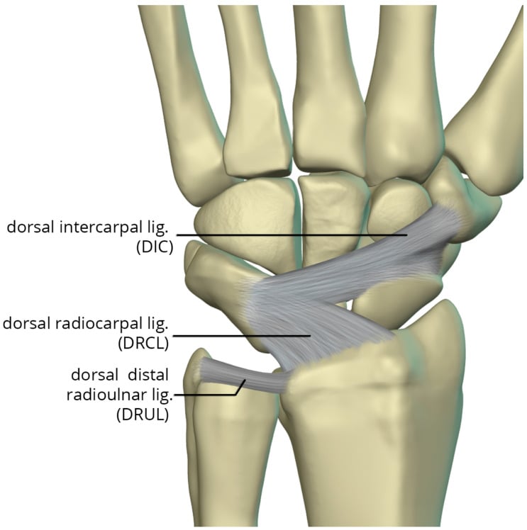
When the ligament fails. The scaphoid and lunate lose their coupling and each goes its own way:
- Scaphoid - loses its connection to the lunate and flexes with the distal row, driven by the trapezium and trapezoid. It rotates into a vertical position (the cortical ring sign on the PA film), and its dorsal lip comes into abnormal contact with the radial styloid.
- Lunate - loses the scaphoid's restraint and extends under the influence of the triquetrum, the DISI pattern. It keeps a normal articulation with the radius because its spherical geometry is preserved.
- Capitate - loses distal support from the flexed scaphoid, migrates proximally into the widened SL gap and makes abnormal contact with the lunate, which degenerates the capitolunate joint progressively.
Why the radiolunate joint survives. This is the key anatomical concept of the topic. The lunate sits in a congruent fossa that is loaded axially, and it keeps its spherical articulation there: the radius of curvature is unchanged, the contact area normal and the pressure distribution even, so the lunate "fits" normally in the lunate fossa despite the arthritis around it. There are no abnormal shear forces, the loading vectors stay normal, intrinsic stability is preserved and cartilage nutrition is adequate, so nothing mechanical drives degeneration at this articulation.
Why that matters. A preserved radiolunate joint makes motion-preserving salvage possible in most patients (Stages I-III):
- Four-corner fusion relies on the intact radiolunate joint for its motion
- Proximal row carpectomy relies on the capitate articulating with an intact lunate fossa
Pathophysiology
The cascade begins. Scapholunate ligament rupture, from acute trauma or chronic attenuation, uncouples the scaphoid from the lunate and starts a carpal instability pattern. Over the following months to years the scaphoid flexes and the lunate extends as described above, and the SL gap widens progressively: over 3mm marks static instability.
Where the load goes. In SLAC the load concentrates abnormally at the styloscaphoid and scaphocapitate joints, while radiolunate loading is unchanged, which explains why that joint is preserved. Load concentrates on the radial styloid and radioscaphoid facet, which is where arthritis appears first.
Stage I, the radial styloid (years 1-5). Repetitive impingement at the styloscaphoid articulation concentrates force as abnormal point loading. Cartilage microtrauma and degradation begin, with synovitis and osteophyte formation ("beaking") at the styloid, and the pain localises to the radial side of the wrist.
Stage II, the radioscaphoid fossa (years 5-10). Loss of the scaphoid's bridge function is now complete and the scaphoid no longer articulates congruently in its fossa. Contact pressure rises, the cartilage degenerates to bone on bone with subchondral sclerosis and cysts, and the capitate begins its proximal migration into the SL gap.
Stage III, the capitolunate joint (years 10-15). Proximal capitate migration exceeds 2-4mm, and the capitate head contacts the lunate abnormally. The normal capitolunate geometry is lost and its cartilage progressively destroyed, with severe carpal collapse that affects grip strength and motion. The radiolunate joint is still preserved, its spherical fit maintained.
Stage IV, pancarpal (rare, over 20 years). Only in very advanced, neglected cases does the arthritis finally reach the radiolunate articulation. The lunate loses its spherical geometry, the carpus collapses completely and no motion-preserving options remain.
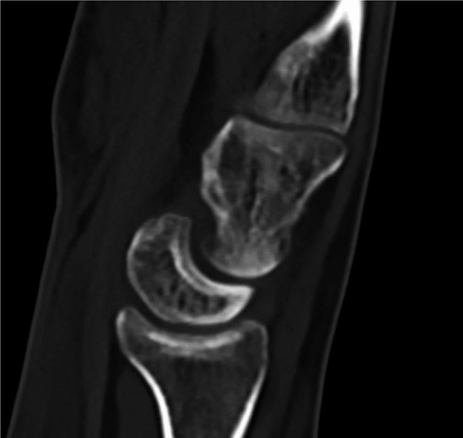
Why progression is inevitable. Once SLAC arthritis begins, progression is inexorable:
- Irreversible cartilage loss - chondrocytes cannot regenerate
- Perpetuating biomechanics - abnormal loading continues with every wrist motion
- Loss of shock absorption - cartilage loss increases bone contact pressure
- Inflammatory cascade - synovitis and cytokine release accelerate degeneration
- Subchondral bone changes - sclerosis and cysts represent end-stage damage
Early intervention with SL ligament repair prevents SLAC. Once arthritis is established, salvage procedures are the only option.
Classification Systems
The Watson classification (1984) is the gold standard staging system for SLAC wrist, based on the radiographic pattern of arthritis. The stage determines the surgical approach and the expected outcome.
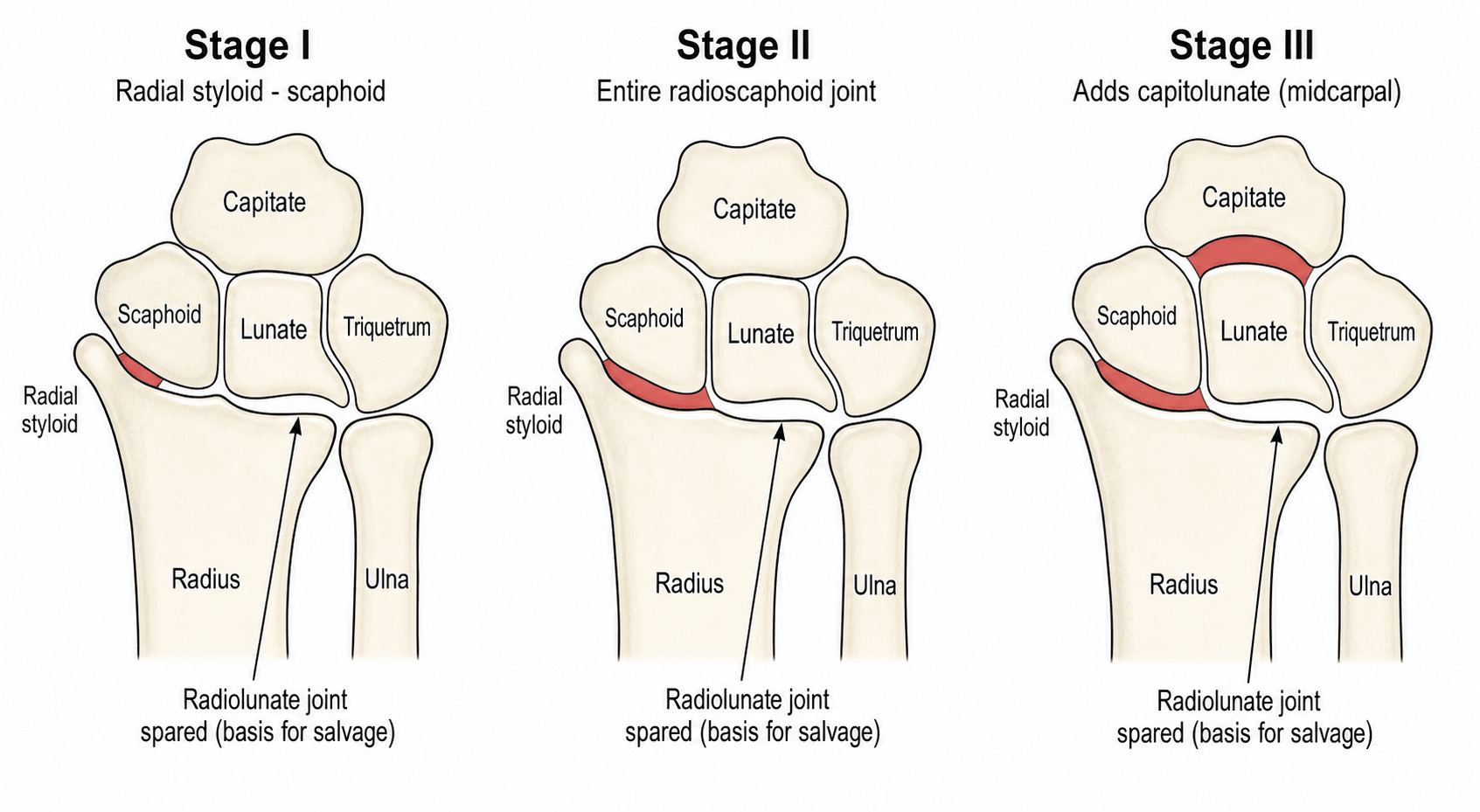
- Arthritis Location
- Radial styloid only
- Radiographic Findings
- Narrowing between scaphoid and radial styloid, sclerosis and osteophyte at the styloid tip (beaking); scaphoid flexed (ring sign), lunate extended; no sclerosis in the scaphoid fossa; capitolunate preserved
- Arthritis Location
- Radioscaphoid fossa
- Radiographic Findings
- Radioscaphoid joint space narrowing and subchondral sclerosis of the scaphoid fossa; proximal scaphoid pole may show cystic change; capitolunate still preserved (key finding); carpal height begins to decrease
- Arthritis Location
- Capitolunate added
- Radiographic Findings
- Proximal capitate migration into the SL gap; capitolunate narrowing, sclerosis and cysts; severe carpal collapse (carpal height ratio under 0.48); radiolunate STILL preserved
- Arthritis Location
- Pancarpal (radiolunate)
- Radiographic Findings
- Radiolunate joint space narrowing and sclerosis; complete carpal collapse
Who presents when. Stage II is the most common presentation (40-50% of patients) and Stage III the second most common (30-40%). Stage I may be managed non-operatively or with styloidectomy. Stage IV is exceedingly rare, under 5% of SLAC cases: most SLAC wrists never reach it.
RSCPSLAC Stage Progression (Watson Classification)
Hook:RSCP: Radial Styloid, Scaphoid fossa, CapitoLunate, Pancarpal - the predictable march of SLAC arthritis!
Modified staging. Some authors subdivide the stages for more granular treatment decisions, to help decide between scaphoid excision alone, four-corner fusion and PRC. Substaging is controversial, and most surgeons use the standard four-stage Watson system.
- Stage IIA - radioscaphoid arthritis without capitate migration
- Stage IIB - radioscaphoid arthritis with early capitate migration (less than 2mm)
- Stage IIIA - capitolunate arthritis, capitate migrated 2-4mm
- Stage IIIB - advanced capitolunate arthritis, capitate migrated over 4mm
Clinical Assessment
History. Dorsal wrist pain, activity-related, with weakness (grip reduced 30-60%) and progressive loss of motion, worsening gradually over years. There may be a wrist sprain years before, but often there is no clear inciting event and the onset is insidious. Manual labour is a common occupation.
Examination. Look for a dorsal prominence and swelling, tenderness over the dorsal wrist and pain with axial loading. Flexion-extension is reduced by 30-60%, and grip on examination is 40-60% of normal. These are late-stage findings, not those of early SL dissociation.
Special tests. The Watson scaphoid shift test is usually negative in a SLAC wrist, because the deformity is chronic and fixed; it may be positive in earlier SL dissociation before arthritis, and should be compared with the other side. The scaphoid compression test applies an axial load along the thumb metacarpal: pain at the radioscaphoid joint is positive and suggests Stage II arthritis.
Symptoms by stage. Pain, stiffness and weakness track the Watson stage:
- Pain Pattern
- Radial-sided, activity-related
- Motion Loss
- Minimal (under 20%)
- Grip Strength
- Near normal
- Dominant Complaint
- Radial styloid pain with radial deviation
- Pain Pattern
- Dorsal wrist, diffuse and constant
- Motion Loss
- Moderate (20-40%)
- Grip Strength
- Reduced 30-40%
- Dominant Complaint
- Stiffness and weakness
- Pain Pattern
- Severe, diffuse
- Motion Loss
- Severe (over 50%)
- Grip Strength
- Reduced over 50%
- Dominant Complaint
- Pain and severe functional limitation
Differential diagnosis. Chronic radial or dorsal wrist pain has several mimics, and imaging differentiates them. Midcarpal instability and extensor tenosynovitis also belong on the list.
- Key distinguishing feature
- Intact scaphoid, widened SL gap over 3mm (Terry Thomas sign), staged radioscaphoid then capitolunate arthritis, radiolunate spared
- Best discriminating investigation
- PA + lateral radiographs; CT for staging
- Key distinguishing feature
- Scaphoid fracture nonunion with sclerosis/cysts; identical arthritis cascade but scaphoid is fractured, not just dissociated
- Best discriminating investigation
- Radiographs/CT showing waist nonunion
- Key distinguishing feature
- Lunate sclerosis, fragmentation and collapse (lunate AVN); SL gap normal
- Best discriminating investigation
- Radiographs; MRI for early (Lichtman I) disease
- Key distinguishing feature
- Ulnar-sided pain, positive ulnar variance, lunate/triquetral subchondral cysts on the ulnar side
- Best discriminating investigation
- PA radiograph (ulnar variance); MRI
- Key distinguishing feature
- Volar-radial pain at scaphotrapeziotrapezoidal joint; arthritis distal to scaphoid, radioscaphoid spared
- Best discriminating investigation
- PA radiograph showing STT joint narrowing
- Key distinguishing feature
- Ulnar-sided pain, painful/limited forearm rotation, DRUJ joint-space loss
- Best discriminating investigation
- Radiographs; CT of DRUJ in pronation/supination
- Key distinguishing feature
- Positive Watson scaphoid shift test, NO established arthritis (pre-SLAC, repairable)
- Best discriminating investigation
- Stress/clenched-fist views; arthroscopy/MRI
Investigations
Plain radiographs first. PA, lateral and scaphoid views. The PA shows the SL gap, scaphoid rotation (the cortical ring sign), joint space narrowing and sclerosis; the lateral shows the SL and capitolunate angles and proximal capitate migration. Measurements quantify the carpal malalignment, confirm the diagnosis and track progression:
- Technique
- PA view, widest SL distance
- Normal
- Under 3mm
- SLAC Finding
- Over 3mm (Terry Thomas sign)
- Technique
- Lateral, scaphoid to lunate axes
- Normal
- 30-60 degrees
- SLAC Finding
- Over 70 degrees (DISI)
- Technique
- Lateral, capitate to lunate
- Normal
- Under 15 degrees
- SLAC Finding
- Over 15 degrees
- Technique
- Lateral, carpal height / 3rd MC length
- Normal
- 0.54 plus or minus 0.03
- SLAC Finding
- Decreased
An SL angle over 70 degrees is the dorsal intercalated segment instability (DISI) pattern, the hallmark of chronic SL incompetence.
Gilula's arcs. Trace them on every wrist film. A step or break in any arc indicates carpal malalignment before arthritis is visible.
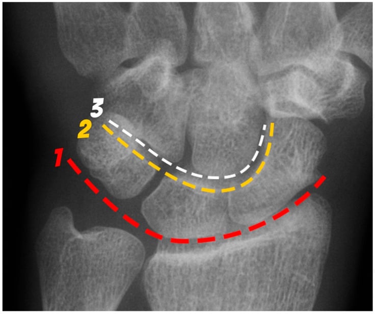
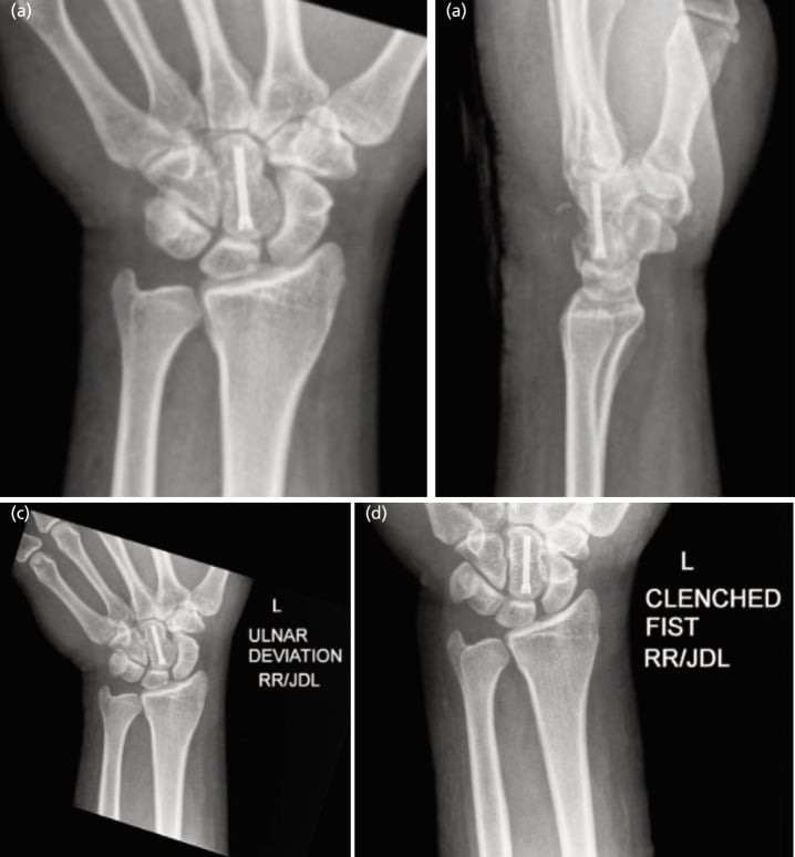
Stress views. Scapholunate widening is dynamic, and a neutral PA film alone will miss it. The clenched-fist PA loads the capitate between scaphoid and lunate, so the SL gap widens with axial loading, and it is the single most useful provocative projection. Compare with the contralateral wrist, since a gap above 3mm is significant only relative to the other side.
When to ask for them. Stress views are useful when static films are normal but clinical suspicion is high, and less helpful in established SLAC, where the deformity is already static. A complete instability series is PA and lateral for static alignment, ulnar deviation to extend the scaphoid, and clenched fist to provoke the gap; order all four when the history suggests instability and the neutral films look normal.
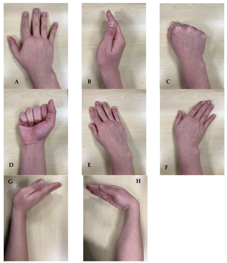
Always obtain a CT scan before four-corner fusion or PRC. It assesses (1) capitate cartilage integrity, which is critical to the PRC decision, (2) the exact extent of arthritis, confirming the stage, (3) bone quality for fusion, and (4) the scaphoid position and proximal pole viability for surgical planning.
MRI is optional, to exclude other pathology: Kienböck disease, TFCC tears and occult fractures. It also assesses cartilage status, and it is less critical when plain films are diagnostic of SLAC.
Management Algorithm
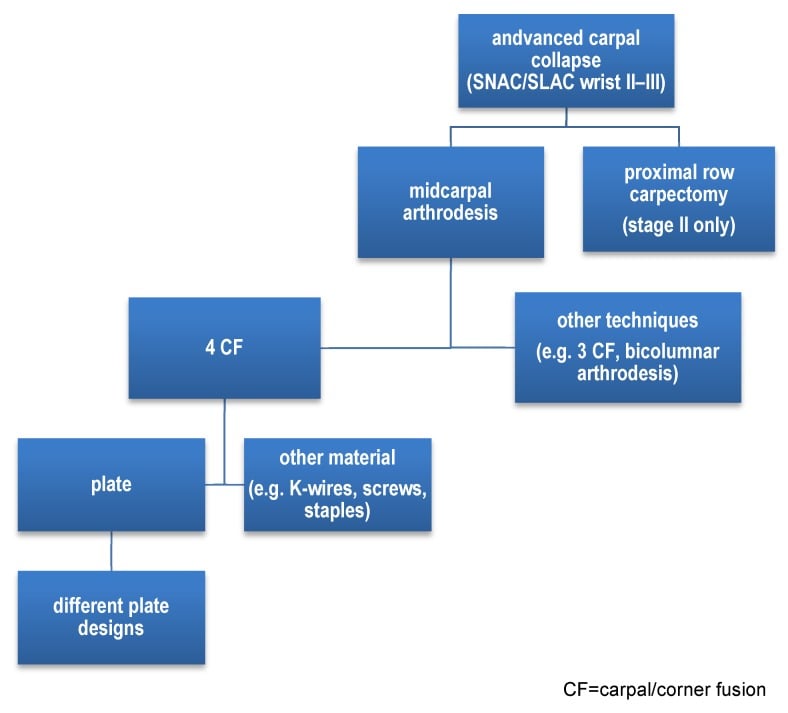
The stage decides. Stage I is treated with radial styloidectomy or denervation, Stages II-III with motion-preserving salvage (four-corner fusion or proximal row carpectomy), and Stage IV with total wrist fusion. Most patients present at Stage II-III.
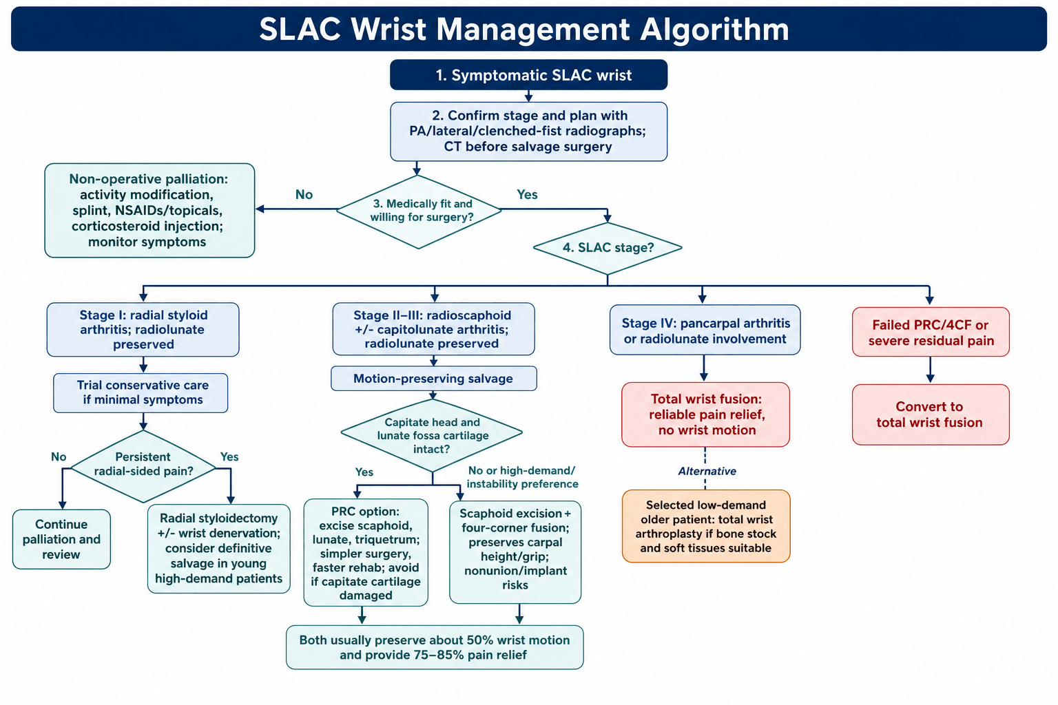
Who. Patients who refuse surgery or whose medical comorbidities prohibit it, the low-demand sedentary patient, and Stage I with minimal symptoms.
Activity modification. Avoid provocative activities, reduce heavy manual work, arrange an ergonomic assessment and modify the job where possible. Success depends heavily on occupation and compliance: a manual worker often cannot comply, which is itself part of the surgical decision.
Wrist orthoses. For symptom flares, a short opponens-style splint worn during aggravating activity, with night use for pain. It rests an arthritic joint; it does not alter the natural history.
Drugs and injection. NSAIDs, topical agents such as diclofenac gel, and paracetamol. Corticosteroid injection gives temporary relief only and has no disease-modifying effect.
What conservative care can do. The three can be used alone or together; they manage symptoms, and none of them touches the disease. The reason is mechanical and irreversible: once the scapholunate ligament has failed, the scaphoid flexes and the capitolunate joint takes load it was never designed for, so cartilage is being worn away by an altered contact pattern that no splint or tablet can correct.
Palliation buys time. Conservative treatment suits the low-demand or elderly patient and anyone not yet ready for surgery, while surgery is the definitive option for most. Say this explicitly when consenting, so the patient understands they are choosing when to operate rather than whether to.
Wrist denervation is more than a Stage I adjunct: it can be offered as a stand-alone procedure at any stage for the patient who wants to keep wrist motion and avoid bony surgery. It works by dividing the articular sensory branches to the wrist — principally the terminal posterior interosseous nerve (PIN) and anterior interosseous nerve (AIN) branches (a limited PIN/AIN neurectomy), with more extensive options dividing additional dorsal and volar sensory branches (the total, Wilhelm-type denervation). Crucially it relieves pain without altering the arthritis, the carpal alignment or the range of motion — nothing is fused or excised, so it burns no bridges to a later four-corner fusion, PRC or total wrist fusion. A diagnostic local-anaesthetic block of the PIN/AIN (temporary pain relief) helps predict who will respond. The trade-offs are that it does not halt disease progression and a proportion lose benefit over time, but for a high-demand worker who needs motion, or as a lower-morbidity option in the elderly or unfit, it is a legitimate motion-preserving choice. The exam point: denervation treats the pain, not the joint.
Surgical Technique Details
Set-up. Both salvage operations are done supine with the arm on a hand table, a tourniquet high on the arm and the forearm pronated.
The dorsal approach. A longitudinal dorsal incision of 8-10cm over Lister's tubercle. Elevate the extensor retinaculum as a radially based flap, enter the wrist between the third and fourth compartments and protect the PIN branches. PRC uses the same approach.
A modified approach. Mark a gently curved incision, raise flaps above the retinaculum, then open the third compartment and step-cut the retinaculum for later closure. Preserving the dorsal capsular ligaments during the capsulotomy protects what stability remains.
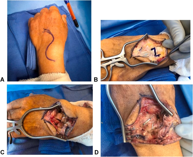
Scaphoid excision. Open the dorsal capsule through a longitudinal T-shaped incision, then:
- Expose and excise the entire scaphoid piecemeal, using rongeurs to remove all fragments
- Protect the radial artery volarly
- Inspect the radioscaphoid joint to confirm the stage
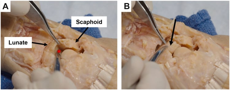
Preparing the fusion. Denude the cartilage from the proximal capitate, distal lunate, proximal hamate and distal triquetrum with a curette, rongeur or burr, down to bleeding subchondral bone. Shape the surfaces for good apposition and pack autograft from the excised scaphoid into the interfaces.
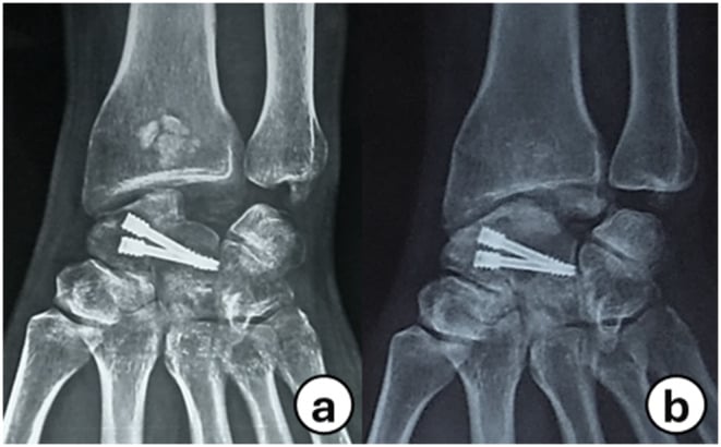
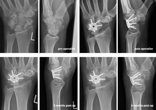
Fixation. A dorsal circular (spider) plate is the most common choice, being low profile and rigid with fewer screws; individual screws or headless compression screws are the alternative.
Reduce before fixing. Correct the lunate out of DISI and into neutral before fixation, because fusing the lunate in extension leaves the capitate impinging on the dorsal radius and limits the extension arc permanently. A joystick wire in the lunate must enter the lunate body and stop short of its concave midcarpal articular surface, or it damages the very cartilage a salvage depends on.
Applying the plate. In order:
- Reduce the carpal alignment (20-30 degrees flexion)
- Stabilise temporarily with K-wires
- Apply the dorsal circular plate
- Verify the screw trajectories under fluoroscopy
- Insert the screws (typically 4-6 in total)
- Confirm there is no radiocarpal or CMC joint penetration
Closure. Repair the dorsal capsule, close the retinaculum over the plate and close the skin. Four-corner fusion is technically demanding but provides reliable outcomes.
Complications
Complication rates are acceptable for both procedures when properly indicated. Early complications are those in the first 6 weeks; late complications come after.
Wound problems (2-5%, either operation). Delayed healing of the thin dorsal skin, superficial infection (redness, drainage), deep infection (rare, under 1%) and dehiscence from tension on closure. Prevent them with careful skin handling, prophylactic antibiotics and early suture removal.
Neurovascular injury (either operation). The radial artery can be injured during scaphoid excision (under 2%): repair it primarily if it is identified, and there are usually no long-term sequelae. PIN neuritis (5%) comes from retraction and traction on the dorsal sensory branch, and dorsal sensory branch injury and superficial radial nerve traction also occur; recognise them early. Nerve injuries are usually temporary, and PIN neuritis is treated with observation and NSAIDs.
Four-corner fusion, early. Three complications are specific to the fusion in the first weeks:
- Malreduction (5%) - improper carpal alignment before plating
- Screw penetration (5%) into the radiocarpal or CMC joints - intraoperative fluoroscopy prevents it; remove and redirect the screw
- Plate prominence (10%) - inadequate soft-tissue coverage
Four-corner fusion, late. Three late complications are specific to the fusion:
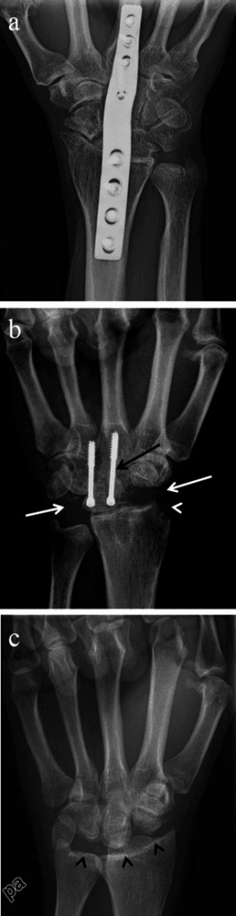
- Nonunion (5-10%) - the most common late complication and the principal failure of the operation. Risk factors are smoking, NSAIDs, diabetes, osteoporosis or poor bone quality, and infection. It may be asymptomatic or painful and is detected on sequential imaging rather than at a single visit; treat it with revision fusion, bone graft and rigid fixation.
- Plate irritation and tendinopathy (10-15%) - dorsal plate prominence irritates the extensor tendons, with painful tenosynovitis over the fourth compartment, usually manifesting 6-18 months postoperatively. After nonunion, dorsal plate impingement is the other recognised cause of persistent pain and loss of extension. Remove the plate after the fusion is solid (12+ months).
- Radiocarpal arthritis (10-20% at 10 years) - progressive degeneration at the radiolunate joint, more common in heavy manual labourers. It may be asymptomatic or cause recurrent pain; observe it if tolerable, total wrist fusion if severe.
Proximal row carpectomy. Five complications are specific to PRC:
- Excessive bleeding (2%, early) - from exposed cancellous surfaces
- Progressive capitate-radius arthritis (10-20% at 10 years) - more common with pre-existing capitate damage, presenting as recurrent pain and reduced motion. Activity modification; convert to total wrist fusion if that fails.
- Ulnar impaction syndrome (5-10%) - carpal height loss causes ulnocarpal abutment, ulnar-sided wrist pain and DRUJ symptoms. Ulnar shortening osteotomy or TFCC debridement.
- Capitate instability or subluxation (rare, under 5%) - usually from excessive soft-tissue and ligament stripping, the capitate migrating dorsally or volarly. Preserve the volar ligaments to prevent it; revise to total wrist fusion.
- Inadequate motion (10%) - less range than expected, often from prolonged immobilisation; an early motion protocol prevents it.
Either operation. Infection (under 2%), tendon adhesions (5-10%) and a need for revision surgery (5-10%). The functional shortfalls:
- Incidence
- 20-30%
- Impact
- Under 30 degrees arc (goal 40-50)
- Management
- Aggressive early physiotherapy
- Incidence
- 30-40%
- Impact
- Under 60% of normal (goal 70-80%)
- Management
- Strengthening programme, occupational modifications
- Incidence
- 15-25%
- Impact
- Activity-limiting discomfort despite surgery
- Management
- Wrist denervation, activity modification, revision surgery
- Incidence
- 3-5%
- Impact
- Disproportionate pain, swelling, stiffness
- Management
- Early recognition, desensitisation, sympathetic blocks
Revision. The indications are a failed salvage with persistent pain (the most common), nonunion of a four-corner fusion, progressive arthritis after PRC, hardware irritation requiring removal, and inadequate function or motion.
- After a failed 4CC - revision fusion with bone graft, conversion to PRC if the radiolunate joint is intact, or total wrist fusion
- Nonunion of a 4CC - revision with iliac crest graft and rigid fixation
- After a failed PRC - total wrist fusion (it cannot be converted to 4CC)
Revision is less predictable than primary surgery, and total wrist fusion is the most reliable option; patient counselling is critical for realistic expectations.
Prevention. Before surgery, smoking cessation 6 weeks beforehand, optimised comorbidities (diabetes control), a CT to assess cartilage and plan the procedure, and patient selection (avoid salvage in end-stage arthritis). In theatre, meticulous soft-tissue handling, fluoroscopic verification of hardware placement, proper carpal alignment before fixation and preservation of the volar ligaments in PRC. Afterwards, an appropriate immobilisation duration, early protected motion when allowed, no NSAIDs until a four-corner fusion is solid, and aggressive hand therapy for motion recovery.
Postoperative Care and Rehabilitation
After four-corner fusion. A short-arm cast for 8-12 weeks, with radiographs at 6 weeks to assess fusion; remove the cast when the fusion is solid, then start therapy for motion and strengthening. Grip strengthening waits until the fusion is radiographically united, usually around three months.
After proximal row carpectomy. A short-arm splint for 2 weeks, then suture removal and gentle motion, progressing to strengthening at 4-6 weeks and full activity at 3 months.
Outcomes over time. Beyond the pain, motion and grip figures in the Management comparison table:
- Four-corner fusion
- 75-85%
- Proximal row carpectomy
- 80-90%
- Four-corner fusion
- 90-95%
- Proximal row carpectomy
- Not applicable
- Four-corner fusion
- -
- Proximal row carpectomy
- 3-4 months
- Four-corner fusion
- 70-80%
- Proximal row carpectomy
- 65-75%
- Four-corner fusion
- Stable or slight decline
- Proximal row carpectomy
- -
- Four-corner fusion
- Revision 5-10%
- Proximal row carpectomy
- Conversion to fusion 5-10%
- Four-corner fusion
- -
- Proximal row carpectomy
- Remains good
Patient factors. Younger patients have better functional outcomes and manual labourers lower satisfaction. Realistic expectations correlate with satisfaction, and compliance with early motion is crucial for motion recovery.
Key Exam Takeaways
Definition and Etiology
- SLAC = Scapholunate Advanced Collapse - most common degenerative wrist arthritis
- Caused by chronic SL ligament incompetence (tear, failed repair, idiopathic)
- Identical pattern to SNAC wrist (scaphoid nonunion) but different etiology
- Progressive over 5-15 years: radial styloid → scaphoid fossa → capitolunate
- Radiolunate joint preserved until very late (Stage IV rare, under 5%)
Watson Classification (Staging)
- Stage I: Radial styloid arthritis only - styloidectomy or denervation
- Stage II: Radioscaphoid fossa arthritis - most common presentation (40-50%)
- Stage III: Capitolunate arthritis added - second most common (30-40%)
- Stage IV: Pancarpal arthritis (radiolunate involved) - rare, under 5%
- Staging determines treatment: I = palliative; II-III = salvage; IV = fusion
Pathophysiology
- Loss of scaphoid bridge function - cannot coordinate proximal-distal rows
- Scaphoid flexes with distal row, lunate extends (DISI pattern)
- SL angle over 70 degrees on lateral X-ray = DISI malalignment
- Abnormal contact: scaphoid dorsal lip impinges radial styloid (Stage I)
- Proximal capitate migration into SL gap causes capitolunate arthritis (Stage III)
Clinical Presentation
- Dorsal wrist pain, progressive over years, activity-related initially
- Weakness: grip strength reduced 30-60% depending on stage
- Stiffness: ROM reduced 30-60% (flexion-extension arc 40-80 degrees)
- Watson test often negative (chronic, fixed deformity unlike acute SL tear)
- Remote wrist injury history in 50%, but often no clear inciting event
Radiographic Findings
- PA view: SL gap over 3mm (Terry Thomas sign), scaphoid ring sign (rotation)
- Lateral: SL angle over 70 degrees (DISI), capitolunate angle over 15 degrees
- Stage-specific arthritis: I = styloid; II = scaphoid fossa; III = capitolunate
- CT scan essential before salvage: assess capitate cartilage (for PRC decision)
- MRI to exclude other pathology: Kienböck, TFCC tear, occult fracture
Treatment Algorithm
- Stage I: Radial styloidectomy (4-6mm) + wrist denervation = 70-80% relief
- Stage II-III: Motion-preserving salvage - 4CC or PRC (both ~50% motion)
- Four-corner fusion: Maintains height, better grip, but nonunion risk 5-10%
- PRC: Simpler, faster recovery, requires intact capitate cartilage
- Stage IV: Total wrist fusion (reliable) or arthroplasty (selected patients)
Surgical Technique Pearls
- 4CC: Excise scaphoid, fuse capitate-lunate-hamate-triquetrum with dorsal plate
- PRC: Excise scaphoid-lunate-triquetrum, capitate articulates with radius
- Critical: Assess capitate cartilage intraoperatively - if degenerated, abort PRC
- 4CC alignment: 20-30 degrees wrist flexion, verify screws under fluoroscopy
- PRC: Preserve volar ligaments to prevent capitate subluxation instability
Outcomes and Complications
- Both 4CC and PRC: 75-85% pain relief, 50% motion, 70-80% satisfaction
- Grip strength: 4CC slightly better (70-80% vs 60-75% normal)
- 4CC complications: Nonunion 5-10%, plate irritation 10-15% (may need removal)
- PRC complications: Progressive arthritis 10-20% at 10 years, ulnar impaction 5-10%
- Conversion to total wrist fusion if salvage fails: 5-10% at long-term
High-Yield Exam Points
- SLAC is MOST COMMON degenerative wrist arthritis pattern
- Radiolunate preserved = allows motion-preserving salvage (key concept)
- SLAC vs SNAC: Same pattern, different cause, same treatment
- 4CC vs PRC: No definitive superiority - equivalent outcomes in meta-analyses
- Stage determines treatment: Do NOT offer styloidectomy for Stage II-III
Guidelines, Registries & Global Practice
Global Epidemiology
SLAC is the single most common pattern of degenerative wrist arthritis worldwide. In Watson and Ballet's defining radiographic review of 4,000 wrists, the SLAC pattern accounted for 57% of all degenerative wrist arthritis (Watson & Ballet, J Hand Surg Am 1984; PMID 6725894), confirmed in Watson and Ryu's later series at 55%, with triscaphe arthritis 26% and combined disease 14% (PMID 3955970). Presentation is typically in the fifth to sixth decade, with a male predominance reflecting the manual-labour occupations (carpentry, construction, mining, agriculture) most affected by chronic scapholunate injury. The latency from the initiating scapholunate ligament injury to symptomatic arthritis is long (commonly 5-15 years), which has medicolegal relevance for occupational-injury causation in any jurisdiction.
Guidelines and Society Guidance (Side by Side)
There is no high-level dedicated society guideline for SLAC salvage; practice is guided by systematic reviews and surgeon consensus. The table summarises how the major bodies and the evidence base frame the key decisions.
- Position on SLAC salvage
- Both 4-corner fusion and PRC endorsed as motion-preserving salvage; shared decision-making; PRC discouraged in patients under ~35y
- Evidence level
- Expert consensus + Level IV
- Position on SLAC salvage
- Stage-based approach; PRC favoured for simplicity and lower complication rate when capitate cartilage intact
- Evidence level
- Expert consensus
- Position on SLAC salvage
- Emphasises preserving the spared radiolunate joint; total wrist fusion reserved for pancarpal (Stage IV) disease or failed salvage
- Evidence level
- Expert consensus
- Position on SLAC salvage
- Equivalence of 4CC and PRC for motion and pain; complication profile differs
- Evidence level
- Level IV meta-analysis
- Position on SLAC salvage
- PRC: better motion, lower conversion-to-fusion (5.2% vs 11%); grip strength equivalent
- Evidence level
- Level IV (PMID 38416092)
Registry and Pooled Evidence
National joint registries (NJR, AOANJRR, AJRR) capture wrist arthroplasty but not the carpal salvage procedures (4CC, PRC, styloidectomy) that dominate SLAC management, so the evidence base is pooled cohort data rather than registry data:
- Largest pooled series (2024): 61 studies, 3,174 wrists - PRC associated with better motion and a lower conversion-to-fusion rate (5.2% vs 11%); grip equivalent; 4CF nonunion 8.9% (PMID 38416092).
- Systematic review (2015): 7 studies, 242 wrists - 4CC gave greater radial deviation and grip (74% vs 67%), PRC gave a wider flexion-extension arc and lower overall complication rate (14% vs 29%) (Saltzman et al., PMID 25294736).
- Long-term PRC cohort: 18% failure at minimum 10 years, concentrated entirely in patients aged 35 or younger (DiDonna et al., PMID 15523004).
Global Practice Variation
- Procedure preference: PRC predominates where simplicity, faster rehabilitation and implant-free salvage are prioritised; 4-corner fusion is favoured by surgeons prioritising carpal-height maintenance and in higher-demand patients, and is mandated when the capitate head cartilage is degenerate.
- Implant choice for 4CC: dorsal circular ("spider") plate versus headless compression screws versus K-wires varies by region and cost setting; circular-plate fixation carries a recognised dorsal-impingement/nonunion profile (Shindle et al.).
- Resource-limited settings: PRC and styloidectomy are attractive where implants, fluoroscopy or hand-therapy access are limited, as they need no internal fixation.
- Surgical-site antibiotic prophylaxis: a single preoperative dose of an intravenous first-generation cephalosporin (e.g. cefazolin) within 60 minutes of incision is the near-universal standard for clean hand surgery, with a glycopeptide alternative for beta-lactam allergy or MRSA risk.
Examination Relevance
SLAC wrist is high-yield across all major boards, particularly in:
- Hand/upper-limb viva (staging, treatment algorithm, surgical technique)
- Clinical stations (chronic wrist pain presentation)
- Radiology viva (DISI deformity, Terry Thomas sign, Watson staging)
Be ready to justify the biomechanical basis for radiolunate preservation and to argue the 4CC-versus-PRC decision from the contemporary evidence above.
Viva Scenarios
Practise clinical reasoning and management decisions out loud
“A 52-year-old male carpenter presents with 2 years of progressive left (dominant) wrist pain and weakness. He recalls a wrist sprain 8 years ago. Examination shows 40 degrees total flexion-extension arc, grip 30kg (right 55kg). Radiographs demonstrate scapholunate gap of 5mm, scapholunate angle 75 degrees, and sclerosis of the radioscaphoid fossa with preserved capitolunate joint. CT scan confirms Stage II SLAC with intact capitate cartilage. How would you manage this patient?”
“You are performing a four-corner fusion for Stage III SLAC wrist. After scaphoid excision and fusion site preparation, you are ready for fixation. The examiner asks: Walk me through your technique for applying a dorsal circular plate. What are the key technical points?”
“A 45-year-old female presents with chronic wrist pain. Radiographs show Stage II arthritis with radial styloid and radioscaphoid involvement. The examiner asks: How would you differentiate SLAC from SNAC wrist? Does it matter for treatment?”
Evidence Base
The Original SLAC Description (Watson & Ballet, 1984)
- Review of 4,000 wrist radiographs; 210 cases of degenerative arthritis analysed
- SLAC (scaphoid-lunate-radius) was the most common pattern at 57%
- Coined the term SLAC and defined the staged radiographic progression
- 18 of 19 operated patients had less pain postoperatively, none requiring analgesia
- Flexion-extension and radioulnar deviation improved after limited salvage fusion
PRC vs Scaphoid Excision + Four-Corner Arthrodesis (Direct Comparison)
- Two matched cohorts of 19 patients (PRC vs scaphoid excision + 4-corner fusion)
- Flexion-extension arc near-identical: 81 degrees (PRC) vs 80 degrees (4CC)
- Grip strength 71% (PRC) vs 79% (4CC) of the opposite wrist
- 4CC group retained greater radial deviation
- Pain relief and patient satisfaction equivalent between procedures
- Two caveats the headline hides: follow-up was UNEQUAL (28 months for the fusion group against 19 for PRC), and the only statistically significant functional difference favouring 4CC was the SF-36 MENTAL health component - not the physical component, and not a wrist-specific score
Meta-analysis: Four-Corner Fusion vs PRC for SLAC/SNAC (3,174 wrists)
- 61 studies, 3,174 wrists (54% PRC, 46% 4CF); mean follow-up 61 months
- PRC gave significantly greater postoperative extension and ulnar deviation
- No significant difference in grip strength between the procedures
- Subsequent arthrodesis required in 5.2% (PRC) vs 11% (4CF)
- 4CF nonunion rate 8.9% (57/640) with 2.2% hardware removal rate
Landmark Series: Scaphoid Excision + Limited Fusion (4CC) for SLAC
- 100 cases treated with scaphoid excision and limited wrist arthrodesis, at an average follow-up of 44 months
- Flexion-extension averaged 72 degrees (53% of the normal opposite wrist)
- Grip strength averaged 80% of the contralateral side
- Radiolunate joint preserved in 98/100 - the basis for motion-sparing salvage
- Nonunion in only 3 cases; capitate-radius impingement (technique-related) in 12%
Long-term PRC Outcomes (minimum 10-year follow-up)
- 22 wrists; average follow-up 14 years (minimum 10 years)
- 4 failures (18%) requiring fusion - all in patients aged 35 years or younger
- Non-failed wrists: flexion-extension arc averaged 72 degrees, grip 91% of opposite side
- Radiocapitate degeneration was seen in 14 of the 17 wrists radiographed yet did NOT correlate with subjective or objective function - though the authors qualify this as holding only 'with the numbers available', which in a series of 22 wrists is a weak negative rather than a demonstrated absence of effect
- Caution advised before performing PRC in patients younger than 35 years
Defining Epidemiology: Evolution of Wrist Arthritis Patterns
- About 95% of wrist degenerative arthritis is periscaphoid in distribution
- SLAC pattern accounted for 55% of cases (the most common pattern)
- Triscaphe (STT) arthritis 26%; combined SLAC + triscaphe 14%
- SLAC progression is driven by scaphoid-radius articular malalignment
- Capitolunate change is secondary to carpal collapse - confirming the staged cascade
- Read the operative half historically: the SLAC procedure as described here was capitate-lunate-hamate-triquetrum fusion PLUS A SILASTIC SCAPHOID IMPLANT, and silicone implants were subsequently abandoned for particulate synovitis. The fusion concept survived; the implant did not
References
-
Watson HK, Ballet FL. The SLAC wrist: scapholunate advanced collapse pattern of degenerative arthritis. J Hand Surg Am. 1984;9(3):358-365.
-
Watson HK, Ryu J. Evolution of arthritis of the wrist. Clin Orthop Relat Res. 1986;(202):57-67.
-
Ashmead D 4th, Watson HK, Damon C, Herber S, Paly W. Scapholunate advanced collapse wrist salvage. J Hand Surg Am. 1994;19(5):741-750.
-
Wyrick JD, Stern PJ, Kiefhaber TR. Motion-preserving procedures in the treatment of scapholunate advanced collapse wrist: proximal row carpectomy versus four-corner arthrodesis. J Hand Surg Am. 1995;20(6):965-970.
-
Krakauer JD, Bishop AT, Cooney WP. Surgical treatment of scapholunate advanced collapse. J Hand Surg Am. 1994;19(5):751-759.
-
DiDonna ML, Kiefhaber TR, Stern PJ. Proximal row carpectomy: study with a minimum of ten years of follow-up. J Bone Joint Surg Am. 2004;86(11):2359-2365.
-
Dacho AK, Baumeister S, Germann G, Sauerbier M. Comparison of proximal row carpectomy and midcarpal arthrodesis for the treatment of scaphoid nonunion advanced collapse (SNAC-wrist) and scapholunate advanced collapse (SLAC-wrist) in stage II. J Plast Reconstr Aesthet Surg. 2008;61(10):1210-1218.
-
Cohen MS, Kozin SH. Degenerative arthritis of the wrist: proximal row carpectomy versus scaphoid excision and four-corner arthrodesis. J Hand Surg Am. 2001;26(1):94-104.
-
Vanhove W, De Vil J, Van Seymortier P, Boone B, Verdonk R. Proximal row carpectomy versus four-corner arthrodesis as a treatment for SLAC (scapholunate advanced collapse) wrist. J Hand Surg Eur Vol. 2008;33(2):118-125.
-
Lumsden BC, Stone A, Engber WD. Treatment of advanced-stage Kienböck's disease with proximal row carpectomy: an average 15-year follow-up. J Hand Surg Am. 2003;28(3):422-430.
-
Saltzman BM, Frank JM, Slikker W, Fernandez JJ, Cohen MS, Wysocki RW. Clinical outcomes of proximal row carpectomy versus four-corner arthrodesis for post-traumatic wrist arthropathy: a systematic review. J Hand Surg Eur Vol. 2015;40(5):450-457.
-
Wall LB, Didonna ML, Kiefhaber TR, Stern PJ. Proximal row carpectomy: minimum 20-year follow-up. J Hand Surg Am. 2013;38(8):1498-1504.
-
Chim H, Moran SL. Long-term outcomes of proximal row carpectomy: a systematic review of the literature. J Hand Surg Eur Vol. 2012;37(9):787-793.
-
Mulford JS, Ceulemans LJ, Nam D, Axelrod TS. Proximal row carpectomy vs four corner fusion for scapholunate (SLAC) or scaphoid nonunion advanced collapse (SNAC) wrists: a systematic review of outcomes. J Hand Surg Eur Vol. 2009;34(2):256-263.
-
Shindle MK, Burton KJ, Weiland AJ, Domb BG, Wolfe SW. Complications of circular plate fixation for four-corner arthrodesis. J Hand Surg Eur Vol. 2007;32(1):50-53.