Volar Angulated Distal Radius | Flexion Mechanism | Volar Plating Standard
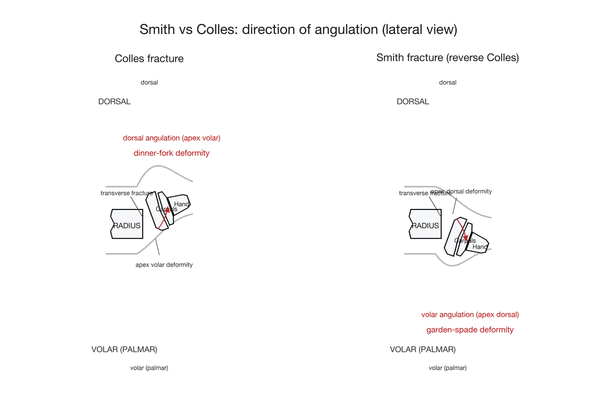
- Volar angulation distinguishes Smith from Colles (dorsal angulation)
- Fall onto the dorsum of a FLEXED wrist, or a direct blow to the dorsal wrist = typical mechanism. Do not call this a FOOSH: an outstretched hand means an EXTENDED wrist, which is the Colles mechanism
- Volar plate fixation is treatment of choice - provides buttress against volar displacement
- Thomas Type II is essentially a volar Barton fracture - highly unstable, needs surgery
- Flexor tendon irritation is main complication - assess at follow-up
- “Smith = volar angulation, Colles = dorsal angulation (garden spade vs dinner fork)
- “Most displaced Smith fractures require surgical fixation - casting usually fails
- “FCR approach protects median nerve and allows direct fracture visualisation
- “Volar plate acts as buttress preventing re-displacement
Overview and Epidemiology
Smith's fracture is a distal radius fracture with volar angulation of the distal fragment (apex dorsal), the "reverse Colles fracture". Strictly, the eponym refers to the volar angulation pattern, and the volar Barton, a rim fracture with carpal subluxation, is distinguished from it; Thomas's classification nonetheless takes in the volar Barton variant as its Type II.
History. The Irish surgeon Robert William Smith, Professor of Surgery at Trinity College Dublin, described the fracture in 1847, 33 years after Abraham Colles described the dorsally angulated variant. His treatise, "A Treatise on Fractures in the Vicinity of Joints and on Certain Forms of Accidental and Congenital Dislocations", set this pattern apart from the Colles fracture.
Who. Smith's fracture accounts for about 5% of all distal radius fractures and is less common than the Colles fracture by approximately 1:10. The distribution is bimodal. Young adults, with a male predominance, break the wrist in high-energy injuries such as sport and motorcycle accidents; the elderly peak is predominantly female and follows low-energy falls on osteoporotic bone, the fragility pattern.
Mechanism. Most commonly a fall onto the dorsum of the hand with the wrist flexed, or a direct blow to the dorsum of the hand that forces the wrist into flexion. This is not a true FOOSH: an outstretched hand implies an extended wrist, which is the Colles mechanism.
Anatomy and Pathophysiology
The normal distal radius. Its normal values are:
- Volar tilt (palmar inclination): 10-15 degrees
- Radial inclination: 22-23 degrees
- Radial height: 11-12mm
- Ulnar variance: usually neutral to slightly negative
The deforming force. The volar radiocarpal ligaments (radioscaphocapitate, long radiolunate and short radiolunate) are strong, and they remain attached to the distal fragment. They pull it volarly, creating the characteristic deformity, and this is why Smith fractures are inherently unstable. Pronator quadratus may contribute to the deforming force.
How it breaks. A fall with the wrist flexed applies a bending moment. The dorsal cortex fails in tension as the wrist is forced into flexion, giving a transverse fracture, while the volar cortex is the compression side and acts as the fulcrum for volar angulation. A direct blow creates a shear force instead, and an oblique fracture.
Why it will not settle. In a Colles fracture, dorsal comminution allows the fragment to settle. A Smith fracture has an intact volar cortex that creates a fulcrum for volar angulation.
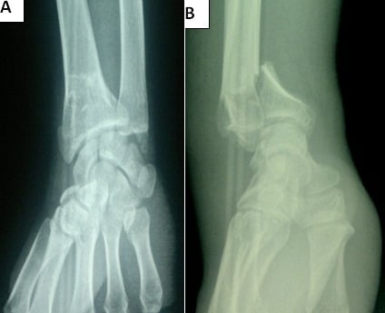
The watershed line. A critical landmark on the volar distal radius, marking the transition from the flat metaphyseal surface to the curved articular surface. A volar plate should not extend beyond it, because a plate there causes flexor tendon irritation.
Classification Systems
Thomas (1957) is the standard classification for Smith fractures. Source: Thomas FB, Reduction of Smith's fracture, J Bone Joint Surg Br 1957;39-B(3):463-70 (PMID 13463033).
- Description
- Extra-articular transverse
- Characteristics
- Simple transverse fracture through metaphysis
- Stability
- Moderate
- Description
- Intra-articular
- Characteristics
- Volar lip fracture with carpal subluxation (volar Barton variant)
- Stability
- Highly unstable
- Description
- Extra-articular oblique
- Characteristics
- Oblique fracture line juxta-articular
- Stability
- Unstable
Thomas Type II is essentially a volar Barton fracture: an intra-articular volar lip fracture with volar subluxation of the carpus. It is the most unstable Smith pattern and ALWAYS requires surgical fixation. The intact volar lip fragment acts as a ramp that the carpus slides volarly upon.
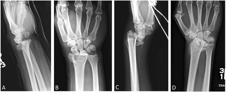
AO/OTA. The AO/OTA classification applies to all distal radius fractures. It is comprehensive, but Thomas remains preferred for Smith fractures specifically because it guides treatment decisions more directly. The codes that concern this topic:
- 23-A2: extra-articular, simple metaphyseal (Thomas Type I)
- 23-B3: partial articular, frontal volar rim (Type II, the volar Barton)
- 23-B1: partial articular, sagittal (the radial styloid), which is not the Smith volar-lip pattern
Clinical Assessment
History. Establish the mechanism (a fall onto the dorsum of a flexed wrist, a direct dorsal blow, or high-energy trauma), hand dominance, occupation and functional demands, previous wrist injuries, and medical comorbidities such as diabetes and osteoporosis.
Examination. The findings, and what each one means, are set out below; compare with the other side.
- Significance
- Characteristic of Smith pattern
- Assessment
- Compare to contralateral side
- Significance
- Garden spade deformity (opposite of dinner fork)
- Assessment
- Assess degree of displacement
- Significance
- Antalgic posture
- Assessment
- Patient avoids extension
- Significance
- At risk with volar displacement
- Assessment
- Test sensation and motor (APB)
- Significance
- Open fracture assessment
- Assessment
- Document any wounds
- Significance
- Associated injury
- Assessment
- Assess DRUJ stability after fixation
Smith = garden spade deformity (volar prominence, the wrist appears flexed). Colles = dinner fork deformity (dorsal prominence, the wrist appears extended). The descriptions help distinguish the fractures clinically before imaging. Smith = Sunrise: the apex points up, dorsally.
The median nerve. It is the structure most at risk, because of the volar displacement. Test sensation in the thumb, index and middle fingers and the motor function of abductor pollicis brevis (APB); carpal tunnel symptoms may develop acutely. Palpate the radial artery at the anatomical snuffbox and the wrist, and check capillary refill in all digits.
Associated injuries. Look for a scapholunate ligament injury, a TFCC injury, DRUJ instability, carpal fractures (especially the scaphoid) and an ulnar styloid fracture.
Differential diagnosis. A volar-prominence deformity with a distal radius injury on plain films can mimic several patterns. The single most discriminating step is the lateral radiograph, for the direction of angulation, together with whether the fracture is articular.
- Distinguishing feature
- Extra-articular distal radius fracture with volar angulation (apex dorsal), garden spade deformity
- How to confirm / refute
- Lateral radiograph shows volar tilt of distal fragment; no carpal subluxation in Types I/III
- Distinguishing feature
- Intra-articular volar rim fracture with volar carpal subluxation; shear mechanism
- How to confirm / refute
- Lateral shows carpus translated volarly with the rim fragment; CT defines articular extent
- Distinguishing feature
- Dorsal angulation (apex volar), dinner fork deformity
- How to confirm / refute
- Lateral shows dorsal tilt - the opposite of Smith
- Distinguishing feature
- Intra-articular dorsal rim fracture with dorsal carpal subluxation; shear mechanism
- How to confirm / refute
- Lateral shows dorsal translation of carpus with dorsal rim fragment
- Distinguishing feature
- Oblique radial styloid fracture, often from scaphoid impaction
- How to confirm / refute
- PA view shows styloid fragment; assess scapholunate interval
- Distinguishing feature
- Salter-Harris pattern through the physis, not a true Smith eponym
- How to confirm / refute
- Age and physeal involvement on radiograph
- Distinguishing feature
- Carpal malalignment without a simple metaphyseal radius fracture
- How to confirm / refute
- Lateral shows lost radius-lunate-capitate collinearity; AP shows crowded carpus
- Distinguishing feature
- Forearm fracture with DRUJ disruption rather than isolated distal radius bending
- How to confirm / refute
- Assess full forearm radiograph and DRUJ stability
Investigations
Radiographs. Three views, each with a job:
- PA: radial inclination, radial height and ulnar variance
- Lateral: critical for the diagnosis, because it shows the volar angulation
- Oblique: additional assessment of articular involvement
The lateral radiograph is ESSENTIAL for diagnosing Smith fractures. The PA view may look similar to a Colles fracture; only the lateral view demonstrates the volar angulation that defines a Smith fracture.
What the films show. Volar angulation of the distal fragment (apex dorsal), volar displacement of the carpus, and loss or reversal of the normal volar tilt. Look for intra-articular extension, which makes it a Type II.
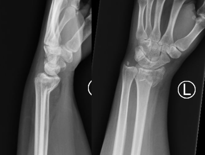
Measure, do not describe. Record the measurements rather than describing alignment as merely "acceptable". They govern both the reduction and the later review of the implant.
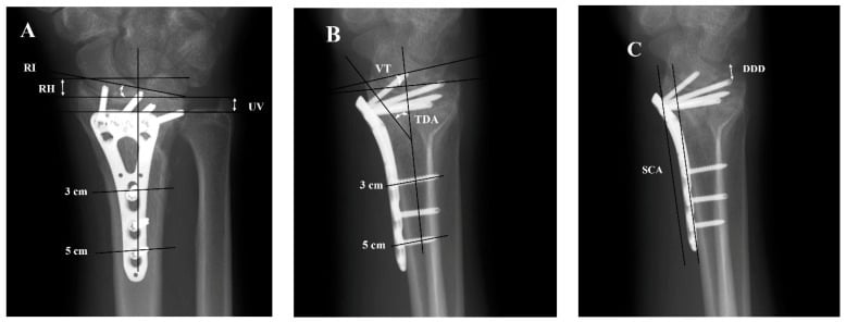
CT. Indicated for intra-articular (Type II) fractures, where it defines the articular step-off and gap and helps plan fragment-specific fixation. It is not routine for a simple Type I fracture.
MRI. Not routine for acute fractures. Consider it if a ligamentous injury is suspected because pain persists, to assess the TFCC and the scapholunate ligament.
Management Algorithm
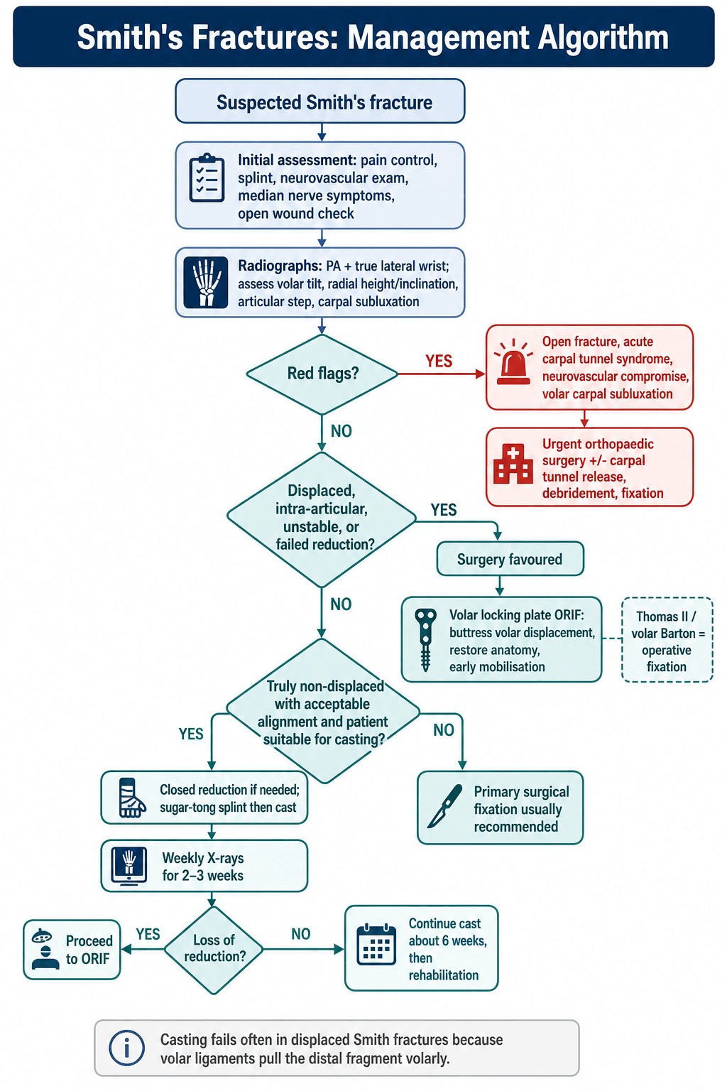
The principle. Most displaced Smith fractures need surgical fixation, because casting fails often: the volar radiocarpal ligaments keep pulling the distal fragment volarly. The modern approach favours early fixation for most displaced fractures, given the predictable results and the benefit of early mobilisation.
How strong is the case. It rests on fracture mechanics rather than trial evidence in Smith fractures (Evidence Base). The Arora trial in patients 65 and older tempers automatic surgery for low-demand elderly extra-articular fractures, but for an unstable, volarly displacing Smith pattern its superior radiographic and grip outcomes support fixation when restoration of alignment matters.
By pattern. An undisplaced Type I is relatively stable and casting can be considered if it is aligned, but this is very rare; most are displaced, and a displaced Type I is a standard surgical indication. A Type III oblique fracture is plated because the obliquity makes casting unreliable. A Type II (volar Barton) is highly unstable: volar plate ORIF is mandatory, and the intra-articular fracture requires anatomic reduction. With an associated carpal injury the instability is complex: address both components, and assess the scapholunate ligament and the DRUJ.
Non-operative management. Reserved for truly non-displaced fractures (rare), for fractures whose acceptable alignment is maintained in the cast, and for patients whose own factors preclude surgery. The technique runs as follows.
- Closed reduction under haematoma block or sedation
- Sugar-tong splint initially, which allows for swelling
- Convert to a long arm cast at 1 week, wrist in extension and forearm supinated
- Immobilise for 6 weeks
Non-operative management of displaced Smith fractures has a high failure rate because of the deforming forces of the volar ligaments. Close follow-up with weekly X-rays for the first 2-3 weeks is essential, and any loss of reduction is an indication for surgery.
Acceptable alignment in a cast. Non-operative treatment accepts:
- Volar tilt neutral or positive, not dorsal
- Radial inclination within 5 degrees of normal
- Radial height within 3mm of normal
- Articular step less than 2mm
Surgical indications. Displaced Smith fractures, the common scenario; Thomas Type II; loss of reduction after casting; polytrauma requiring early mobilisation; and bilateral fractures, where one hand needs to be functional.
Absolute indications. Surgery is absolute for:
- Intra-articular displacement greater than 2mm
- Volar carpal subluxation
- Open fracture
- Associated carpal instability
- Acute carpal tunnel syndrome requiring release
Relative indications. A young, active patient with any displacement, high functional demands, or an inability to maintain reduction in a cast.
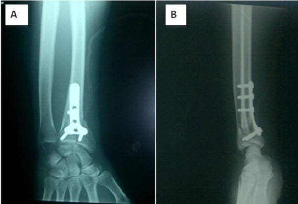
Fixation. The volar locking plate is the standard of care and the gold standard, chosen for its reliable outcomes and low complication rates. It is the most common fixation method, an angular-stable construct that provides a direct buttress against volar displacement and allows early range of motion.
- K-wires and cast: historic, rarely used now
- External fixation: for severe soft tissue injury
- Fragment-specific fixation: for complex articular patterns
- Dorsal plate: not appropriate for a Smith fracture (wrong side)
In a child the same flexion mechanism produces a volarly displaced distal radius physeal (Salter-Harris II) or metaphyseal fracture rather than the adult eponymous Smith, and the management philosophy is the opposite. Because of the enormous remodelling potential of the distal radial physis (which contributes roughly 75% of the growth of the radius, the distal radius and ulna together accounting for about 75-80% of forearm growth), most are treated by closed reduction and a well-moulded cast, not a plate; sagittal-plane (volar/dorsal) angulation in particular remodels reliably in the young child. The reduction reverses the deformity and the cast holds it. Open reduction or fixation is reserved for an irreducible fracture, an open fracture, neurovascular compromise, or the older adolescent near skeletal maturity, where remodelling can no longer be relied upon. Beware the Salter-Harris growth-arrest risk of repeated forceful manipulation, and counsel families that a degree of residual angulation in a young child is acceptable because it will correct with growth.
Surgical Technique
Set-up. The patient lies supine with the arm on an arm table or hand table, a tourniquet on the upper arm at 250-280 mmHg, and the image intensifier coming in from the opposite side.
The FCR approach. It gives excellent exposure of the volar distal radius while protecting the neurovascular structures.
- Incision: longitudinal over the FCR tendon, 6-8cm proximally from the wrist crease
- Identify FCR: palpate the tendon at the wrist crease and incise its sheath along the radial border of the tendon
- Retract FCR ulnarly, which protects the median nerve lying ulnar to it
- Incise the floor of the FCR sheath to expose flexor pollicis longus (FPL)
- Retract FPL ulnarly to expose pronator quadratus; avoid forceful radial retraction of FPL, which risks denervation
- Elevate pronator quadratus through an L-shaped incision from its radial border
- Expose the fracture for direct visualisation of the volar radius
The median nerve lies ULNAR to FCR, so retracting FCR ulnarly protects it. This is the key safety principle of the approach: never dissect ulnar to FCR without identifying the nerve.
Reduction. Evacuate the haematoma from the fracture site and assess the pattern of the fragments. Reduce with traction, manipulation and direct reduction, hold the reduction provisionally with K-wires, and confirm it on PA and lateral fluoroscopy.
Plate application. In order:
- Select a plate sized to fit the distal radius
- Position it proximal to the watershed line: as distal as safely possible to support the fragment, but NEVER beyond the line
- Fix the shaft first, through the oval hole that allows the plate to be adjusted
- Confirm the position on fluoroscopy before locking
- Insert the distal locking screws in subchondral position, 2-4mm from subchondral bone on the lateral view
- Complete the proximal fixation with the remaining shaft screws
The watershed line is the most distal extent of the flat volar surface. The plate must NOT extend beyond this line or it will cause flexor tendon irritation and rupture. Use lateral fluoroscopy to confirm plate position.
The dorsal side. The distal locking pegs are angular-stable and are deliberately kept just short of the dorsal cortex, to avoid extensor tendon irritation and EPL rupture.
Fluoroscopic views. The goal is anatomic restoration of volar tilt, radial inclination and radial height.
- PA: radial inclination, radial height, screw length
- Lateral: volar tilt restoration, plate position, screw position
- Tilted lateral (20 degrees): confirms the screws are not in the joint
The small volar-rim fragment. Volar marginal fragments can be too distal or too small for standard plate screws, and a spring wire may be used to secure the fragment; the wire must hold it without entering the radiocarpal joint. A more distal plate may improve capture but increases flexor tendon risk.
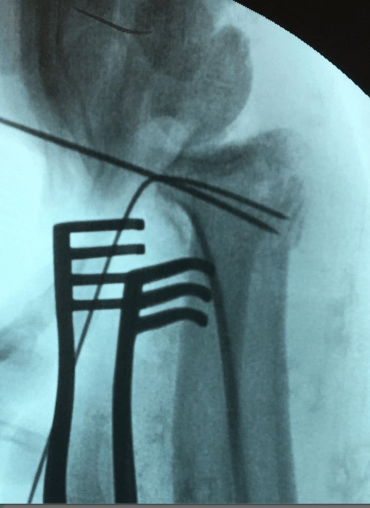
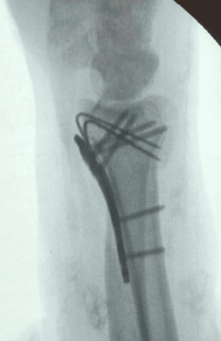
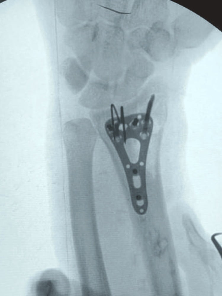
Closure. In order:
- Repair pronator quadratus over the plate if possible
- Close the FCR sheath loosely
- Close the subcutaneous layer with absorbable sutures
- Close the skin with sutures or staples
- Apply a bulky supportive dressing and a volar wrist splint in neutral
Before leaving theatre. Check DRUJ stability after the radius is fixed, and address the ulnar styloid if there is a large base fragment with DRUJ instability. The radial styloid reduction can be used as a landmark for rotation. The intraoperative errors to avoid are a plate beyond the watershed, screws in the radiocarpal joint, an inadequate reduction and a missed articular injury; meticulous technique and fluoroscopic confirmation of hardware position prevent them.
Complications
- Incidence
- 5-10%
- Prevention/Management
- Plate position below watershed line, PQ repair
- Incidence
- Varies with treatment
- Prevention/Management
- Anatomic reduction, stable fixation
- Incidence
- 5-8%
- Prevention/Management
- Protect during approach, avoid excessive retraction
- Incidence
- 10-15%
- Prevention/Management
- Proper plate sizing, consider removal at 1 year
- Incidence
- 5-10%
- Prevention/Management
- Address ulnar styloid base, repair TFCC
- Incidence
- 5%
- Prevention/Management
- Release if acute, monitor if subacute
- Incidence
- 10-20%
- Prevention/Management
- Early mobilisation, hand therapy
- Incidence
- 2-5%
- Prevention/Management
- Early recognition, multimodal treatment
The incidence figures in this table are conventional estimates from clinical experience and small series, not cohort measurements specific to Smith fractures. The measured numbers on this page come from the Soong studies of volar-plated distal radius fractures generally (Evidence Base): flexor tendon irritation about 4% and frank FPL rupture under 1% in the low-profile-plate cohort, rising to 4% rupture with a prominent plate design - plate position, not the fracture eponym, drives the risk.
FPL rupture is the most concerning complication of volar plating. The risk factors are a plate extending beyond the watershed line, prominent screw heads and failure to repair pronator quadratus. The patient loses interphalangeal joint flexion of the thumb, and the treatment is tendon reconstruction (FDS ring finger transfer).
Plate prominence and the tendons. On the lateral radiograph, increasing plate prominence (the Soong grades) predicts flexor tendon irritation and rupture. A plate placed too distal on the watershed region lies beneath flexor pollicis longus and the other flexors, and pronator quadratus coverage cannot reliably neutralise severe prominence, so capture the rim with the least-prominent safe construct. New volar pain, crepitus or weakness of thumb flexion after union requires urgent tendon assessment and consideration of plate removal.
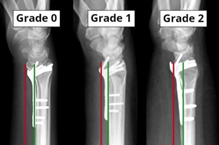
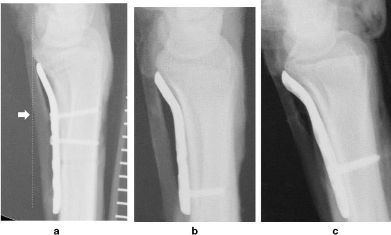
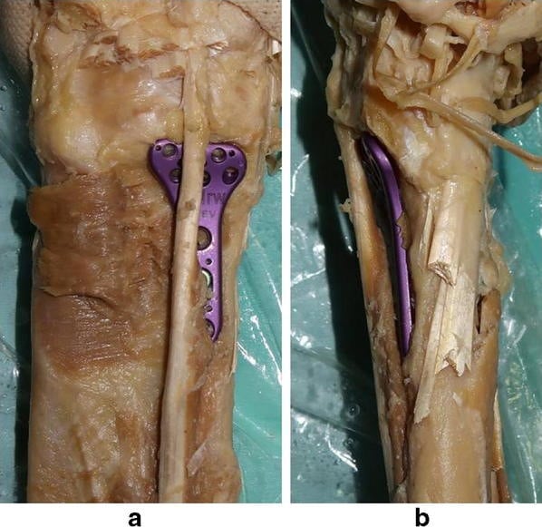
Malunion. A volar malunion is better tolerated than a dorsal (Colles pattern) one, although loss of volar tilt affects wrist mechanics. A symptomatic malunion may require corrective osteotomy.
Median nerve injury. It can come from the initial injury, through the volar displacement, or from the surgical approach. Most are neuropraxic and recover; an acute carpal tunnel syndrome may require release.
Postoperative Care and Rehabilitation
- Volar splint in neutral position
- Elevation above heart level
- Active finger ROM immediately
- Ice and analgesia
- Wound check at 48-72 hours
- Remove splint, convert to removable wrist brace
- Begin active wrist ROM (flexion/extension, radial/ulnar deviation)
- Suture removal at 10-14 days
- Hand therapy referral
- Continue finger and elbow ROM
- Progress active wrist ROM
- Begin forearm rotation
- Oedema control (compression, elevation)
- Scar massage
- Light functional use
- X-ray at 6 weeks to confirm healing
- Discontinue brace
- Progressive strengthening
- Grip strengthening exercises
- Return to light duties
- Full ROM goal by 8-10 weeks
- Return to full activities
- Sports clearance at 3 months if healed
- Consider hardware removal if symptomatic
- Final outcome assessment
Early motion. The advantage of volar locked plating is that range of motion can begin within the first week, which prevents stiffness and improves outcomes; traditional casting required 6 weeks of immobilisation. Strengthening waits until the fracture has healed at 6 weeks, and the patient needs education on activity restrictions.
Therapy. Hand therapy optimises outcomes, and the timeline above refers at 1-2 weeks. The Cochrane review (Evidence Base) found that after volar plating a home exercise programme, preceded by therapist instruction, performed at least as well as routine supervised physiotherapy.
Outcomes and Prognosis
With modern treatment. Volar locked plating of Smith fractures produces excellent results in most patients, and functional outcomes are comparable to or better than those of historical non-operative management. Young age, an isolated injury and an anatomic reduction are favourable; intra-articular involvement, an associated carpal injury and an elderly patient with osteoporosis are unfavourable.
Function. These are conventional estimates from distal radius series generally, as no Smith-specific cohort exists; Orbay's volar-plated series measured grip at 79% of contralateral (Evidence Base).
- Functional range of motion in most patients: 60 degrees of flexion/extension and 30 degrees of radial/ulnar deviation
- Grip strength typically recovers to 80-90% of contralateral
- Return to work: 4-8 weeks for desk work, 8-12 weeks for manual labour
- Return to sport: 3-4 months
In the long term. Post-traumatic arthritis follows in intra-articular (Type II) fractures. Hardware is removed if symptomatic: a 15-20% removal rate is the conventional figure, though measured rates vary widely with plate prominence and patient age. Overall the prognosis with modern fixation is excellent.
For the older, low-energy half of the bimodal distribution, fixing the wrist is only half the job. A distal radius fracture from a standing-height fall is a sentinel fragility (osteoporotic) fracture and one of the earliest predictors of a future hip or vertebral fracture. Beyond fracture care you must trigger a bone-health pathway, ideally a Fracture Liaison Service (FLS) referral, comprising a falls-risk assessment, calcium and vitamin D optimisation, a DEXA / FRAX-based fracture-risk assessment, and antiresorptive or anabolic therapy where indicated. Missing this "second fracture prevention" step is a recognised quality-of-care and medicolegal failing; the wrist fracture is the warning shot for the hip fracture to come, so treat the patient and the bone, not just the radius.
Guidelines, Registries & Global Practice
Global epidemiology:
Distal radius fractures are among the commonest fractures worldwide, with a bimodal age distribution - a peak in children and young adults from higher-energy mechanisms and a larger peak in older (predominantly female, osteoporotic) patients from low-energy falls. Prevalence has risen over four decades in both groups. Smith (volar-displacing) fractures are far less common than the dorsally displacing Colles pattern, but the volar pattern is over-represented among younger, higher-energy injuries (sport, motorcycle, direct dorsal blow), which is why mechanism shapes the demographic. The figures below are PubMed-verifiable population data, not country-specific framing.
- Pattern
- Among the commonest fractures globally; rising prevalence over 40 years
- Source / Note
- Nellans 2012 epidemiology review
- Pattern
- Bimodal: young high-energy peak and larger elderly fragility peak
- Source / Note
- Nellans 2012
- Pattern
- Smith is the minority volar-displacing pattern; Colles dominates overall
- Source / Note
- Volar pattern skews to younger, higher-energy injury
- Pattern
- Wrist/hand fractures are the commonest sport fractures; male predominance in young adults
- Source / Note
- Court-Brown 2008 sports-fracture epidemiology
Major guidelines, side by side:
There is no Smith-specific international guideline; recommendations come from the broader distal radius literature and society guidance. The unifying message across bodies is that an unstable, displaced, volarly translating distal radius fracture is poorly controlled by cast alone and is a strong indication for fixation, while low-demand elderly extra-articular fractures may be managed conservatively.
- Position relevant to Smith / volar shear
- Surgery favoured for fractures with post-reduction radial shortening, dorsal/volar tilt or articular step beyond accepted thresholds; in over-55s evidence does not strongly favour one fixation method
- Evidence basis
- Guideline synthesis, mostly moderate/limited strength
- Position relevant to Smith / volar shear
- Definitive fixation within accepted time windows; surgery for unstable or irreducible patterns; shared decision-making, especially in older adults
- Evidence basis
- Consensus + RCT evidence
- Position relevant to Smith / volar shear
- Volar shear (reverse Barton / Thomas II) is intrinsically unstable and is a surgical lesion; buttress/locked volar plate is the standard construct
- Evidence basis
- Mechanistic + outcome evidence
- Position relevant to Smith / volar shear
- Anatomic reduction and stable fixation for displaced unstable patterns; early mobilisation after rigid fixation
- Evidence basis
- Consensus + RCT/meta-analysis
Registry and trial evidence:
National joint registries (NJR, AJRR, AOANJRR, SHAR and others) primarily capture arthroplasty rather than fracture fixation, so distal radius implant-survival data come chiefly from trials and large cohorts rather than registries. The key high-level evidence is therefore:
- Arora 2011 RCT - volar plating gives better early function, grip strength and radiographic restoration than casting in patients 65 and older, but equivalent patient-rated outcomes at 12 months with more complications; this is the central plate-versus-cast trial.
- Soong 2011 - defined the watershed-line plate-prominence grading that governs flexor tendon rupture risk and is now a global benchmark for plate position.
- Handoll & Elliott 2015 Cochrane - no rehabilitation regimen is clearly superior; coached home exercise after stable plating performs at least as well as routine supervised physiotherapy.
Global practice variation:
- High-resource settings: volar locking plate fixation is the default for displaced/unstable Smith and volar-shear (Thomas II) fractures, enabling early motion.
- Limited-resource settings: closed reduction with sugar-tong/cast or K-wire fixation remains common where locking plates or image intensifiers are scarce; for a truly unstable volar-shear pattern this carries a higher redisplacement risk, so buttress fixation is prioritised when available.
- Older, low-demand patients: more variation - acceptance of a degree of malunion in exchange for avoiding surgery is reasonable for extra-articular patterns, but volar-shear instability with carpal translation still favours fixation in any setting.
Implant note (vendor-neutral):
Multiple anatomically pre-contoured volar locking plate systems are available worldwide, with fragment-specific options for complex articular patterns. The principle (a fixed-angle buttress proximal to the watershed line) matters far more than the manufacturer.
Smith fractures are a favoured topic across boards because they test: (1) distinguishing Smith from Colles on the lateral radiograph, (2) the Thomas classification and why Type II (volar Barton) is a surgical lesion, (3) the FCR approach and median-nerve safety, and (4) plate-position complications via the Soong grading. Expect a probe on plate-versus-cast evidence in the elderly (Arora) and on flexor tendon rupture mechanism (watershed line).
MCQ Practice Points
Q: What distinguishes a Smith fracture from a Colles fracture? A: Smith fracture has volar angulation (apex dorsal), Colles has dorsal angulation (apex volar). Smith = reverse Colles.
Q: A distal radius fracture with volar subluxation of the carpus and fracture of the volar rim is classified as: A: Thomas Type II Smith fracture (also known as volar Barton fracture). This is the most unstable Smith fracture pattern.
Q: What is the typical mechanism for a Smith fracture? A: Fall on outstretched hand with wrist in flexion, or direct blow to the dorsum of the hand. This contrasts with Colles which occurs with wrist in extension.
Q: In the FCR approach for volar plating, which structure is retracted ulnarly? A: The FCR tendon is retracted ulnarly. This protects the median nerve which lies ulnar to FCR.
Q: What is the most significant complication of volar plating related to plate position? A: Flexor pollicis longus rupture due to plate extending beyond the watershed line. Presents with loss of thumb IP flexion.
Clinical Decision Scenarios
Practise clinical reasoning and management decisions out loud
“A 32-year-old motorcyclist presents after low-speed fall onto his flexed left wrist. X-rays show a distal radius fracture with volar angulation. The fracture is extra-articular. How would you manage this patient?”
“A 45-year-old woman presents after a fall with a distal radius fracture. The lateral X-ray shows volar angulation with the carpus subluxated volarly. The fracture involves only the volar rim of the distal radius. What is your diagnosis and management?”
“You see a 55-year-old patient 8 months after volar plating of a Smith fracture. She reports sudden loss of ability to bend her thumb at the IP joint. Examination confirms no active FPL function. What is your diagnosis and management?”
DEFINING FEATURE
- Distal radius fracture with VOLAR angulation (apex dorsal)
- Reverse Colles - Smith = Sunrise (apex up)
- Less common than Colles (approximately 5% of distal radius fractures)
- Mechanism: fall onto the dorsum of a flexed wrist, or a direct dorsal blow (not a FOOSH - that is Colles)
THOMAS CLASSIFICATION
- Type I: Extra-articular transverse
- Type II: Intra-articular volar Barton (most unstable)
- Type III: Extra-articular oblique
- Type II ALWAYS needs surgery
MANAGEMENT
- Most displaced Smith fractures require surgical fixation
- Volar locking plate is standard treatment
- FCR approach: retract FCR ulnarly (protects median nerve)
- Plate provides BUTTRESS against volar displacement
KEY TECHNICAL POINTS
- Plate MUST sit proximal to the watershed line
- Distal locking pegs stay SHORT of the dorsal cortex (extensor tendon risk)
- Confirm with lateral fluoroscopy
- Repair pronator quadratus over plate
COMPLICATIONS
- FPL rupture (plate beyond watershed line)
- Median nerve injury (protect during approach)
- Malunion (ensure anatomic reduction)
- Hardware irritation (consider removal if symptomatic)
EXAM TRAPS
- Confusing Smith with Colles - CHECK LATERAL X-RAY
- Recommending casting for displaced Smith - high failure rate
- Missing Type II (volar Barton) pattern
- Plate placed too distal causing tendon problems
Evidence Base
Read this section knowing what it is not. There is no randomised trial of Smith's fractures. Every outcome study below recruited dorsally displaced or unselected distal radius fractures - Orbay's landmark series is explicitly of dorsally displaced fractures, and Arora's RCT enrolled displaced fractures in patients over 65, a population dominated by Colles patterns. The recommendation to plate a Smith's rests on fracture mechanics rather than direct trial evidence: the volar displacing force has no bony buttress to work against, so a plate applied volarly resists it while a cast does not. That reasoning is sound and universally accepted, but quote it as a mechanical argument supported by extrapolated series, not as trial-proven superiority in this fracture pattern. The classification itself comes from Thomas's 1957 descriptive paper (PMID 13463033), which predates structured abstracts and outcome scoring entirely.
Orbay & Fernandez. Volar fixation for dorsally displaced fractures of the distal radius: a preliminary report
- Landmark series of 29 patients (31 fractures) treated through a single volar approach with a new fixed-angle (locking) plate. Final volar tilt averaged 5 degrees, radial inclination 21 degrees, radial shortening 1mm, articular incongruity 0mm, grip strength 79% of contralateral. All Gartland and Werley scores were excellent or good. Preservation of dorsal soft tissues gave rapid healing with a low incidence of tendon problems.
Arora et al. A prospective randomized trial comparing nonoperative treatment with volar locking plate fixation for displaced and unstable distal radial fractures in patients 65 years and older
- RCT of 73 patients: 36 volar locking plate versus 37 closed reduction and cast. Plating gave better early DASH/PRWE and significantly better grip strength at every time point, plus better radiographic restoration (dorsal tilt, radial inclination, radial shortening). However ROM, pain and patient-rated scores were equivalent at 12 months, and complications were higher in the operative group (13 versus 5).
Soong et al. Volar locking plate implant prominence and flexor tendon rupture
- Two-cohort comparative study (168 plated radii) that defined the Soong grading of plate prominence relative to the volar critical (watershed) line: Grade 0 (proximal/not volar to the line), Grade 1 (volar to line but proximal to rim) and Grade 2 (on or distal to the rim). All flexor tendon ruptures occurred with the more prominent plate design, and Grade 2 prominence was strongly over-represented in the rupture cases.
Soong et al. Fracture of the distal radius: risk factors for complications after locked volar plate fixation
- Cohort of 594 (later 321) volar-plated distal radius fractures. Plate-attributable complications were relatively uncommon; the commonest late problem was flexor tendon irritation (one frank FPL rupture), with intra-articular screws and tendon rupture as the major complications (about 1.3% early, less than 1% late). Higher-energy injury and ipsilateral elbow injury predicted early complications; plate design and surgeon familiarity influenced later ones.
Nellans, Kowalski & Chung. The epidemiology of distal radius fractures
- Distal radius fractures are among the commonest fractures worldwide, with a bimodal age distribution: a peak in children/young adults from higher-energy trauma and a larger peak in older (predominantly female, osteoporotic) patients from low-energy falls. Prevalence has trended upward over four decades in both the paediatric and elderly populations.
Handoll & Elliott. Rehabilitation for distal radial fractures in adults (Cochrane systematic review)
- Cochrane review of 26 RCTs (1269 patients). Evidence for any specific rehabilitation regimen was low or very low quality; after volar plate fixation, a structured home exercise programme preceded by therapist instruction performed at least as well as routine supervised physiotherapy, and accelerated rehabilitation gave only a short-term benefit.