Diastematomyelia | Pang Types I-II | Tethered Cord | Pre-Scoliosis MRI Mandate
- Pang Type I has separate dural sacs and bony septum; Type II shares one dural sac with fibrous band
- Cutaneous marker (hairy patch) highly suggestive; always image the whole spine
- Up to 80 percent coexist with congenital scoliosis or kyphosis
- MRI is mandatory before any corrective surgery for congenital spinal deformity
- Progressive neurological deficit or bladder dysfunction indicates need for detethering
- “Hairy patch at lumbar level = split cord until proven otherwise
- “Type I septum is bony and can cause cord compression or tether
- “Always obtain full-spine MRI with contrast before scoliosis correction
- “Tethered cord signs may appear after growth spurts or after deformity surgery if missed
Overview and Epidemiology
Split cord malformation (SCM) is an occult spinal dysraphism that frequently coexists with congenital scoliosis. Missed before a corrective osteotomy or instrumentation, it can produce catastrophic cord injury when the hemicords are stretched across a fixed septum. Early recognition allows safe detethering and dramatically reduces operative risk.
The mandate. MRI is mandatory before any instrumentation or osteotomy in every child with congenital spinal deformity, even when the neurological examination appears normal. It is obtained before the operating theatre is booked.
Who. SCM is rare, estimated at 1 in 5,000-10,000 live births, with a slight female predominance. It presents anywhere from infancy to adolescence, and many cases are diagnosed during a scoliosis work-up.
Embryology. The error is a failure of primary neurulation: a split notochord, or an adhesion between ectoderm and endoderm. Pang's unified theory holds that both types result from a similar embryological error at days 18-25. Associated anomalies (hemivertebra, block vertebra, rib fusions) lie at the same level as the split or at a remote one.
The scoliosis association, in the right direction. About 60-80% of patients with an SCM have a congenital scoliosis. The reverse runs much lower: across 251 congenital scoliosis patients, 18.3% had an occult intraspinal anomaly, most often a split cord. That risk is pattern-dependent and peaks at 52% only for a unilateral unsegmented bar with a contralateral hemivertebra in the lower thoracic or thoracolumbar spine. Quoting 60-80% as the yield of screening a scoliotic spine reads the figure backwards.
Pathophysiology
Tethering. The split creates two hemicords that are mechanically tethered, and the split itself acts as a tether point. In Type I the rigid bony septum fixes the cord and prevents its normal ascent during growth; in Type II the fibrous band produces similar tethering. A thickened filum terminale or scar may add further tethers.
From stretch to deficit. Progressive stretch produces ischaemia, axonal loss and syringomyelia. Growth spurts, or spinal lengthening during scoliosis surgery, increase the tension and produce an acute or progressive deficit.
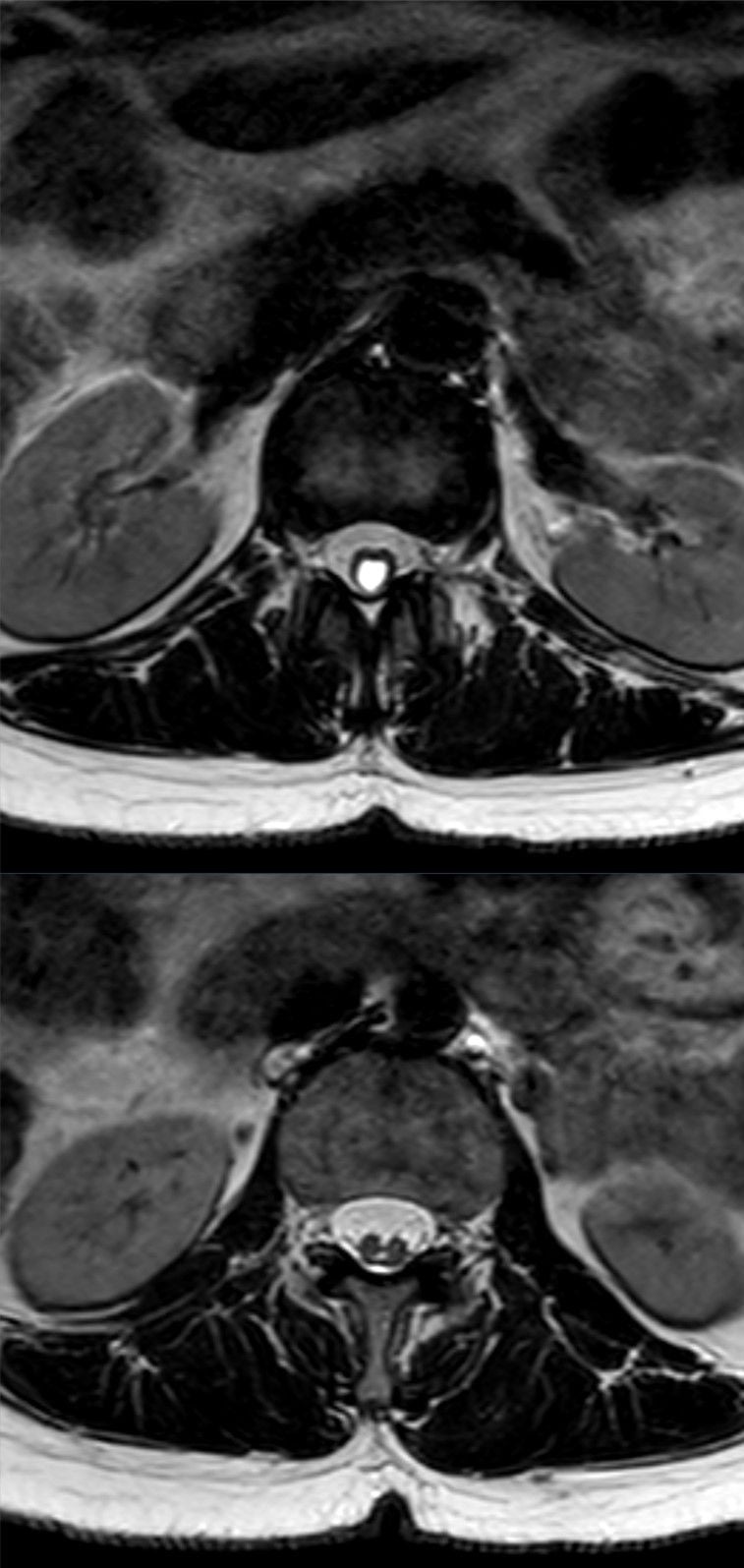
The second tether. A low-lying conus (below L2) or a thickened filum over 2 mm is common, and how common depends sharply on level. In Pang's series every lumbar SCM had both a low-lying conus and at least one additional tethering lesion, whereas only 1 of 7 cervical and high thoracic lesions did.
Why that matters. A lumbar split cord obliges you to hunt for the second lesion and plan to deal with it. Releasing the septum alone leaves the cord tethered.
Why deformity correction is dangerous. Correction of congenital scoliosis often requires distraction, derotation or vertebral column resection. An unrecognised bony septum acts as a fulcrum and tears the hemicords, and even a Type II fibrous band can produce significant stretch injury once the spine is lengthened. Any manoeuvre that lengthens the column, distraction or osteotomy, can acutely worsen the tether if the septum has not been removed first.
Where SCM Sits: the Classification of Spinal Dysraphism
Examiners expect you to place split cord malformation within the wider spinal-dysraphism framework rather than treat it in isolation. The first division is open versus closed, and the closed (occult) group is then split by whether there is a subcutaneous mass.
- Skin
- Neural tissue exposed / not skin-covered
- Examples
- Myelomeningocele, myelocele - present at birth, need urgent closure
- Skin
- Skin-covered lump
- Examples
- Lipomyelomeningocele, posterior meningocele, myelocystocele
- Skin
- Skin-covered, often only a cutaneous stigma
- Examples
- Split cord malformation (diastematomyelia), tethered/thickened filum, filum lipoma, dorsal dermal sinus, caudal regression
SCM is a closed (occult) dysraphism without a subcutaneous mass. There is no lump to prompt imaging and the cord is fully skin-covered, which is exactly why the subtle cutaneous stigmata (hairy patch, sinus, dimple) matter so much. The unifying functional theme across the whole closed group is tethering of the cord.
Classification and Types
Pang's classification is built on the dural tubes. Type I has two hemicords in two separate dural sacs, divided by a rigid bony or osteocartilaginous septum. Type II has two hemicords in a single shared dural sac, divided only by a fibrous band. The split usually occupies one level.
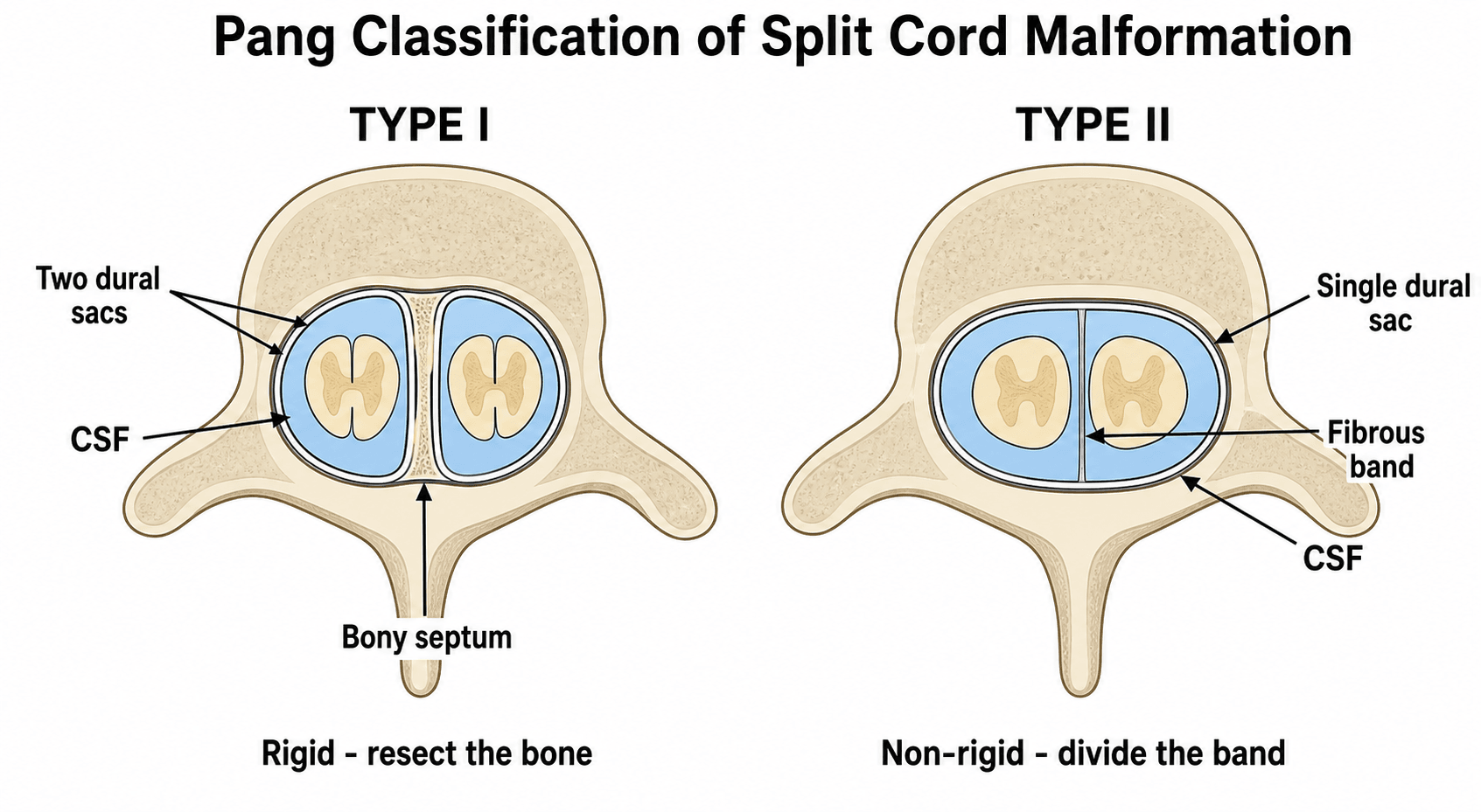
- Type I (Diastematomyelia)
- Two separate sacs
- Type II (Diplomyelia)
- Single shared sac
- Type I (Diastematomyelia)
- Bony or (osteo)cartilaginous
- Type II (Diplomyelia)
- Fibrous band only
- Type I (Diastematomyelia)
- Hemicords widely separated
- Type II (Diplomyelia)
- Hemicords close together
- Type I (Diastematomyelia)
- Rigid bony fixation
- Type II (Diplomyelia)
- Flexible fibrous tether
- Type I (Diastematomyelia)
- Thoracolumbar
- Type II (Diplomyelia)
- Any level
- Type I (Diastematomyelia)
- Resect septum, close dual sacs
- Type II (Diplomyelia)
- Divide band, preserve single sac
- Type I (Diastematomyelia)
- Higher - requires bone removal
- Type II (Diplomyelia)
- Lower - band division only
Type predicts the operation, not the course. It is tempting to assume the rigid bony septum of Type I is the more dangerous lesion, and examiners like that assumption. Pang's series does not support it: symptomatic progression was independent of lesion type, with a Type I to Type II ratio of 13:11, and independent of level, at 67% for cervical against 64% for thoracolumbar lesions.
What type does decide. Type I is technically harder and carries slightly higher surgical morbidity. It is more dangerous still when an oblique septum has divided the cord asymmetrically into one large and one small, delicate hemicord.
What decides the risk of deterioration. Age and symptoms, not type. All 8 adults in the series were symptomatic at diagnosis, against only 16 of 31 children.
Type II still tethers. The hemicords remain tethered by the band, tethered-cord signs may develop later, and the cord may appear unified on axial views if the images are not read carefully.

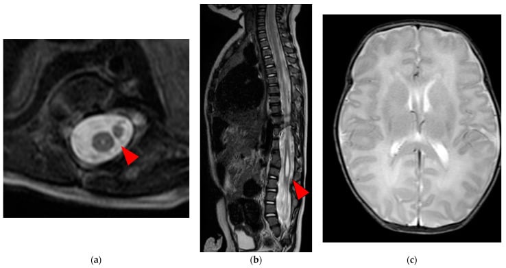
Associated anomalies. Other lesions travel with the split; the table gives their frequency and what each means for the operation.
- Frequency
- 60-80 percent
- Clinical Impact
- Progressive curve, rib fusion
- Management Note
- Must image before correction
- Frequency
- Greater than 70 percent
- Clinical Impact
- Bladder, leg weakness
- Management Note
- Section filum at same sitting
- Frequency
- 40-60 percent
- Clinical Impact
- May extend into hemicords
- Management Note
- Monitor or shunt if symptomatic
- Frequency
- 20-30 percent
- Clinical Impact
- Additional tether mass
- Management Note
- Requires careful debulking
Clinical Assessment
The skin. Hypertrichosis (a hairy patch, classically over the lumbar spine) is the classic sign and the best single predictor, but quote it honestly: it was present in 56% of Pang's 39 patients, so nearly half had no hairy patch. What is close to universal is a cutaneous marker of some kind: occult or open dysraphic stigmata were present in all but 3 of the 39. Look also for a dermal sinus, lipomatous mass, capillary malformation or an asymmetric gluteal cleft.
A normal back does not clear the spine. Any midline cutaneous lesion below L1 demands MRI, yet a normal-looking back does not exclude an SCM. The 3 of 39 with completely normal skin are about 8 percent, and later series report higher rates.
The neurology. Look for asymmetric leg weakness, cavus feet or bladder dysfunction in a child with scoliosis; the foot may be cavus or equinovarus on one side. The neurological level often does not match the level of the bony deformity.
History. The answers that point to a split cord:
- A cutaneous lesion noted at birth or during growth
- Leg weakness, numbness or a change in gait, and whether it is progressing
- Early bladder or bowel dysfunction: incontinence, recurrent UTI, constipation
- A curve noticed by the parents or at school screening
- Any previous spinal procedure performed without an MRI
Examination. Beyond the skin:
- Legs: asymmetric atrophy, cavus feet, trophic ulcers
- Neurology: asymmetric reflexes, a sensory level, Babinski sign
- Spine: the congenital curve, kyphosis, local tenderness
Any child with congenital scoliosis plus one of the following requires MRI:
- Hairy patch
- Leg-length discrepancy
- Cavus foot
- Early-onset progressive deficit
- Neurogenic bladder
- Typical Level
- Lumbosacral
- Associated Risk
- SCM Type I or II
- Next Step
- Full-spine MRI
- Typical Level
- Any level
- Associated Risk
- Infection, dermoid, tether
- Next Step
- MRI + surgical excision
- Typical Level
- Lumbosacral
- Associated Risk
- Lipomyelomeningocele
- Next Step
- MRI + neurosurgical referral
- Typical Level
- Gluteal crease
- Associated Risk
- Low risk if above coccyx
- Next Step
- Ultrasound or MRI if atypical
Investigations
MRI makes the diagnosis. No plain film or CT finding replaces it. Plain radiographs show the scoliosis and vertebral anomalies but never confirm or exclude SCM.
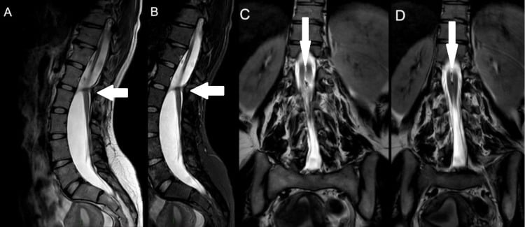
The protocol. Image the entire spine, foramen magnum to sacrum, because multiple levels or additional anomalies (syrinx, lipoma, dermal sinus) are common. Sagittal and axial T1, T2, STIR and post-contrast T1 are standard. Read them for the hemicords, the septum and whether it is bony or fibrous, the conus level, filum thickness, and a syrinx, lipoma or Chiari.
Read the axials. The question is whether the hemicords lie in separate or shared dural sacs, and whether a bony, cartilaginous or fibrous septum is present. Axial images are essential: sagittal views alone can miss the split.
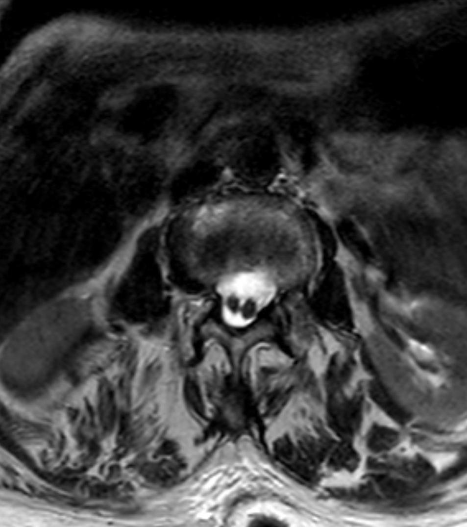
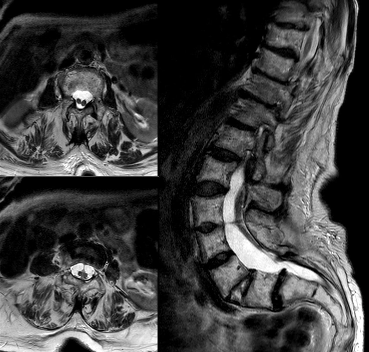
Type on the dural tubes, not on the septum you can see. The two septa are not equally visible. In Pang's series every Type I bone septum was identified before surgery, but only 5 of the 18 Type II fibrous septa appeared on preoperative imaging, while at operation a fibrous septum or other tethering bands were found in every Type II case. A single dural tube with two hemicords and no visible septum is therefore still a Type II SCM with something tethering it, and it should be counselled and explored as one. The number of dural tubes is the criterion that never fails; the septum is the one that hides.
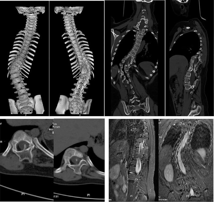
CT is for surgical planning of a bony septum, not diagnosis. It shows the location and extent of the bony bar and the associated vertebral anomalies, and it helps anticipate a difficult dural closure.
Urodynamics are indicated for any bladder symptom, including a history of incontinence, and before scoliosis surgery. They may show detrusor-sphincter dyssynergia, high-pressure voiding or reflux, and they give a baseline for monitoring neurological recovery after surgery.
Neonatal spinal ultrasound. In the neonate and young infant, up to about 3 to 6 months, the posterior vertebral elements are incompletely ossified and leave an acoustic window, which makes ultrasound the first-line screen for a cutaneous stigma (sacral dimple, hairy patch, lipoma, dermal sinus). It can show the conus level, cord pulsation and movement, a thickened filum and even the hemicords of an SCM, with no sedation and no radiation.
When ultrasound hands over to MRI. A confidently normal study in a low-risk marker can avoid MRI. An abnormal, equivocal or high-risk finding, and any child beyond the ossification window, proceeds to MRI, which remains the definitive test. This newborn-clinic pathway for an isolated skin marker is distinct from the pre-deformity-surgery MRI mandate.
Management
Asymptomatic split cord with scoliosis. The goal is to protect the cord during planned deformity correction, and the plan follows the type:
- Type I bony septum: plan simultaneous or staged detethering and septum resection
- Type II fibrous band: divide the band during the same anaesthetic as the scoliosis correction
- Always section the filum terminale if it is thickened or the conus is low
- Intraoperative neuromonitoring is mandatory
Progressive neurological deficit or bladder dysfunction. The goal is to release every tethering point before the damage becomes irreversible. The sequence:
- Positioning. Prone on a Jackson table, head neutral, all pressure points padded, Foley catheter.
- Exposure. Midline incision; expose the laminae one level above and below the split; laminectomy preserving the facets.
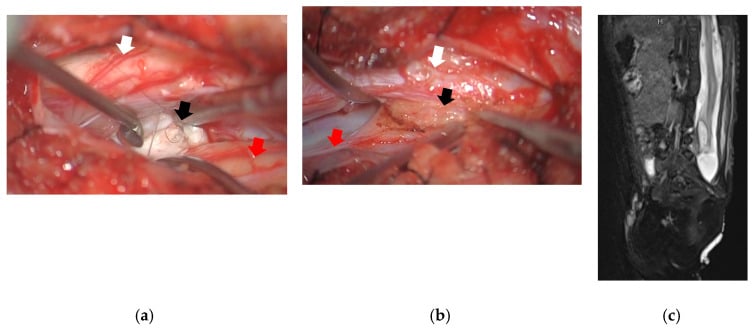
- Dural opening. Open the dura in the midline and identify both hemicords and the septum or band.
- Septum. Type I: resect the bony septum with a high-speed burr and Kerrison, protecting both hemicords. Type II: divide the fibrous band sharply under magnification.
- Tether release. Section the filum terminale, release any scar or lipoma, and confirm free cord movement with a Valsalva manoeuvre.
- Closure. Watertight dural closure, layered wound closure, and a drain if there is a large dead space.
Never apply distraction or correction to the spine until the septum is removed and the cord is fully detethered. Even gentle distraction across a fixed bony septum can produce immediate cord transection.
- Diagnosis
- MRI shows Type I or II SCM
- Action
- Close observation or prophylactic detether
- Key Pearl
- Cutaneous marker alone justifies imaging
- Diagnosis
- SCM with tethered cord on MRI
- Action
- Surgical detethering before or with correction
- Key Pearl
- Delay risks irreversible deficit
- Diagnosis
- No pre-op MRI performed
- Action
- Cancel surgery until MRI obtained
- Key Pearl
- Never instrument without MRI clearance
Complications
Complications arise from the natural history of an untreated split cord, from a missed diagnosis during deformity surgery, and from the detethering operation itself.
Of the untreated or missed split cord. The single most important complication to prevent is acute cord injury from operating on congenital scoliosis without a preoperative whole-spine MRI. Distraction or derotation across an unrecognised bony septum can transect the hemicords and cause immediate, permanent paraplegia: the feared, preventable disaster of congenital-scoliosis surgery. The others:
- Progressive tethered-cord syndrome: worsening leg weakness, sensory loss, cavus foot deformity and gait deterioration with growth
- Neurogenic bladder and bowel: detrusor-sphincter dyssynergia, recurrent UTI, incontinence, which may be irreversible if late
- Syringomyelia extending into the hemicords
- Curve progression while definitive correction is deferred
Of detethering and septum resection.
- New or worsened neurological deficit (motor, sensory, bladder) from manipulation of the hemicords
- CSF leak or pseudomeningocele, especially after the difficult dual-tube dural closure in Type I
- Wound infection or meningitis
- Re-tethering from scar, which may present as late deterioration after initial improvement
- Incomplete release leaving a residual tether (an unsectioned thick filum, for example) with persistent symptoms
Guidelines, Registries & Global Practice
- SCM prevalence is consistent across ethnic groups; most series come from high-volume pediatric spine centers in North America, Europe, India and East Asia
- Congenital scoliosis association: 60-80 percent of patients WITH an SCM have a congenital scoliosis. There is no SCM registry, so this range comes from surgical series rather than population data - and it must not be read backwards as the yield of screening scoliosis, which is 18.3 percent overall and 52 percent at its worst pattern
- Missed diagnosis remains the leading cause of catastrophic intraoperative cord injury in congenital deformity surgery worldwide
- High-resource centers: routine whole-spine MRI with contrast for every congenital curve; intraoperative ultrasound and neuromonitoring
- Limited-resource settings: MRI access may be delayed; plain film and clinical markers still drive referral, but outcomes suffer when imaging is unavailable
- Universal principle: any child with congenital scoliosis and a cutaneous marker or neurological asymmetry must reach an MRI-capable center before surgery
- MRI Indication
- Mandatory whole-spine MRI before any congenital scoliosis correction
- Surgical Sequence
- Detether and resect septum before or with deformity correction
- Registry Evidence
- No dedicated SCM registry; captured in congenital scoliosis databases
- MRI Indication
- MRI recommended for all congenital curves; urgent if cutaneous stigmata
- Surgical Sequence
- Neurosurgical input before orthopaedic correction
- Registry Evidence
- National registry data show reduced neurological complications with protocolized MRI
- MRI Indication
- MRI standard of care; CT for bony anatomy only
- Surgical Sequence
- Single-stage or staged release then correction
- Registry Evidence
- European registries emphasize avoidance of distraction across unrecognized tether
- MRI Indication
- High volume experience supports early prophylactic detether in asymptomatic SCM
- Surgical Sequence
- Combined neurosurgery-orthopaedics teams common
- Registry Evidence
- Large published series confirm safety of aggressive release protocols
There is no international SCM-specific registry. Data are derived from congenital scoliosis registries (NJR, AJRR, AOANJRR) and large single-center series. The consistent message across all sources is that preoperative MRI changes the operative plan in one-third of congenital scoliosis cases and prevents the majority of intraoperative neurological catastrophes.
Record in every congenital scoliosis patient:
- Presence or absence of cutaneous markers
- Neurological examination findings
- MRI performed (date, findings, who reviewed)
- Multidisciplinary discussion with neurosurgery
- Informed consent discussion of SCM risk
A missed SCM leading to postoperative paraplegia is a recurring source of claims worldwide. The standard of care in 2026 is full-spine MRI before booking the operating theatre.
Controversies & Areas of Uncertainty
Some centers advocate early release of every identified SCM even without deficit, arguing that growth or future surgery will eventually produce symptoms. Others observe selected asymptomatic patients with serial MRI and neurology. No randomized data exist; decision is individualized by age, curve magnitude, and family preference.
When both detethering and major osteotomy are required, some teams prefer two separate anesthetics to reduce operative time and blood loss. Others perform both in one sitting with two teams. Outcomes appear similar when neuromonitoring is used; choice depends on institutional resources and surgeon experience.
Ultrasound can confirm cord pulsation and free movement after release, but adds time and requires an open dural field. Its routine use is not universal; many experienced teams rely on direct visualization and neuromonitoring alone.
Rare adult presentations with new deficit after decades of compensation exist. Whether to operate on a stable adult with incidentally discovered SCM is controversial; most reserve surgery for progressive symptoms or when major spinal surgery is planned for other reasons.
MCQ Practice Points
Q: What is the key difference between Pang Type I and Type II split cord malformation? A: Type I has two separate dural sacs separated by a bony or cartilaginous septum; Type II has two hemicords within a single dural sac separated by a fibrous band only. The dural architecture and septum composition dictate the surgical approach and risk profile.
Q: When is MRI mandatory in a child with congenital scoliosis? A: Before any corrective surgery or even definitive bracing decision. Greater than 60 percent of congenital curves harbor occult SCM. An unrecognized bony septum can cause cord transection during distraction or osteotomy. Full-spine contrast MRI is the minimum standard.
Q: What cutaneous finding most strongly suggests split cord malformation? A: Hypertrichosis (hairy patch) over the lumbosacral spine. While not 100 percent sensitive, a hairy patch in a child with spinal deformity is an absolute indication for MRI. Other markers include dermal sinus, lipoma, and asymmetric gluteal cleft.
Q: Should detethering occur before, during, or after scoliosis correction? A: Before or at the same anesthetic as correction, never after. The cord must be free before any lengthening or derotation maneuver. Performing detethering after correction risks stretching an already injured cord across a residual septum.
Q: What additional finding is present in the majority of split cord cases and must be addressed surgically? A: Tethered cord from a thickened filum terminale or low conus. Even after septum resection, failure to section the filum leaves the cord under tension and allows late deterioration. Always inspect and release the filum at the same procedure.
Exam Viva Scenarios
Practise clinical reasoning and management decisions out loud
“A 9-year-old girl is referred with a 45-degree congenital thoracic scoliosis. On examination she has a prominent hairy patch over the lumbar spine. She has no neurological symptoms but her mother reports occasional bed-wetting. What is your next step and why?”
“A 12-year-old boy undergoes posterior instrumentation for congenital scoliosis. On the second postoperative day he develops bilateral leg weakness and urinary retention. Review of the preoperative workup shows no MRI was performed. What happened and how do you manage it now?”
Pang Classification
- Type I: two dural sacs, bony septum - resect bone and close dual tubes
- Type II: single dural sac, fibrous band - divide band only
- Both types frequently tethered by thick filum or scar
- Always release every tethering element at the index procedure
Cutaneous and Clinical Red Flags
- Hypertrichosis (hairy patch) is the strongest marker
- Asymmetric cavus feet or leg atrophy in a scoliotic child
- Early bladder dysfunction or recurrent UTI
- Any midline skin lesion below L1 demands MRI
Pre-Operative Mandate
- Full-spine contrast MRI in every congenital scoliosis case
- MRI changes surgical plan in greater than 30 percent of patients
- Never instrument or distract without MRI clearance
- Coordinate neurosurgery and spine teams before booking
Surgical Principles
- Position prone, expose one level above and below the split
- Resect bony septum under magnification protecting both hemicords
- Divide fibrous band and section thickened filum
- Confirm free cord movement before any spinal lengthening
Complications to Avoid
- Acute cord transection from distraction across unrecognized septum
- Incomplete release leaving residual tether
- CSF leak from difficult dural closure in Type I
- Late retether from scar formation
Evidence Base
Split cord malformation: Part I: A unified theory of embryogenesis for double spinal cord malformations
- Seminal paper establishing the embryologic basis for both Type I and Type II SCM from a single developmental error
- Described the spectrum of associated vertebral and cutaneous anomalies
- Provided the classification system still used worldwide for surgical decision-making
Split cord malformation: Part II: Clinical syndrome
- 39 patients (8 adults, 31 children): 19 Type I, 18 Type II, 2 composite with both lesion types in tandem. 31 were lumbar, 6 cervical, 2 thoracic
- Symptomatic progression was INDEPENDENT OF LESION TYPE - the Type I:Type II ratio was 13:11 - and independent of level, with 67% of cervical versus 64% of thoracolumbar lesions progressing
- Age, not type, separated the presentations: all 8 adults had pain and progressive sensorimotor deficit at diagnosis, whereas only 16 of 31 children had any symptoms
- Left-right functional discrepancy was prominent in 8 children - the feature that distinguishes SCM from tethered cord of other causes
- Hypertrichosis was by far the best predictor, present in 56%; some cutaneous marker of an occult or open dysraphic state was present in all but 3 of the 39
- Every Type I bone septum was identified before surgery, but only 5 Type II fibrous septa were seen on preoperative imaging - yet a fibrous septum or other tethering bands were found at surgery in EVERY Type II case
- All lumbar SCMs had a low-lying conus and at least one additional tethering lesion; only 1 of 7 cervical and high thoracic lesions did
Split cord malformations
- Review consolidating the unifying SCM classification: Type I = two dural sacs with a bony/fibrocartilaginous spur; Type II = a single dural sac with intradural fibrous bands
- The intervening mesenchymal elements tether the cord and drive progressive neurological, urological and orthopaedic deterioration
- The natural history supports an early, aggressive operative approach to untether the cord BEFORE clinical deterioration begins
Occult intraspinal anomalies and congenital scoliosis
- Of 251 patients with congenital scoliosis, occult intraspinal anomalies were found in 18.3% (46 patients); diastematomyelia was the commonest (41 patients)
- The risk was highly pattern-dependent: a unilateral unsegmented bar with a contralateral hemivertebra in the lower thoracic/thoracolumbar spine carried the highest rate (52%)
- Neural deterioration could be halted by excising the anomaly, supporting myelography (now MRI) in every congenital scoliosis case before correction