Ulnar collateral ligament injury of thumb MCP joint
- Stener lesion: UCL flips over adductor aponeurosis - blocks healing
- Stress test: greater than 30° absolute laxity OR greater than 15° vs contralateral side
- Complete tear with Stener lesion MUST have surgical repair
- Test in extension (assesses accessory collateral) AND flexion (proper collateral)
- “Gamekeeper's (chronic) vs Skier's (acute) - both UCL injuries
- “Always compare to contralateral side for stress testing
- “MRI or ultrasound can identify Stener lesion preoperatively
- “No endpoint on stress test = complete tear
Overview
The ulnar collateral ligament (UCL) of the thumb metacarpophalangeal (MCP) joint is one of the most commonly injured ligaments in the hand. A valgus force, directed radially, stretches or ruptures the UCL complex.
Two names for one ligament. "Gamekeeper's thumb" comes from Scottish gamekeepers, who broke the necks of rabbits and attenuated the UCL over time, so it names the chronic injury. The acute traumatic tear is now commonly called "skier's thumb", given its high incidence in skiing falls with the thumb in a pole strap.
The Stener lesion. In approximately 70% of complete tears the torn UCL displaces to lie superficial to the adductor pollicis aponeurosis, which then sits between the ligament and its insertion on the proximal phalanx. The ligament cannot heal to bone across it, even with prolonged immobilisation, so surgery is mandatory for complete functional recovery.
Anatomy and Biomechanics
The joint. The thumb MCP is a condyloid (biaxial) joint. Flexion-extension is its primary motion, with abduction-adduction and rotation secondary, and it is less constrained than the finger MCP joints.
The UCL. It arises from the ulnar condyle of the metacarpal head and inserts on the volar-ulnar base of the proximal phalanx. It has two components, taut in opposite positions:
- Origin
- Dorsal metacarpal head
- Insertion
- Volar base of proximal phalanx
- Taut in
- Flexion
- Origin
- Volar metacarpal head
- Insertion
- Volar plate
- Taut in
- Extension
The volar plate attaches to the accessory collateral and provides additional stability.
The adductor aponeurosis. Adductor pollicis arises from the third metacarpal, capitate and trapezoid and inserts on the proximal phalanx at the ulnar sesamoid. Its aponeurosis lies superficial to the UCL insertion, which makes it central to the pathoanatomy of the Stener lesion.
Acute injury (skier's thumb). A forced valgus (radial deviation) stress, in any of these settings:
- A skiing fall with the thumb caught in the pole strap
- Ball-handling sports such as football and basketball, the thumb caught during a catch
- A fall onto the outstretched or abducted thumb
- Motor vehicle accidents
- Manual labour
Chronic injury (gamekeeper's thumb). Repetitive valgus stress, originally occupational in gamekeepers, attenuates the ligament gradually, and the instability is progressive.
How a Stener lesion forms.
- Forced valgus stress ruptures the UCL, which tears from its insertion on the proximal phalanx.
- The MCP joint subluxates briefly.
- As the joint reduces, the ligament flips over the adductor aponeurosis.
- The aponeurosis now lies between ligament and bone, and healing is impossible without surgical relocation.
The lesion is more frequent with greater initial displacement, and it cannot be definitively diagnosed clinically.
Classification
Clinical grade (Heyman). The grade comes from the stress test and guides the initial treatment.
- Injury
- Sprain (partial tear): ligament stretched but intact
- Laxity
- Less than 15°
- Endpoint
- Firm
- Treatment
- Conservative
- Injury
- Partial tear: some fibres torn
- Laxity
- 15-30°
- Endpoint
- Soft but present
- Treatment
- Usually conservative
- Injury
- Complete rupture
- Laxity
- Greater than 30°, or greater than 15° vs contralateral
- Endpoint
- No firm endpoint
- Treatment
- Surgical, especially if Stener lesion
Where the ligament tears.
- Distal avulsion from the proximal phalanx - most common
- Midsubstance tear within the ligament body
- Proximal avulsion from the metacarpal - less common
Is there a Stener lesion? If yes, surgery is mandatory. If no, consider conservative or surgical treatment. If uncertain, obtain an MRI or explore surgically. Location and the presence of a Stener lesion together determine the surgical approach.
Bony avulsion. Fragment size and displacement guide the fixation strategy:
- Non-displaced - may be treated conservatively
- Displaced greater than 2mm - consider ORIF
- Displaced into the joint - requires surgery
- Large fragment, greater than 20% of the articular surface - ORIF preferred
Clinical Presentation
History. Pain at the ulnar aspect of the thumb MCP after one of the mechanisms above, with swelling over the thenar region. The patient notices weakness of pinch grip, a sense of instability in key pinch and difficulty with fine motor tasks.
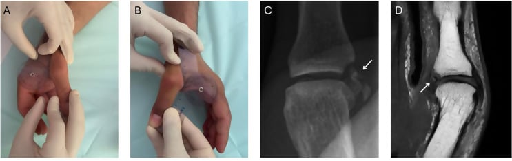
Look and feel. Swelling at the ulnar MCP joint and ecchymosis over the ulnar thumb; an angular deformity may be visible in severe cases. The tenderness is over the UCL insertion at the volar-ulnar base of the proximal phalanx. A palpable mass at the ulnar MCP is the sign of a Stener lesion, the rolled-up ligament sitting superficial to the aponeurosis, and it is highly specific but not sensitive. Range of motion is usually full, limited by pain; compare with the contralateral side.
Stress testing. This is the critical examination.
- Anaesthesia: local infiltration or a digital block if needed.
- Stabilise the metacarpal firmly with one hand.
- Test in extension: a radial-deviation (valgus) force applied to the proximal phalanx assesses the accessory collateral.
- Test in 30° flexion: valgus stress assesses the proper collateral.
- Evaluate both the degree of laxity, in degrees, and the quality of the endpoint, firm or soft.
- Compare with the contralateral thumb, always.
Read the result against the Heyman grades. Laxity of greater than 30° absolute or greater than 15° more than the other thumb indicates a complete tear, as does the absence of a firm endpoint, and with no firm endpoint a Stener lesion is likely.
X-ray before stress testing to exclude a fracture; if there is an avulsion fracture, stress testing is contraindicated. Avoid forceful repeated testing, which may convert a partial tear to a complete one.
Pinch and function. Key pinch, thumb against the side of the index finger, is reduced with UCL insufficiency; compare with the other side. The functional assessment covers writing, buttoning, turning a key and opening a jar.
Differential diagnosis. Several conditions mimic an ulnar-sided thumb injury, and the common trap is to attribute all base-of-thumb pain to a UCL tear.
- 1
- Ulnar MCP tenderness, valgus laxity, weak key pinch; palpable mass if Stener
- 1
- Radial-sided MCP pain and varus laxity
- 1
- Pain at thumb base/CMC, positive grind test, radiographic CMC changes - not MCP
- 1
- Radial wrist/first-dorsal-compartment pain, positive Finkelstein, no MCP laxity
- 1
- Intra-articular fracture of thumb metacarpal base on X-ray; bony, not ligamentous
- 1
- Hyperextension mechanism, volar tenderness, no ulnar valgus laxity
- 1
- Catching/locking at thumb IP/MCP, A1 pulley nodule, no instability
- 1
- Anatomical snuffbox tenderness, axial load pain, distinct from MCP injury
The thumb's radial collateral ligament is the under-recognised counterpart to the UCL and is genuinely examinable:
- Far less common than UCL injury (roughly a 1:4 ratio), caused by a forced ADDuction (ulnar-deviation) force - the mirror of the abduction force that tears the UCL.
- Presents with radial-sided MCP pain/swelling and varus laxity on stress testing, often with volar and ulnar subluxation/rotation of the proximal phalanx (the metacarpal head becomes prominent dorsoradially) and a weak pinch.
- A true Stener-type lesion is rare on the radial side, because the abductor/extensor expansion is broader and less discrete than the adductor aponeurosis - so the displaced-ligament mechanical block that defines the UCL Stener lesion is much less of a feature; RCL tears more often retract or avulse without that interposition.
- Management mirrors the UCL: immobilise partial/stable tears, repair (suture anchor) complete or displaced tears, and reconstruct chronic/irreparable cases. Always stress both sides of the MCP so a radial injury is not missed while you focus on the ulnar side.
Investigations
Radiographs. Three views of the thumb:
- PA - shows a bony avulsion if present
- Lateral - subluxation
- Oblique - additional view of the fragment
Look for a bony avulsion fragment at the base of the proximal phalanx, a proximally displaced fragment (a radiographic sign of a Stener lesion), and subluxation, the proximal phalanx radially deviated on the metacarpal.
Stress radiographs are controversial. Some advocate them, but they risk displacing a partial tear, and MRI is preferred if the diagnosis is uncertain.
MRI. The indications:
- Equivocal clinical examination
- Identifying a Stener lesion preoperatively
- Assessing associated injuries
- Differentiating a partial from a complete tear
Reading the MRI. The normal UCL is low signal on all sequences, with a smooth contour from metacarpal to proximal phalanx. A partial tear shows thickening and increased signal within a ligament whose continuity is maintained. A complete tear with a Stener lesion shows a discontinuous UCL displaced proximally and superficially, the "yo-yo on a string" appearance, with the adductor aponeurosis visibly interposed.
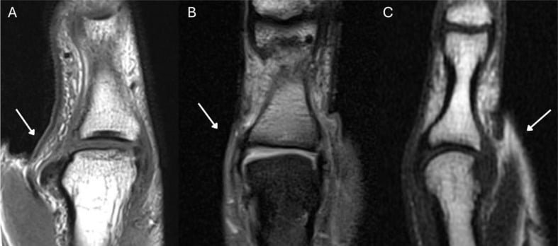
Ultrasound. A dynamic study that can visualise the stress test, cost-effective and free of radiation, and becoming more popular. It identifies the displaced ligament and can visualise a Stener lesion, but it is operator-dependent.
CT has a limited role, mainly for complex avulsion fractures and for planning ORIF, with 3D reconstruction to assess the fragment.
Management
The decision. The grade, the presence of a Stener lesion, any bony fragment and the time since injury settle each scenario below.
- Decision
- Thumb spica 4-6 weeks
- Rationale
- Heals with immobilisation
- Decision
- Consider conservative vs surgery
- Rationale
- May heal if stable
- Decision
- Surgical repair
- Rationale
- Cannot heal - blocked by aponeurosis
- Decision
- Likely surgical
- Rationale
- High suspicion for complete tear
- Decision
- Likely surgical
- Rationale
- Even if less than 30° absolute
- Decision
- Depends on displacement
- Rationale
- Fix if displaced greater than 2mm
- Decision
- Ligament reconstruction
- Rationale
- Primary repair often not possible
- Decision
- ORIF + ligament repair
- Rationale
- Address both components
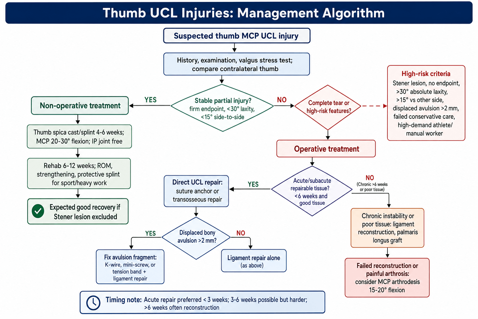
Who. Grade I and II injuries, a complete tear without a Stener lesion, and non-displaced avulsion fractures. The examination findings that support it are less than 30° absolute laxity, less than 15° difference from the other side and a firm endpoint. The Stener lesion must be excluded first.
The protocol.
- 0-2 weeks - thumb spica cast or splint, MCP in 20-30° flexion, IP joint free; elevation and ice
- 2-6 weeks - thumb spica for a total of 4-6 weeks, which may convert to a removable splint at 4 weeks; serial clinical assessment
- 6-12 weeks - gentle range of motion, progressive strengthening, and splinting for sport and heavy activities; return to full activity at 10-12 weeks
Surgical Technique
Setup. Supine, the arm on a hand table and a tourniquet on the upper arm. General anaesthesia or a regional block (axillary or supraclavicular), often with local infiltration added. The kit is 2.0-2.4mm suture anchors, typically one or two, or the equipment for transosseous sutures, with fine hand instruments and loupe magnification at 2.5-3.5x.
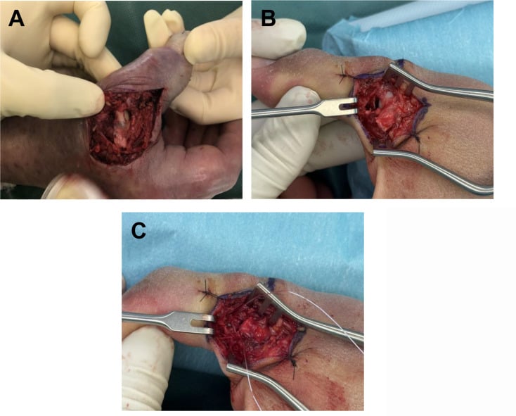
- Incision. Mark it over the ulnar MCP joint: a chevron (inverted V) or a curvilinear incision along the ulnar border, approximately 2-3 cm long.
- Superficial dissection. Identify and protect the dorsal sensory branches of the radial nerve; multiple small branches lie in the field, and injury causes a painful neuroma and numbness. Dissect the subcutaneous layer carefully.
- Expose the adductor aponeurosis, a white fibrous band running longitudinally. In a Stener lesion the rolled-up UCL is palpable as a mass superficial to it.
- Incise the aponeurosis longitudinally. The normal UCL lies deep to it.
- Identify the UCL: the proximal stump, usually retracted and rolled up, and the distal insertion site on the base of the proximal phalanx. Assess tissue quality and remove any scar tissue between ligament and bone.
Beyond the standard suture-anchor repair above, the modern refinement is suture-tape "internal brace" augmentation - a strong, low-profile non-absorbable tape anchored in the metacarpal head and the proximal phalanx base, spanning the repaired UCL like a check-rein. Its purpose is not to replace the biological repair but to protect it:
- The tape carries valgus load while the native ligament heals, so the construct is immediately more stable.
- This permits an accelerated rehabilitation protocol - earlier protected motion and earlier return to sport - which is why it is popular in athletes (return-to-play rates remain very high).
- It can also augment a reconstruction or a tenuous repair in poor-quality tissue.
Caveats: it adds hardware and cost, over-tensioning the tape can over-constrain and stiffen the MCP, and it does not substitute for correctly relocating a Stener lesion deep to the adductor aponeurosis. The examinable point is the concept of "ligament repair + internal brace = earlier motion," not the brand.
Complications
Early.
- Wound - infection (rare), haematoma, dehiscence
- Nerve injury - the dorsal sensory branches of the radial nerve, with numbness over the dorsal thumb; usually a neurapraxia that recovers
- Fixation failure - suture anchor pull-out or refracture of the avulsion fragment; may need revision
Late.
- Chronic instability - the most significant complication, from a missed Stener lesion or an inadequate repair; may need reconstruction
- Stiffness - common after prolonged immobilisation; hand therapy is essential, and it usually resolves with time
- Post-traumatic arthritis - uncommon with anatomical repair, more common after chronic instability; may need arthrodesis
- Persistent weakness - a pinch strength deficit that usually improves over 6-12 months and may be permanent in chronic cases
- Cold intolerance - common in the first year, usually improving with time
Postoperative Care
Days 0-14. The rigid thumb spica splint is maintained full-time and kept clean and dry, with elevation above heart level for the first 48-72 hours and ice packs around the splint without getting it wet. The first dressing change at 48-72 hours inspects for haematoma and infection, and the sutures come out at 10-14 days.
Analgesia. Expect moderate pain for the first 24-48 hours.
- Paracetamol 1g four times daily
- Ibuprofen 400mg three times daily, if there are no contraindications
- A short course of opioids if needed, 3-5 days maximum
What to watch for. Excessive pain raises concern for compartment syndrome, which is rare. Increasing pain, fever or drainage suggest infection. Numbness in the radial nerve distribution is expected and is monitored.
Weeks 2-6: protected immobilisation. The spica stays on full-time with the MCP in 20-30° flexion; it may convert to a removable splint at week 4, at the surgeon's preference, and from weeks 4-6 it is removable but taken off only for gentle washing. The IP joint remains free, with active IP flexion and extension encouraged; adjacent finger range-of-motion exercises are added in weeks 2-4, and the IP joint exercises continue through weeks 4-6. The repair takes no stress, though very gentle active motion may begin at 4-6 weeks if the surgeon permits. No lifting, gripping, pinching or driving. Showering needs a waterproof cover, and desk work is acceptable with the splint on.
Weeks 6-8: early mobilisation. The splint comes off 3-4 times daily for gentle active MCP flexion and extension, therapist-supervised if possible, and is worn part-time between sessions, at night and for any potentially stressful activity. No passive stretching and no valgus stress.
- Thumb opposition to each fingertip
- Gentle fist making (light grip)
- Tabletop slide exercises
- Tendon gliding exercises
Light daily activities, computer work and writing are allowed; sport and heavy lifting are not.
Weeks 8-12: strengthening. Grip activity increases gradually, with putty progressing from soft to medium to firm and pinch strengthening (key, tip and lateral pinch), resistance advancing as tolerated. The goals are full active range of motion, MCP flexion of 50-60°, and extension to neutral or near-neutral, with no extension lag acceptable. Work- and sport-specific tasks are simulated with a graded return to daily tasks; taping may support the thumb during activities, and the splint is worn at night only. Most daily activities, light recreation and a gradual return to manual work resume, but not contact sport.
Week 12 onward: return to sport. The criteria:
- Pain-free range of motion
- Strength at least 80% of the contralateral side
- No instability on stress testing
- Patient confidence in stability
- Surgeon clearance
A gradual return-to-sport protocol follows, with position-specific training for athletes and possibly protective taping or a brace initially; the splint is used as needed for sport. Full clearance usually comes 3-4 months after surgery. Strength may continue improving for up to 12 months; review annually in the first year and monitor for late instability or arthritis.
Red flags needing immediate review.
- Sudden increase in pain
- Loss of motion after initial gains
- Recurrent sensation of instability
- Signs of infection
- Persistent weakness at 3 months
- Donor site problems, if a graft was used
Outcomes and Prognosis
Acute repair. Return to full activities is 85-90%, and patient satisfaction is greater than 90%.
- 1
- 85-95% recovery
- 1
- 90-100% recovery
- 1
- Near-full to full ROM
- 1
- Less than 10° laxity vs contralateral
- 1
- 3-4 months
- 1
- 3-4 months
- 1
- 4-6 months
What predicts success. Early recognition and treatment, with under 3 weeks optimal; accurate diagnosis of the Stener lesion; a secure anatomical repair; a protected rehabilitation protocol; and patient compliance with the restrictions.
Chronic reconstruction. Return to the previous level of activity is 60-70%. Mild instability may persist, a slight loss of motion (a 10-15° MCP flexion deficit) is common, and there is occasional weather-related discomfort.
- Acute repair
- 90-95% with appropriate patient selection
- Chronic reconstruction
- 70-80% good to excellent
- Acute repair
- 85-95%
- Chronic reconstruction
- Typically 70-85%
- Acute repair
- Expected
- Chronic reconstruction
- Often reduced
- Acute repair
- Usually full
- Chronic reconstruction
- Often a slight deficit
- Acute repair
- 3-4 months
- Chronic reconstruction
- 4-6 months
- Acute repair
- Less than 5%
- Chronic reconstruction
- 10-15%
- Acute repair
- Low
- Chronic reconstruction
- Moderate
Conservative treatment. For appropriate indications, partial tears without a Stener lesion, success is 85-90%, and full recovery, including full strength, is expected with compliant immobilisation. The 10-15% that fail may have had a missed Stener lesion. Failure is predicted by:
- Initial laxity greater than 25°, even if less than 30°
- A soft or absent endpoint
- A palpable mass suggesting a Stener lesion
- A high-demand occupation or sport
Return to sport depends on the sport.
- Typical Return
- 8-12 weeks
- Considerations
- May return with taping
- Typical Return
- 12-16 weeks
- Considerations
- Ensure full strength, consider bracing
- Typical Return
- 12-16 weeks
- Considerations
- Higher reinjury risk, protective equipment
- Typical Return
- 16-20 weeks
- Considerations
- High stress on thumb, gradual progression
The overall reinjury rate is 5-10%, higher in contact sports (10-15%) and reduced by protective taping or bracing; most reinjuries occur in the first year.
Long term, beyond 5 years. Post-traumatic MCP arthritis develops in 10-20% at 10 years, with a higher risk after an intra-articular fracture component, chronic instability before treatment, or diagnosis delayed beyond 6 weeks. Mild weather-related discomfort persists in 20-30% and cold intolerance in 15-20%; persistent radial nerve numbness is rare (5-10%), and clinically significant instability affects less than 5% after proper repair. The overall revision rate is less than 10%, most commonly for persistent instability, with reconstruction or arthrodesis as salvage.
- Poor prognosis
- Chronic injury (greater than 12 weeks)
- Poor prognosis
- Multiple previous injuries
- Poor prognosis
- Poor tissue quality
- Poor prognosis
- Non-compliance with rehabilitation
- Poor prognosis
- Heavy manual labour or contact sport
- Poor prognosis
- Smoking (impairs healing)
Counselling. Full recovery takes 4-6 months at minimum. Mild aching in cold weather may persist long term, and a small area of numbness is common but usually improves. Excellent stability is expected with proper treatment, and the arthritis risk is present but low. Protective taping may be needed for high-risk sports; avoid activities that stress the thumb into valgus in the first year, return to full activity gradually, and listen to symptoms during the return to sport.
Guidelines, Registries & Global Practice
Guidelines, Registries and Global Practice
Global Epidemiology
The thumb UCL is the most frequently injured ligament at the base of the thumb. A 2025 systematic review reported that UCL injuries account for approximately 86% of all base-of-thumb injuries (Assefa 2025, PMID 40741552). Acute "skier's thumb" remains one of the commonest upper-limb injuries in alpine sports, while ball-handling and contact sports (AFL, rugby codes, basketball, American football) generate a year-round burden. The Stener lesion complicates a substantial proportion of complete tears; modern radiographic series put the displaced-fleck (high-grade) subgroup at around 7.5% of all UCL presentations (Daryoush 2025, PMID 39831878).
Guideline and Society Positions (Side-by-Side)
There is no single high-level clinical-practice guideline (CPG) specific to thumb UCL injury; practice is driven by hand-society consensus and systematic-review evidence. The table summarises the prevailing position of major bodies.
- Position on complete tear / Stener
- Surgical repair for complete tear with Stener or gross instability; conservative for partial tears with firm endpoint
- Preferred imaging
- Ultrasound or MRI; plain film first to exclude avulsion
- Evidence basis
- Society consensus + Level I-III reviews
- Position on complete tear / Stener
- Same operative threshold; ultrasound increasingly first-line in hand units
- Preferred imaging
- Ultrasound-led pathway, MRI for equivocal cases
- Evidence basis
- Consensus + meta-analysis
- Position on complete tear / Stener
- Repair complete/displaced tears; reconstruction or arthrodesis for chronic/irreparable
- Preferred imaging
- MRI or high-resolution ultrasound
- Evidence basis
- Consensus + systematic review
- Position on complete tear / Stener
- Acute primary/suture-anchor repair; ORIF for displaced bony avulsion; reconstruction when chronic
- Preferred imaging
- Stress views/CT for bony avulsion
- Evidence basis
- Technique consensus
Registry and High-Level Evidence
Thumb UCL repair is a soft-tissue procedure and is not captured by arthroplasty joint registries (AOANJRR / NJR / AJRR), so registry survivorship data do not exist. The best population-level evidence is therefore from pooled systematic reviews:
- Population
- 616 thumbs, 24 studies
- Headline result
- Stable thumb: primary repair 95%, suture-anchor 95%, reconstruction 81%
- Population
- 311 athletes
- Headline result
- Return-to-play 98.1%; complication rate 10.3%
- Population
- 593 injuries, 17 studies
- Headline result
- Ultrasound for Stener: sensitivity 0.96, specificity 0.90
Practice Variation
International practice varies chiefly in first-line imaging (ultrasound-led in many UK/European hand units versus MRI in many North American centres) and in fixation choice (transition from transosseous sutures to suture anchors, and now suture-tape internal-brace augmentation enabling earlier motion). The threshold for surgery is more consistent: complete tear with a Stener lesion, gross instability, no firm endpoint, or a displaced bony avulsion.
Viva Scenarios
Viva Scenarios
Clinical Decision Scenarios
Practise clinical reasoning and management decisions out loud
“A 28-year-old female presents after falling while skiing. She has pain and swelling at the base of her thumb on the ulnar side. Describe your assessment and management.”
“Explain the anatomy of the Stener lesion and why it prevents healing.”
“A patient presents 3 months after a thumb injury with persistent instability and weak pinch. What are your options?”
MCQ Practice Points
MCQ Practice Points
High-Yield Exam Facts
Q: What is the Stener lesion and why does it mandate surgery? A: UCL displaced superficial to adductor aponeurosis - Occurs in ~70% of complete tears. The aponeurosis creates a mechanical block preventing ligament-to-bone contact, making healing impossible without surgical reduction.
Q: What are the stress test thresholds for complete UCL tear? A: Greater than 30° absolute laxity OR greater than 15° difference - Must test in BOTH extension (accessory collateral) AND flexion (proper collateral). No firm endpoint is the most specific sign.
Q: What is the difference between Gamekeeper's and Skier's thumb? A: Mechanism and Chronicity - Gamekeeper's is chronic attrition (historic rabbit neck breaking), Skier's is acute trauma (pole strap). Both affect the UCL of the thumb MCP.
Q: What are the absolute indications for UCL surgery? A: Stener lesion, greater than 30° laxity, no endpoint, displaced avulsion (greater than 2mm) - Any evidence of complete tear or Stener lesion requires repair as these do not heal with immobilization.
Q: How does timing affect UCL treatment options? A: Acute (less than 3 weeks) = Repair; Chronic (greater than 6 weeks) = Reconstruction - Primary repair success drops significantly after 3-4 weeks due to tissue retraction. Chronic cases require tendon graft.
Q: Which UCL component is tight in flexion? A: Proper Collateral Ligament - Taut in flexion (test at 30°). Accessory collateral is taut in extension. Stener lesion occurs when UCL flips superficial to adductor aponeurosis.
- Gamekeeper's thumb = chronic (repetitive injury from rabbit neck breaking)
- Skier's thumb = acute (traumatic fall with pole strap)
- Both refer to same anatomic injury (UCL of thumb MCP)
- UCL inserts on volar-ulnar base of proximal phalanx
- Proper collateral: dorsal origin, taut in flexion
- Accessory collateral: volar origin, taut in extension
- Adductor aponeurosis runs superficial to UCL insertion
- Acute repair: less than 3 weeks (best results)
- Subacute: 3-6 weeks (repair still possible)
- Chronic: greater than 6 weeks (reconstruction usually needed)
- Conservative: thumb spica 4-6 weeks
- Return to sport: 12-16 weeks post-op
- Acute repair success: 90-95%
- Chronic reconstruction success: 70-80%
- Conservative treatment (partial tears): 85-90%
- Pinch strength recovery: 85-95% after acute repair
- Long-term arthritis risk: 10-20% at 10 years
- RCL injury (radial collateral) - much less common than UCL
- IP joint instability - different entity
- De Quervain's tenosynovitis - radial wrist pain, not MCP
- CMC joint arthritis - base of thumb, not MCP
- Trigger thumb - different pathology
- X-ray to exclude bony avulsion (mandatory before stress testing)
- MRI: high sensitivity for Stener lesion; ultrasound pooled sensitivity 0.96, specificity 0.90 (Raheman 2021, PMID 33156740)
- Ultrasound: dynamic assessment possible, operator-dependent
- Stress radiographs controversial (risk converting partial to complete)
- Approach: chevron or curvilinear incision over ulnar MCP
- Must protect dorsal sensory branches of radial nerve
- Incise adductor aponeurosis to access UCL
- Fixation: suture anchors (2.0-2.4mm) most common
- Alternative: transosseous sutures through bone tunnels
- Graft for reconstruction: palmaris longus first choice
- Most common: temporary numbness (radial nerve branches)
- Most significant: chronic instability (from missed Stener)
- Late complication: post-traumatic arthritis (10-20%)
- Surgical: nerve injury, infection, anchor pullout
Common Exam Scenarios
Scenario 1: Acute Injury with Stener
- Young skier, fall with pole strap
- Palpable mass, greater than 30° laxity, no endpoint
- Management: Surgical repair with suture anchors
- Key teaching point: Stener lesion cannot heal conservatively
Scenario 2: Partial Tear vs Complete
- Patient with 25° laxity, 10° vs contralateral, firm endpoint
- Management: Conservative with thumb spica 4-6 weeks
- Key teaching point: Firm endpoint suggests partial tear
Scenario 3: Chronic Presentation
- 3-month history, persistent instability, weak pinch
- Management: Ligament reconstruction with tendon graft
- Key teaching point: Greater than 6 weeks = primary repair usually not possible
Scenario 4: Bony Avulsion
- X-ray shows 3mm displaced avulsion fragment
- Management: ORIF with mini-screw or K-wire
- Key teaching point: Greater than 2mm displacement = operative indication
Exam Cheat Sheet
Exam Day Cheat Sheet
Terminology
- Gamekeeper's thumb = chronic (repetitive attrition)
- Skier's thumb = acute (traumatic)
- Both = UCL injury of thumb MCP joint
- Stener lesion = UCL superficial to adductor aponeurosis
Anatomy
- Proper UCL: taut in flexion
- Accessory UCL: taut in extension
- Test BOTH positions for complete assessment
- Adductor aponeurosis lies superficial to UCL insertion
Stress Testing
- Greater than 30 degrees absolute laxity = complete tear
- Greater than 15 degrees vs contralateral = complete tear
- No firm endpoint = complete tear
- ALWAYS compare to other side
Treatment
- Partial tear: thumb spica 4-6 weeks
- Complete with Stener: surgical repair mandatory
- Acute repair: suture anchors to proximal phalanx
- Chronic (over 6 weeks): ligament reconstruction
Outcomes
- Acute repair: 90-95% success
- Chronic reconstruction: 70-80% success
- Early treatment is key
- Missed Stener = poor outcome
Evidence and Guidelines
Evidence Base
The Stener Lesion - Pathoanatomy and Defining Concept
- The Stener lesion, first described by Bertil Stener in 1962, is a displaced UCL tear in which the adductor pollicis aponeurosis becomes interposed between the retracted ligament and its insertion, mechanically preventing natural healing
- Recognition mandates surgical repair or reconstruction; the same interposition mechanism (Stener-like lesions) can occur at other hand, knee and foot ligaments
The Displaced Fleck Sign - Plain-Film Predictor of Stener Lesion
- In 228 patients, a 'displaced fleck sign' (avulsion fragment from the ulnar proximal phalanx base displaced proximal to the MCP joint line) was present in 7.5% with excellent interobserver reliability (kappa 0.94)
- The sign had a positive predictive value of 100% for a Grade III rupture and 94.1% for a Stener lesion - all positive cases were grossly unstable with no endpoint
Ultrasound Diagnostic Accuracy - Meta-analysis
- Bivariate meta-analysis of 17 studies (593 UCL injuries): for the Stener lesion, ultrasound pooled sensitivity 0.96 (95% CI 0.89-0.99) and specificity 0.90 (95% CI 0.81-0.94), AUC 0.98
- For complete ruptures without a Stener lesion, sensitivity 0.82 and specificity 0.94