DHS or Cephalomedullary Nail? Intertrochanteric Fracture Fixation Decoded
DHS vs intramedullary nail for intertrochanteric fractures: what Cochrane, NICE, AAOS and the lateral wall say about which implant suits which fracture pattern.
By OrthoVellum Editorial TeamPublished 12 min read
Educational content for clinicians, not medical advice. Editorial policy
Key points
- For a stable two-part fracture with an intact lateral wall, either implant works. The 2022 Cochrane review (76 studies, 10,979 participants) found probably little or no difference in mortality at 4 or 12 months.
- Nails trade fewer non-unions (RR 0.55) and superficial infections (RR 0.71) for more implant-related femoral fractures: RR 2.94 intra-operatively and 3.62 later, one extra fracture per 67 patients, not reduced by newer designs.
- Reverse obliquity, subtrochanteric extension and an incompetent lateral wall (under 20.5 mm) are the patterns in which a sliding hip screw is prone to fail and a nail is used.
- The 2018 AO/OTA revision redefined 31-A2 by lateral wall thickness, so 'A2' in older trials and in current papers does not describe the same fracture.
- Whichever implant you choose, a tip-apex distance under 25 mm, a centre or inferior-centre screw and a reduction that is not in varus decide whether it holds.
On this page11 sections
An older patient with a trochanteric hip fracture will almost always be fixed, and the real choice is the implant: a dynamic (sliding) hip screw or a cephalomedullary nail. The DHS vs intramedullary nail question has a clearer answer than the volume of argument around it suggests. For a stable two-part fracture with an intact lateral wall, either works, and the DHS is cheaper and breaks fewer femurs. For reverse obliquity, subtrochanteric extension or an incompetent lateral wall, use a nail. Between those two, the reduction and the screw position matter more than the device. The clinical background is on the intertrochanteric fractures topic page.
Why is DHS versus nail still argued about?
Because practice moved faster than the evidence. Anglen and Weinstein searched the case lists that American Board of Orthopaedic Surgery Part II candidates submit, and found that the share of intertrochanteric fractures fixed with a nail rose from 3% in 1999 to 67% in 2006. In that database, plate-fixed patients had slightly less pain and deformity, function and satisfaction did not differ, and nailed patients had more procedure-related complications, particularly fracture. The authors noted that the change happened "despite a lack of evidence in the literature supporting the change".
NICE and the AAOS still read the same trials differently.
| Guideline | Stable intertrochanteric | Unstable intertrochanteric | Reverse oblique or subtrochanteric |
|---|---|---|---|
| NICE CG124 (UK, 2011, amended 2023) | Sliding hip screw in preference to a nail | Same: sliding hip screw for fractures above and including the lesser trochanter | Reverse oblique is excluded from the sliding hip screw preference; intramedullary nail for subtrochanteric fractures |
| AAOS (US, 2021) | Either device (strong) | Cephalomedullary device (strong) | Cephalomedullary device (strong) |
NICE's recommendation (opens in a new tab) does not divide trochanteric fractures by stability at all; the AAOS does. Knowing that difference, and why it exists, is most of the viva.
What makes an intertrochanteric fracture unstable?
Stability is whether the reduced fracture can rest on its own cortex and take load. The landmarks are:
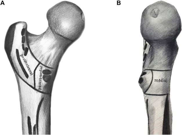
- The posteromedial cortex (calcar). It resists varus and shortening; comminution here marks an unstable pattern.
- The lesser trochanter. A separate lesser trochanter fragment means the medial buttress has gone.
- Reverse obliquity. The line runs from medial proximal to lateral distal, and muscle forces medialise the shaft.
- Subtrochanteric extension. The fracture continues into a high-stress zone below the lesser trochanter (see subtrochanteric fractures).
- The lateral wall. Strictly this selects the implant rather than grading the fracture: a thin wall breaks under a sliding hip screw.
Why does "A2" mean different things in different papers?
The AO/OTA classification of trochanteric fractures (31-A) was revised in the 2018 compendium, and the revision changed what A2 means.
| Older AO/OTA usage | AO/OTA 2018 | |
|---|---|---|
| 31-A1 | Simple two-part pertrochanteric | Pertrochanteric with an intact lateral wall (over 20.5 mm); A1.3 includes a posteromedial fragment |
| 31-A2 | Multifragmentary pertrochanteric (posteromedial comminution) | Multifragmentary pertrochanteric with an incompetent lateral wall (under 20.5 mm) |
| 31-A3 | Reverse oblique or transverse intertrochanteric | Intertrochanteric (reverse obliquity) |
So a fracture with a posteromedial fragment but a thick lateral wall was an A2 in Palm's 2007 series and is an A1.3 today. When a trial says "A2 fractures did better with a nail", check which edition it used before applying it to your patient.
Measuring the wall. The AO reference measures from a point 3 cm below the innominate tubercle of the greater trochanter, upwards at 135° to the fracture line on the AP radiograph. It recommends a traction view in neutral rotation, which is often hard to get before surgery, so the wall should be reassessed on fluoroscopy after closed reduction and before the implant is chosen. A 3D CT shows the planes in detail when the films are unclear.
How do a DHS and a nail hold the fracture?
The dynamic hip screw. A lag screw in the head slides within the barrel of a side plate fixed to the lateral cortex. Under load the head-neck fragment impacts on the shaft in a controlled way, so the fracture is compressed as it heals. That only works if there is a medial cortex to impact on and an intact lateral wall to buttress the construct; without one, the fracture keeps sliding and collapses. In Palm's series of 214 fractures fixed with a 135° sliding hip screw, 74% of the lateral wall fractures happened during the operation itself. The step-by-step technique is in the DHS operative guide.
The cephalomedullary nail. The nail sits inside the canal, with a shorter moment arm than a side plate, and a lag screw or helical blade passes through it into the head. It resists varus collapse even without medial support, while still letting the fracture slide and settle. Its costs are an entry point through the abductor insertion, a longer implant in the canal and, as below, more femoral fractures. The set-up and short-versus-long decision are in the cephalomedullary nail operative guide.
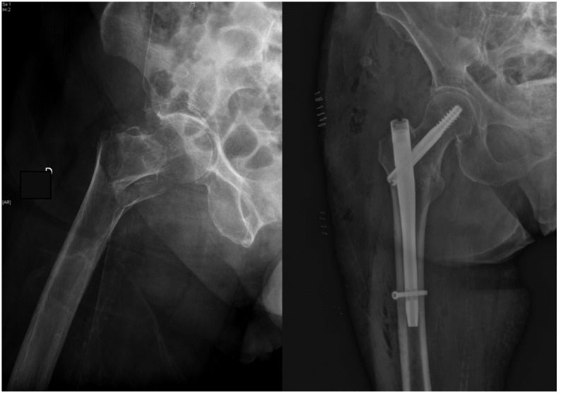
Both implants allow immediate full weight-bearing, so weight-bearing is not a reason to choose one over the other.
What does the evidence show?
The best summary is the 2022 Cochrane review by Lewis and colleagues: 76 randomised and quasi-randomised studies, 10,979 participants, comparing cephalomedullary nails with extramedullary implants, mostly the sliding hip screw.
| Outcome | Nail compared with extramedullary implant | Certainty |
|---|---|---|
| Mortality within 4 months | RR 0.96 (95% CI 0.79–1.18) | Moderate |
| Mortality at 12 months | RR 0.99 (95% CI 0.90–1.08) | Moderate |
| Unplanned return to theatre | RR 1.15 (95% CI 0.89–1.50), imprecise | Low |
| Superficial infection | RR 0.71; one fewer per 303 patients | Moderate |
| Non-union | RR 0.55 | Moderate |
| Intra-operative implant-related fracture | RR 2.94 | Moderate |
| Later implant-related fracture | RR 3.62 | Moderate |
Taking intra-operative and later fractures together, a nail causes one extra implant-related fracture per 67 patients.
Three findings matter most. First, the extra femoral fractures were not reduced by newer nail designs. Second, the subgroup analyses found no difference by fracture stability, by nail length or by nail generation. Third, more than half the studies predate 2010, and few measured what matters to a frail patient: activities of daily living, quality of life, mobility and delirium. The authors' conclusion is that extramedullary devices "yield very similar functional outcomes".
The 2010 version of the review, before the newer trials, was harsher on the early nail: the Gamma nail had more operative and later femoral fractures and a higher reoperation rate than the sliding hip screw, while three trials (394 participants) of the proximal femoral nail found no difference in fixation complications or reoperation.
The largest single trial is Parker's randomised comparison of the sliding hip screw and the Targon PF nail in 600 trochanteric fractures (mean age 82). Operating time was 46 minutes against 49, revision or conversion to arthroplasty 9 cases against 3 (not significant, p = 0.14), and mortality, pain and shortening were similar. Mobility at one year was better with the nail (p = 0.01). Both implants, in the authors' words, "produced comparable results".
When is a nail the right answer?
The case for the nail is strongest in three patterns, and it rests partly on mechanics and pattern-specific series, because the Cochrane subgroups did not show it.
Reverse obliquity. Haidukewych and colleagues found 55 reverse obliquity fractures among 2,472 hip fractures at the Mayo Clinic (2%). The sliding hip screw failed in 9 of 16, and 95° fixed-angle devices did significantly better (p = 0.023). The series held only three cephalomedullary nails and three intramedullary hip screws, so the routine citation of it as evidence for nailing rests on six patients. Two later studies fill that gap. Sadowski's randomised trial of 39 elderly patients with 31-A3 fractures found implant failure or non-union in 7 of 19 with a 95° screw-plate against 1 of 20 with a proximal femoral nail, with shorter operations, fewer transfusions and shorter stays in the nail group. In the Norwegian Hip Fracture Register (2,716 transverse or reverse oblique trochanteric and subtrochanteric fractures), one-year reoperation was 6.4% with a sliding hip screw and 3.8% with a nail (p = 0.011), which is observational evidence.
An incompetent lateral wall. Palm found reoperation within six months in 3% of patients whose lateral wall was intact after surgery against 22% when it was fractured. Wall fractures occurred in 3% of A1 and A2.1 fractures against 31% of A2.2 and A2.3 fractures (older AO usage). Hsu, in 208 fractures fixed with a DHS, derived the 20.5 mm threshold below which a secondary wall fracture became likely. Hsu had no nail arm, so the claim that a nail would have prevented those fractures is an inference, and the threshold was derived and tested on the same patients.
Subtrochanteric extension. NICE and the AAOS agree on an intramedullary device here. A long nail is used so that its tip does not sit as a stress riser at the level of the fracture.
The AAOS rates its recommendation for nailing unstable intertrochanteric fractures as strong, from high and moderate quality studies, while noting that varied classification systems and implant designs make the literature hard to interpret. Put that beside the Cochrane finding of no stability-subgroup difference and the honest position is: the nail is the logical implant for these patterns, and the trials have not proven it superior.
Short nail or long nail?
A short nail (about 170–240 mm) ends in the metaphysis and suits an isolated intertrochanteric fracture with an intact lateral wall. A long nail (about 340–420 mm) ends 3–4 cm above the knee and is used for a thin or broken lateral wall, subtrochanteric extension, reverse obliquity and pathological fractures. A short nail in a wide canal can toggle into varus, so canal width is part of the decision. The Cochrane review found no difference between nail lengths, but only one of its 76 studies was confined to subtrochanteric fractures, so it does not test the patterns for which a long nail is chosen.
Does the screw position matter more than the implant?
Often, yes. Cut-out of the lag screw through the femoral head is the commonest mechanical failure with either implant, and its main controllable cause is the tip-apex distance (TAD).
Baumgaertner studied 198 peritrochanteric fractures fixed with a sliding hip screw. Of 19 failures, 16 were cut-outs. Mean TAD was 24 mm in fractures that healed and 38 mm in those that cut out (p = 0.0001), and none of the 120 screws with a TAD of 25 mm or less cut out. That is a zero numerator over 120: the true risk is low, not nil, and the threshold was derived for a sliding hip screw and applied to nails by extension.
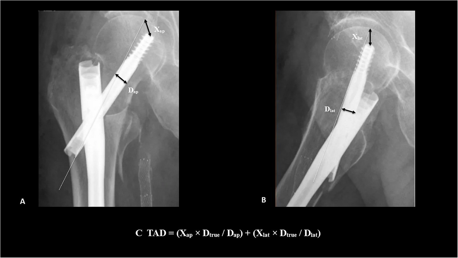
The rest of the checklist. Place the screw centre-centre or inferior-centre on both views; a superior screw carries the highest cut-out risk. Accept anatomic or slight valgus alignment, never varus. Check the anteromedial cortex on the post-reduction films: positive or neutral cortical support is acceptable, and a negative medial buttress predicts collapse and should be re-reduced.
Haidukewych's series makes the point in numbers: 80% of fractures with a poorly placed implant failed, against 26% with an ideally placed one (p = 0.023). Failure after non-anatomic reduction was 46% against 17%, a difference that did not reach significance (p = 0.060). The right implant badly placed still fails.
Where does arthroplasty fit?
Rarely as a first choice. Primary arthroplasty is sometimes proposed for a severely comminuted unstable fracture or a hip with established arthritis, but the calcar is often deficient, early morbidity is higher, and it is not standard practice. Its main role is salvage after failed fixation or cut-out. This is the opposite of the intracapsular fracture, where a displaced fracture in an older patient is replaced (see Garden vs Pauwels and the neck of femur fracture page), and the choice between hemiarthroplasty and total hip replacement is the real decision.
A pathological fracture is a contraindication to a DHS. It needs staging and biopsy planning first, then a long nail or, where bone stock is too poor for fixation, arthroplasty; see pathological fractures of the femur. Every fragility fracture also needs a bone-health assessment and osteoporosis treatment.
DHS vs nail at a glance
| Dynamic hip screw | Cephalomedullary nail | |
|---|---|---|
| Fracture it suits | Stable, two-part, intact lateral wall (2018 31-A1) | Reverse obliquity, subtrochanteric extension, incompetent lateral wall (2018 31-A2), pathological |
| Mortality and function | Probably no difference (Cochrane 2022) | Probably no difference (Cochrane 2022) |
| Femoral fracture | Fewer | RR 2.94 intra-operative, 3.62 later |
| Non-union and superficial infection | More | Fewer (RR 0.55 and 0.71) |
| Exposure | Open lateral approach splitting vastus lateralis | Percutaneous entry at the greater trochanter |
| Weight-bearing | Full, immediately | Full, immediately |
| Guideline position | NICE's default for trochanteric fractures | AAOS strong recommendation for unstable patterns |
In the exam
The examiner wants the stability assessment first, the implant second, and the technical rules for either device last. Name the lateral wall and the reverse obliquity pattern unprompted.
Frequently asked questions
Is a DHS or an intramedullary nail better for an intertrochanteric fracture?
Neither is better across the board. The 2022 Cochrane review found probably little or no difference in mortality and very similar function. Nails gave fewer non-unions and superficial infections but about three times as many implant-related femoral fractures. A DHS suits a stable two-part fracture with an intact lateral wall; a nail is used for reverse obliquity, subtrochanteric extension and a thin or broken lateral wall.
When should a sliding hip screw not be used?
Avoid a sliding hip screw in a reverse oblique fracture, a fracture with subtrochanteric extension, a fracture with a lateral wall under about 20.5 mm or already broken, and a pathological fracture. In Haidukewych's reverse obliquity series the sliding hip screw failed in 9 of 16 hips, and in Palm's series reoperation rose from 3% to 22% when the lateral wall was fractured.
How is lateral wall thickness measured?
On the AP radiograph, start at a point 3 cm below the innominate tubercle of the greater trochanter and measure upwards at 135° to the fracture line. A wall under 20.5 mm is classed as incompetent (AO/OTA 31-A2 since 2018). The AO recommends measuring on a traction view in neutral rotation; because that view is hard to obtain before surgery, reassess the wall on fluoroscopy after closed reduction, before choosing the implant.
Can patients weight-bear fully after a DHS or a nail?
Yes. Both a sliding hip screw and a cephalomedullary nail allow immediate full weight-bearing, so the usual instruction after stable internal fixation is to weight-bear as tolerated from the day after surgery. Restricting weight-bearing is hard for older patients to follow and is not needed with either implant when the reduction and screw position are satisfactory.
What is the tip-apex distance and why does it matter?
The tip-apex distance is the distance from the lag screw tip to the apex of the femoral head on the AP plus the same distance on the lateral, corrected for magnification. In Baumgaertner's 198 fractures none of the 120 screws with a distance of 25 mm or less cut out. It was derived for sliding hip screws and is applied to nails as a reasonable default.
References
- Lewis SR, Macey R, Gill JR, Parker MJ, Griffin XL. Cephalomedullary nails versus extramedullary implants for extracapsular hip fractures in older adults. Cochrane Database Syst Rev 2022;1(1):CD000093. DOI (opens in a new tab)
- Parker MJ, Handoll HH. Gamma and other cephalocondylic intramedullary nails versus extramedullary implants for extracapsular hip fractures in adults. Cochrane Database Syst Rev 2010;(9):CD000093. DOI (opens in a new tab)
- Parker MJ, Bowers TR, Pryor GA. Sliding hip screw versus the Targon PF nail in the treatment of trochanteric fractures of the hip: a randomised trial of 600 fractures. J Bone Joint Surg Br 2012;94(3):391-397. DOI (opens in a new tab)
- Anglen JO, Weinstein JN. Nail or plate fixation of intertrochanteric hip fractures: changing pattern of practice. A review of the American Board of Orthopaedic Surgery Database. J Bone Joint Surg Am 2008;90(4):700-707. DOI (opens in a new tab)
- Palm H, Jacobsen S, Sonne-Holm S, Gebuhr P. Integrity of the lateral femoral wall in intertrochanteric hip fractures: an important predictor of a reoperation. J Bone Joint Surg Am 2007;89(3):470-475. DOI (opens in a new tab)
- Hsu CE, Shih CM, Wang CC, Huang KC. Lateral femoral wall thickness. A reliable predictor of post-operative lateral wall fracture in intertrochanteric fractures. Bone Joint J 2013;95-B(8):1134-1138. DOI (opens in a new tab)
- Meinberg EG, Agel J, Roberts CS, Karam MD, Kellam JF. Fracture and Dislocation Classification Compendium-2018. J Orthop Trauma 2018;32 Suppl 1:S1-S170. DOI (opens in a new tab)
- Haidukewych GJ, Israel TA, Berry DJ. Reverse obliquity fractures of the intertrochanteric region of the femur. J Bone Joint Surg Am 2001;83(5):643-650. DOI (opens in a new tab)
- Sadowski C, Lübbeke A, Saudan M, et al. Treatment of reverse oblique and transverse intertrochanteric fractures with use of an intramedullary nail or a 95° screw-plate: a prospective, randomized study. J Bone Joint Surg Am 2002;84(3):372-381. PubMed (opens in a new tab)
- Matre K, Havelin LI, Gjertsen JE, et al. Sliding hip screw versus IM nail in reverse oblique trochanteric and subtrochanteric fractures. A study of 2716 patients in the Norwegian Hip Fracture Register. Injury 2013;44(6):735-742. DOI (opens in a new tab)
- Baumgaertner MR, Curtin SL, Lindskog DM, Keggi JM. The value of the tip-apex distance in predicting failure of fixation of peritrochanteric fractures of the hip. J Bone Joint Surg Am 1995;77(7):1058-1064. DOI (opens in a new tab)
- O'Connor MI, Switzer JA. AAOS Clinical Practice Guideline Summary: Management of Hip Fractures in Older Adults. J Am Acad Orthop Surg 2022;30(20):e1291-e1296. DOI (opens in a new tab)
Study the full topic
Included in Pro
Go deeper with Pro
- Operative guide: dynamic hip screw for intertrochanteric fracture
- Operative guide: cephalomedullary nail for intertrochanteric fracture
Share this article
Cite this article
OrthoVellum Editorial Team. DHS or Cephalomedullary Nail? Intertrochanteric Fracture Fixation Decoded [Internet]. OrthoVellum; 2026 Nov 6 [cited 2026 Nov 6]. Available from: https://www.orthovellum.com/blog/dhs-vs-intramedullary-nail
About this content
Prepared by the OrthoVellum Editorial Team from cited sources, under our editorial policy.
For education and exam preparation; not medical advice or a substitute for clinical judgement and local guidance.
Spotted an error? Tell us; every report is read.
Get new articles by email
New clinical explainers, landmark-trial summaries and exam guidance from the OrthoVellum blog. No account needed; unsubscribe from any email.