Neer Classification of Proximal Humerus Fractures: Parts, Displacement Rules and Interobserver Reality
The Neer classification for clinicians: Codman's four segments, the 1 cm and 45° rule, why observers disagree, and what the part count changes in treatment.
By OrthoVellum Editorial TeamPublished 12 min read
Educational content for clinicians, not medical advice. Editorial policy

Key points
- Neer counts displaced segments, not fracture lines: a segment is a 'part' only if it is separated by more than 1 cm or angulated more than 45°. Neer set the thresholds when a journal editor insisted on defined criteria, and he accepted they were somewhat arbitrary.
- Interobserver agreement is moderate at best. A systematic review of 11 observer studies found mean kappa values of 0.17–0.52, and experience, CT and simplifying the system did not reliably improve it.
- Formal training does help: in a randomised trial, kappa rose from 0.27 to 0.62 after two sessions, and to 0.79 among specialists.
- Surgeons agree more on treatment than on the category, so a disputed part count often does not change the plan.
- Part count is a weak guide to the blood supply. Calcar length and the medial hinge (Hertel) predicted intraoperative head ischaemia better than the number of fragments.
On this page11 sections
Every displaced proximal humerus fracture is described in parts: "a three-part fracture in a 70-year-old", "a four-part with a head split". The Neer classification supplies that language, and it is the system examiners expect. The question is how much weight a part count can carry. The short answer: Neer's four-segment idea explains how these fractures displace and groups them usefully, but observers agree on the category only moderately, so the count should start the treatment discussion, not settle it. This post covers the parts, the displacement rule, the reliability data and the decisions that follow. The clinical overview is on the proximal humerus fractures topic page.
What are the four parts?
Neer built on an observation of Codman's: every proximal humerus fracture is made up of four segments, the articular segment (the head), the greater tuberosity, the lesser tuberosity and the shaft. Neer's 1970 paper drew on the radiographs and operative findings of 300 fractures he treated in New York between 1953 and 1967 (Carofino and Leopold's summary of the original).
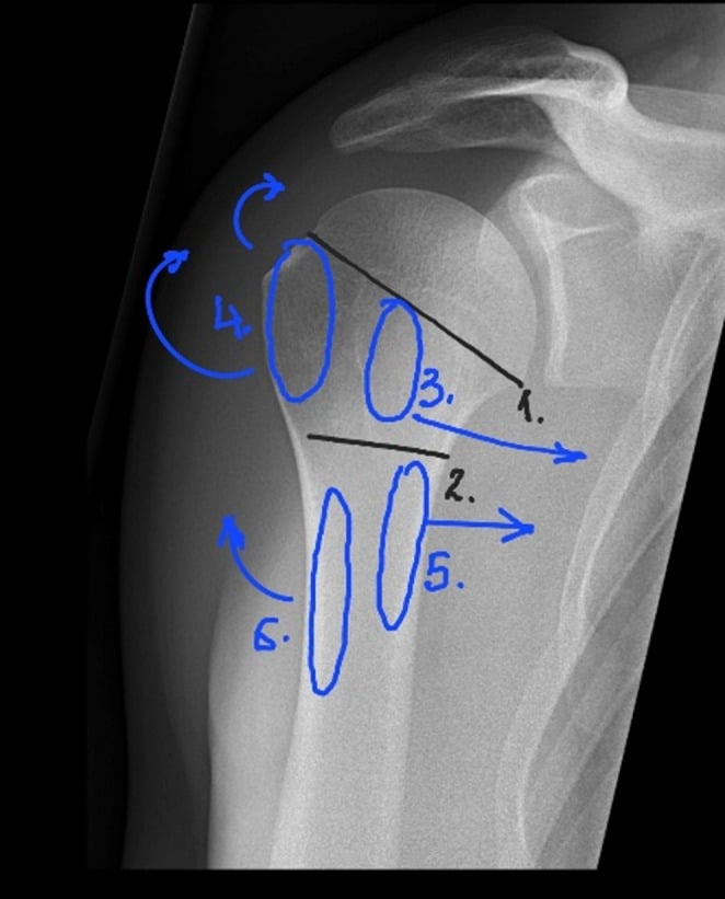
The segments matter because of what is attached to them. The muscle attachments predict the direction of displacement, which is how you read a film:
- Greater tuberosity: supraspinatus, infraspinatus and teres minor; supraspinatus pulls it superiorly.
- Lesser tuberosity: subscapularis, which pulls it into internal rotation.
- Shaft: pectoralis major pulls it medially, and the deltoid may displace it laterally.
- Head: no muscle of its own; its position follows the impact or the direction of dislocation, and it carries the blood-supply question.
How does the Neer classification count parts?
A segment is a separate part only if it is displaced by more than 1 cm or angulated by more than 45°. You count displaced parts, not fracture lines or fragments. A shattered-looking film in which nothing passes the threshold is a one-part fracture, and a comminuted surgical neck with one displaced greater tuberosity is two-part.
| Neer category | What is displaced | What it looks like |
|---|---|---|
| One-part | Nothing meets the threshold | Any number of fracture lines, however comminuted it looks |
| Two-part | One segment: surgical neck, anatomical neck, greater tuberosity or lesser tuberosity | Surgical neck is the common one; anatomical neck is rare |
| Three-part | Surgical neck plus one tuberosity | The tuberosity left attached rotates the head |
| Four-part | Both tuberosities, head and shaft | Classic form: head laterally displaced and out of contact with the glenoid |
| Valgus-impacted four-part | Head driven down into valgus between splayed tuberosities | Head keeps contact with the glenoid; added by Neer in 2002 |
The full system also has separate groups for fracture-dislocations (anterior and posterior) and articular-surface fractures (head-splitting and impression fractures). Neer separated them because they carry a different prognosis and treatment: dislocations are more severe injuries, with more avascular necrosis and heterotopic ossification. With those groups, the scheme has 16 categories, as numbered by Sidor and colleagues.
The three-part rotation. In a three-part fracture the tuberosity left attached to the head keeps its cuff, so the head is pulled into a rotational deformity by whichever tendons remain. That is why a three-part fracture can look odd on an AP alone, and why the shoulder fracture-dislocation and articular groups need the axillary view before anyone counts parts.
Where did 1 cm and 45° come from?
Not from outcome data. When the paper went to the Journal of Bone and Joint Surgery, the editor, Thornton Brown, insisted on defined criteria for when a segment counts as displaced. Neer set 1 cm of separation and 45° of angulation in response, and he later acknowledged that the numbers were somewhat arbitrary while accepting that some definition was needed for the system to be used at all. The thresholds have become the most quoted part of the classification. In 2002 Neer revisited the system in a paper titled "Four-segment classification of proximal humeral fractures: purpose and reliable use", which is where the valgus-impacted four-part category was added.
Measuring is harder than it sounds. The PROFHER investigators, preparing to classify 250 trial radiographs, concluded that a centimetre cannot be measured accurately on a plain film without a calibration object at the fracture, and that the PACS scale is lost when images are anonymised. They noted that the stems of the standard L and R cassette markers are 9 mm long, a rough ruler on some films. Angulation depends on the projection and on how clearly the fragment edges show.
The greater tuberosity: the exception to the rule
The 1 cm rule defines a displaced part; it is not the threshold for fixing an isolated greater tuberosity. The topic page gives the fixation threshold as over 5 mm, and over 3 mm in an overhead athlete or manual worker, because even small superior displacement causes subacromial impingement and loss of external rotation. A greater tuberosity displaced 7 mm is a one-part fracture by Neer's definition and still a candidate for screws or sutures. Calling it undisplaced is the commonly missed error.
How reliable is the Neer classification?
Moderately at best, and the finding has been repeated for three decades. Kappa runs from 0 (agreement no better than chance) to 1 (complete agreement); the studies below describe values around 0.4–0.5 as fair to moderate.
| Study | Material and observers | Interobserver agreement (kappa) |
|---|---|---|
| Sidor 1993 | 50 fractures, full trauma series; shoulder specialist, traumatologist, skeletal radiologist, two residents | 0.48 and 0.52 (two viewings); all five agreed on 32% and 30% of fractures |
| Siebenrock and Gerber 1993 | 95 fractures; five shoulder surgeons | Neer 0.40; AO/ASIF 0.53 |
| Bernstein 1996 | 20 fractures, radiographs then radiographs plus CT; four observers | 0.52 radiographs; 0.50 with CT |
| Majed 2011 | 96 complex fractures as 3D-printed models from CT; four senior observers | Neer 0.33; AO 0.11; Codman-Hertel 0.44 |
| Brorson 2008 (systematic review) | 11 observer studies, 88 observers, 468 cases | Mean values 0.17–0.52 |
| Handoll 2016 (PROFHER) | 250 trial fractures; two trained consultant shoulder surgeons | 0.48 (68% agreement) |
Intraobserver agreement is better but still imperfect. In Sidor's study it ranged from 0.83 for the shoulder specialist to 0.50 for the radiologist, with a mean of 0.66.
The four-part boundary is the weak point. In PROFHER the two independent raters disagreed most on the four-part category: one called 30 fractures four-part and the other 4. After a consensus meeting they settled on 11 (4.4%), while recruiting surgeons had recorded 64 (25.6%) as involving both tuberosities. "Both tuberosities involved" and "four-part" are not the same claim, and the gap shows how loosely the term is used in clinic.
Does CT, a simpler scheme or experience fix it?
Mostly not.
- CT. In Bernstein's study, adding CT raised intraobserver reliability slightly (0.64 to 0.72) but not interobserver agreement (0.52 to 0.50). The authors concluded that even experts cannot uniformly agree on which fragments are fractured. Majed's group gave senior surgeons physical models printed from CT, and Neer agreement was still only 0.33.
- Fewer categories. Sidor collapsed the 16 categories to six, and agreement did not significantly improve. Bernstein ignored displacement and scored only which segments were fractured, and interobserver kappa moved from 0.52 to 0.56.
- Experience. The systematic review found that selecting experienced observers did not improve agreement, and the authors could not support the belief that experts disagree less than non-experts. Individual studies show at most a small edge: in Sidor's series attendings scored 0.52 against 0.48 for residents.
Training does. In a randomised trial, Brorson and colleagues gave 14 doctors either two training sessions or none before they classified 42 pairs of radiographs. In the trained group mean kappa rose from 0.27 to 0.62, and among specialists from 0.30 to 0.79. One trial of 42 cases is a small base, but it was the only randomised test of an intervention the systematic review found, and it worked.
If surgeons disagree on the category, does it matter?
Less than the kappa values suggest. Brorson's 2012 study asked five experienced shoulder surgeons to classify 193 consecutive fractures twice, three months apart, and to recommend non-operative treatment, a locking plate or hemiarthroplasty for each. Agreement on the treatment (kappa 0.48 and 0.52) was significantly higher than on the Neer category (0.33 and 0.36). Observers changed category in 36% of repeat readings, but changed their treatment recommendation in only 34% of those. Agreement on treatment still did not exceed moderate.
The PROFHER analysis points the same way. Neither of the trial's subgroup analyses, by tuberosity involvement or by Neer category, supported choosing surgery on the basis of fracture type for the Oxford Shoulder Score; the trial itself is covered in our post on the PROFHER trial. The practical reading: surgeons decide on a fuller picture than the part count, and the count is a summary of that picture rather than its basis.
Does the part count predict avascular necrosis?
Neer expected it to. He reasoned that in four-part fractures the blood supply to the head had been severed, reported avascular necrosis in more than half of them and advocated arthroplasty (as summarised by Carofino and Leopold). Later work has not confirmed that a four-part pattern on its own predicts necrosis.
Two findings explain the gap.
- The head's main supply. The textbook vessel, the arcuate artery from the anterior circumflex humeral artery, is disrupted in about 80% of proximal humerus fractures, yet osteonecrosis is uncommon. Cadaver MRI work by Hettrich and colleagues, in uninjured shoulders, found that the posterior circumflex humeral artery supplies about 64% of the head, which may explain why a head can survive an obviously disrupted arcuate artery. The detail is on the proximal humerus fractures page.
- What predicts ischaemia. Hertel assessed perfusion at operation in 100 intracapsular fractures. The best predictors were a calcar (dorsomedial metaphyseal head extension) shorter than 8 mm (accuracy 0.84), a disrupted medial hinge (0.79) and the basic fracture pattern, particularly the anatomical neck. Neer's own variables did worse: a four-fragment fracture had an accuracy of 0.67, head angulation over 45° 0.62 and tuberosity displacement over 10 mm 0.61. Combining anatomical neck, short calcar and disrupted hinge gave positive predictive values of up to 97%.
Hold Hertel loosely as well. The perfusion test was drill-hole backflow at surgery, the series came from one institution, and ischaemia is not necrosis: many ischaemic heads revascularise, and Carofino and Leopold note that these markers of poor perfusion did not predict later necrosis either. Read the calcar and the hinge as reasons to plan for possible replacement, as the humeral head fractures page puts it, not as a prediction that the head will collapse.
Neer or AO/OTA?
The AO/OTA system is more comprehensive. In its 2018 revision (opens in a new tab) it absorbed Neer's part count: 11-A is extra-articular, unifocal and two-part, 11-B extra-articular, bifocal and three-part, and 11-C articular or four-part. It is not clearly more reproducible. Siebenrock and Gerber found the earlier AO system slightly ahead at the main level (kappa 0.53 against 0.40 for Neer), but called both fair or poor, and agreement worsened as AO groups and subgroups were added. Majed's model study gave AO the lowest agreement of the four systems tested (0.11). Know both. Use Neer to talk about displacement and treatment, and give the AO code when a trial or registry asks for it.
How does the classification feed the decision?
The part count is weighed against age, functional demand, comorbidity and bone quality, and the pattern details Neer does not grade: calcar length, the medial hinge, varus or valgus head position and articular involvement. Get a CT for three- and four-part fractures and any head-split pattern. The topic page's algorithm runs as follows.
| Pattern and patient | Usual direction | The deciding detail |
|---|---|---|
| One-part, any age | Sling and early motion | Check the greater tuberosity against the 5 mm threshold |
| Two-part surgical neck, older or low-demand | Non-operative is the default | PROFHER found no benefit from surgery in displaced surgical-neck fractures |
| Two-part surgical neck, young or active | Plate or intramedullary nail | Functional demand; diaphyseal extension favours the nail |
| Two-part greater tuberosity over 5 mm | Screws or suture fixation | Over 3 mm in overhead athletes and manual workers |
| Three-part, young or active | Locking plate | Restore the medial column with a calcar screw |
| Valgus-impacted four-part | Fixation if the medial hinge is intact | Hinge and calcar predict a perfused head |
| Displaced three- or four-part, older patient | Reverse shoulder arthroplasty if surgery is chosen | Bone quality and tuberosity healing |
| Head-split or unsalvageable head | Arthroplasty, usually reverse | The articular segment cannot be rebuilt |
Two cautions travel with the table. PROFHER randomised adults with displaced fractures involving the surgical neck; fracture-dislocations and open fractures were excluded, and the Cochrane review records insufficient evidence for patients under 60, high-energy injuries, two-part tuberosity fractures, fracture-dislocations and articular-surface fractures. And surgery in PROFHER meant fixation or humeral head replacement, not reverse arthroplasty, so the preference for a reverse prosthesis in older patients rests on other evidence. The fixation technique is in the locking plate operative guide, and the arthroplasty side on the reverse total shoulder arthroplasty topic page.
In the exam
Examiners want the parts counted correctly, the threshold stated, the weakness acknowledged and a decision that does not rest on the count alone. For learning the categories, see how to memorise orthopaedic classifications.
Frequently asked questions
What are the four parts in the Neer classification?
The four segments described by Codman: the articular segment (humeral head), the greater tuberosity, the lesser tuberosity and the humeral shaft. A segment counts as a separate part only when it is displaced more than 1 cm or angulated more than 45°. A one-part fracture can therefore have several fracture lines, as long as nothing meets the displacement threshold.
What is the difference between a three-part and a four-part proximal humerus fracture?
In a three-part fracture the surgical neck and one tuberosity are displaced; the other tuberosity stays attached to the head and rotates it. In a four-part fracture both tuberosities, the head and the shaft are all displaced, and in the classic form the head is out of contact with the glenoid. Neer added the valgus-impacted four-part fracture as a separate category in 2002.
How reliable is the Neer classification?
Moderately at best. Sidor and colleagues found mean interobserver kappa of 0.48 to 0.52, and a systematic review of 11 observer studies reported mean values from 0.17 to 0.52. Experienced observers, CT and simplifying the categories did not reliably improve agreement. A randomised trial found that formal training raised kappa from 0.27 to 0.62.
Is a displaced greater tuberosity judged by the 1 cm rule?
Not for treatment. Neer's 1 cm threshold defines a displaced part, but an isolated greater tuberosity fragment is usually fixed at a much smaller displacement: over 5 mm, or over 3 mm in an overhead athlete or manual worker. Small superior displacement causes subacromial impingement and loss of external rotation, so the fragment should not be dismissed as undisplaced.
References
- Neer CS 2nd. Displaced proximal humeral fractures. I. Classification and evaluation. J Bone Joint Surg Am 1970;52(6):1077-1089. DOI (opens in a new tab)
- Neer CS 2nd. Four-segment classification of proximal humeral fractures: purpose and reliable use. J Shoulder Elbow Surg 2002;11(4):389-400. DOI (opens in a new tab)
- Carofino BC, Leopold SS. Classifications in brief: the Neer classification for proximal humerus fractures. Clin Orthop Relat Res 2013;471(1):39-43. DOI (opens in a new tab)
- Sidor ML, Zuckerman JD, Lyon T, et al. The Neer classification system for proximal humeral fractures: an assessment of interobserver reliability and intraobserver reproducibility. J Bone Joint Surg Am 1993;75(12):1745-1750. DOI (opens in a new tab)
- Siebenrock KA, Gerber C. The reproducibility of classification of fractures of the proximal end of the humerus. J Bone Joint Surg Am 1993;75(12):1751-1755. DOI (opens in a new tab)
- Bernstein J, Adler LM, Blank JE, et al. Evaluation of the Neer system of classification of proximal humeral fractures with computerized tomographic scans and plain radiographs. J Bone Joint Surg Am 1996;78(9):1371-1375. DOI (opens in a new tab)
- Majed A, Macleod I, Bull AM, et al. Proximal humeral fracture classification systems revisited. J Shoulder Elbow Surg 2011;20(7):1125-1132. DOI (opens in a new tab)
- Brorson S, Hróbjartsson A. Training improves agreement among doctors using the Neer system for proximal humeral fractures in a systematic review. J Clin Epidemiol 2008;61(1):7-16. DOI (opens in a new tab)
- Brorson S, Bagger J, Sylvest A, Hrøbjartsson A. Improved interobserver variation after training of doctors in the Neer system: a randomised trial. J Bone Joint Surg Br 2002;84(7):950-954. DOI (opens in a new tab)
- Brorson S, Olsen BS, Frich LH, et al. Surgeons agree more on treatment recommendations than on classification of proximal humeral fractures. BMC Musculoskelet Disord 2012;13:114. DOI (opens in a new tab)
- Handoll HHG, Brealey SD, Jefferson L, et al. Defining the fracture population in a pragmatic multicentre randomised controlled trial: PROFHER and the Neer classification of proximal humeral fractures. Bone Joint Res 2016;5(10):481-489. DOI (opens in a new tab)
- Hertel R, Hempfing A, Stiehler M, Leunig M. Predictors of humeral head ischemia after intracapsular fracture of the proximal humerus. J Shoulder Elbow Surg 2004;13(4):427-433. DOI (opens in a new tab)
Study the full topic
Included in Pro
Go deeper with Pro
- Operative guide: proximal humerus ORIF with a locking plate
- Operative guide: reverse shoulder arthroplasty for proximal humerus fracture
Share this article
Cite this article
OrthoVellum Editorial Team. Neer Classification of Proximal Humerus Fractures: Parts, Displacement Rules and Interobserver Reality [Internet]. OrthoVellum; 2026 Nov 4 [cited 2026 Nov 4]. Available from: https://www.orthovellum.com/blog/neer-classification-proximal-humerus-fractures
About this content
Prepared by the OrthoVellum Editorial Team from cited sources, under our editorial policy.
For education and exam preparation; not medical advice or a substitute for clinical judgement and local guidance.
Spotted an error? Tell us; every report is read.
Get new articles by email
New clinical explainers, landmark-trial summaries and exam guidance from the OrthoVellum blog. No account needed; unsubscribe from any email.