Bankart | Hill-Sachs | Bone Loss | Bankart vs Latarjet
- Bankart lesion (labral tear) occurs in 90%+ of traumatic anterior dislocations
- Hill-Sachs lesion (humeral head defect) in 65-70% of first dislocations
- Glenoid bone loss has TWO thresholds: above 20-25% redislocation rises sharply, but function is already measurably worse from 13.5% even in shoulders that never redislocate
- Engaging Hill-Sachs increases recurrence - Latarjet or remplissage needed
- Age is strongest predictor of recurrence - 67% in under 20 years old
- “ISIS score predicts recurrence - greater than 6 suggests bony procedure
- “On-track vs off-track concept for Hill-Sachs engagement
- “Latarjet provides bone augmentation, sling effect, and capsular repair
- “Arthroscopic Bankart has higher recurrence than open in young athletes
Overview and Epidemiology
The glenohumeral joint is the most commonly dislocated major joint, with an incidence of 24 per 100,000 per year. Anterior dislocations make up 95% of the total and posterior dislocations 2-4%. The peak is in males aged 15-25, and contact sports are overrepresented.
Mechanism. A force applied to the abducted, externally rotated arm, a direct blow to the posterior shoulder, or a fall on the outstretched hand. In sport the injury comes from tackling and overhead throwing. In the hyperlax shoulder the dislocation may be atraumatic.
Recurrence. Age at first dislocation is the strongest predictor. Under 20 years the recurrence rate is 67%; between 20 and 40 it is 30-40%; over 40 it falls to 10-15%, though the older patient carries a higher risk of rotator cuff tear. Young athletes in contact sports have the highest recurrence after conservative treatment, and young age with contact sport indicates early surgical consideration.
Pathophysiology and Mechanisms
Static restraints. The glenohumeral ligaments are the primary static stabilisers. The inferior glenohumeral ligament (IGHL) is the most important: its anterior band resists anterior translation with the arm in abduction and external rotation. The middle glenohumeral ligament has variable anatomy and resists anterior translation in the mid-range, and the superior glenohumeral ligament resists inferior translation. The labrum deepens the glenoid by 50% and is the attachment for the ligaments.
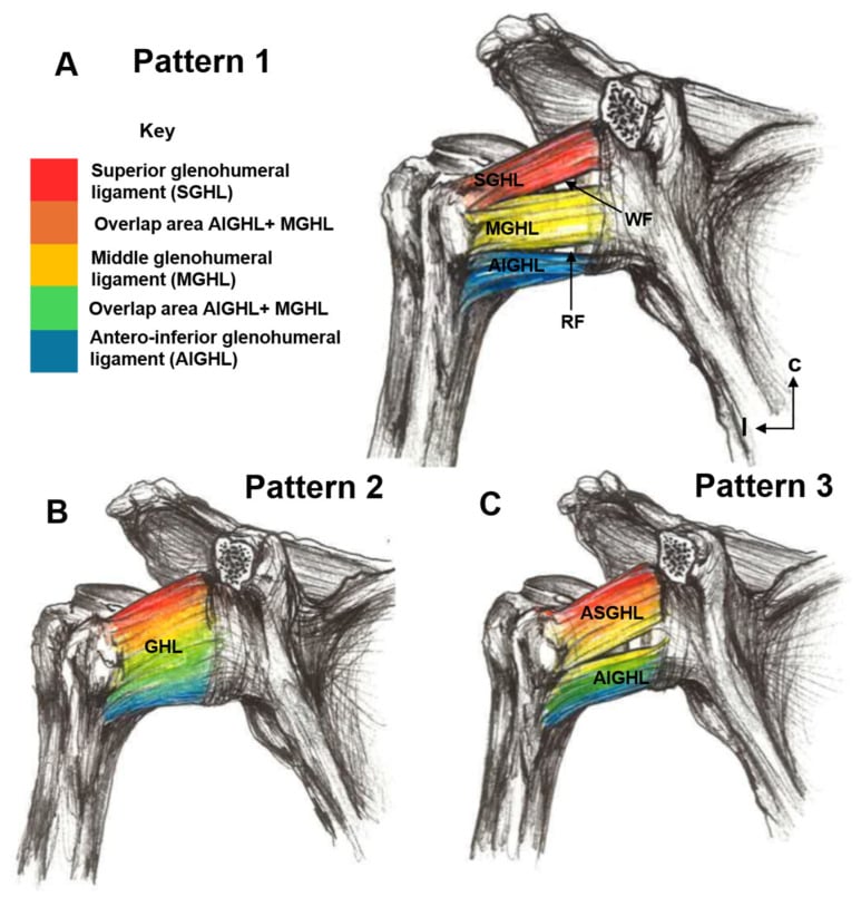
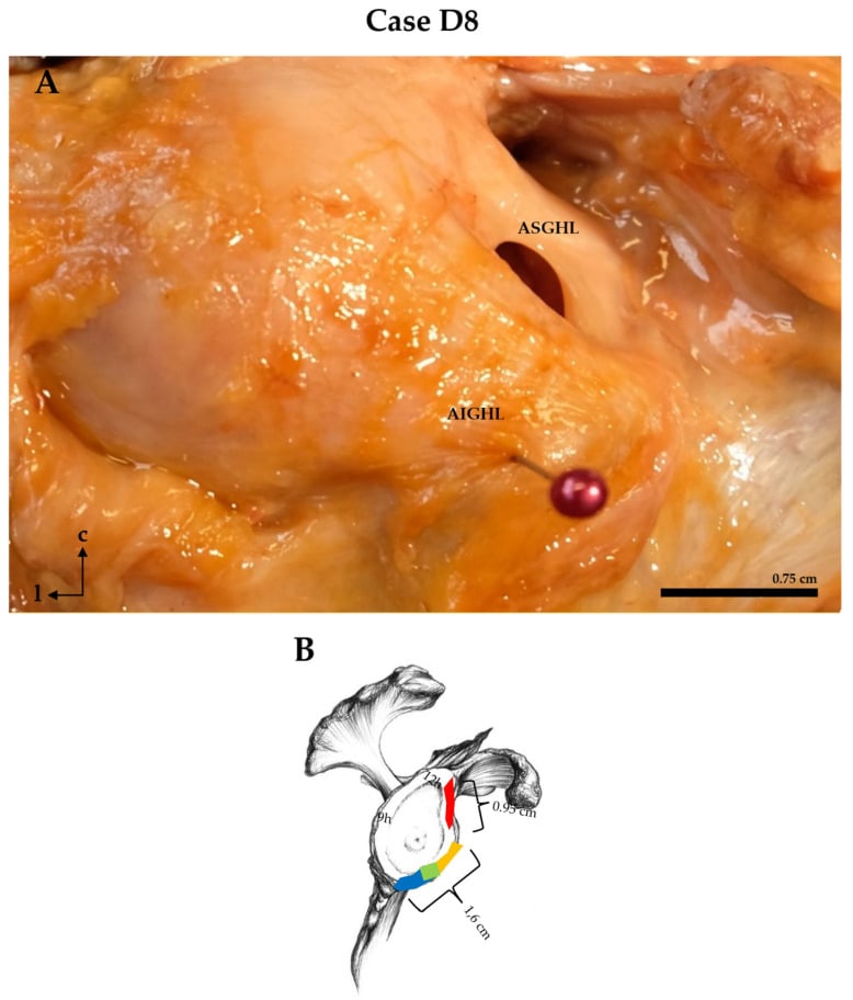
Dynamic restraints. The rotator cuff compresses the humeral head into the glenoid (concavity-compression), and subscapularis is the most important dynamic anterior stabiliser. The long head of biceps is a secondary restraint, and the periscapular muscles maintain the position of the glenoid.
The lesions. The key pathological lesions of anterior instability are:
- Bankart lesion: anteroinferior labral tear with avulsion of the IGHL, present in over 90%
- Bony Bankart: glenoid rim fracture with labral avulsion
- Hill-Sachs lesion: posterolateral humeral head compression fracture, present in 65-70% of first dislocations
- HAGL: humeral avulsion of the glenohumeral ligaments, less common
- ALPSA: anterior labroligamentous periosteal sleeve avulsion, with the labrum displaced medially
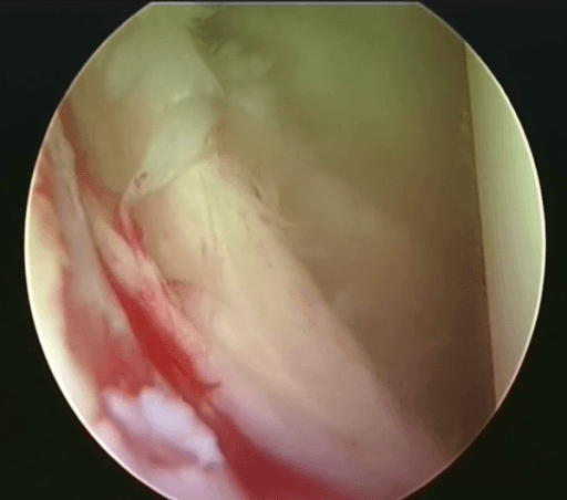
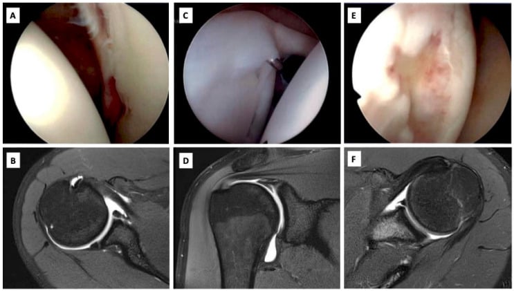
Classification Systems
Glenoid Bone Loss Measurement
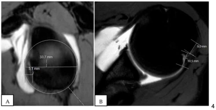
The best-fit circle. A 3D CT reconstruction with the humeral head subtracted is viewed en face. A circle is fitted to the intact inferior two-thirds of the glenoid, whose margin is reliably circular, and the missing segment is expressed as a percentage of that circle. The full measurement technique, its alternatives and the disagreement between linear, area and surface-based methods are set out in glenoid bone loss in instability.
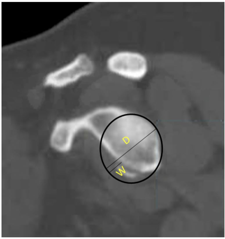
Treatment thresholds. The percentage measured places the shoulder in one of three bands:
- Less than 13.5%: arthroscopic Bankart likely sufficient
- 13.5-20%, the "subcritical" band: Bankart holds in most, but function is measurably worse, so augmentation is increasingly favoured, especially with an off-track Hill-Sachs, contact sport or a high ISIS
- Greater than 20-25%: Latarjet or bone graft
Why Three Different Numbers Are Quoted for the Same Decision
The 13.5%, 20% and 25% figures are not competing estimates of one threshold. They answer two different questions, and which one applies depends on what you are protecting the patient from.
In a cohort of 73 shoulders undergoing isolated arthroscopic Bankart repair (evidence level 3: a single-institution cohort of 72 patients with a mean follow-up of 48 months), divided into quartiles by bone loss (mean 2.8%, 10.4%, 16.1% and 24.5%), the two outcomes separated:
- Redislocation rose sharply only in the top quartile. Failure was 7.3% across the lowest three quartiles combined against 27.8% above roughly 20%. That is where the classic "critical" 20-25% figure comes from, and for the question "will the repair hold?" it is the right number.
- Patient-reported function deteriorated much earlier. WOSI worsened significantly at every quartile step, and at 13.5% it crossed into the range consistent with a poor clinical outcome. Critically, this held even among shoulders that never redislocated: increasing bone loss predicted worse function independently of recurrence.
The practical consequence is that "the repair did not fail" is not the same as "the patient did well". Counting only redislocation makes 25% look like the point at which bone loss starts to matter; asking the patient makes it about 13.5%. A 15-25% defect is therefore no longer a comfortable "grey zone where you might consider" something, and measurement method matters at these magnitudes because a few percentage points now change the recommendation.
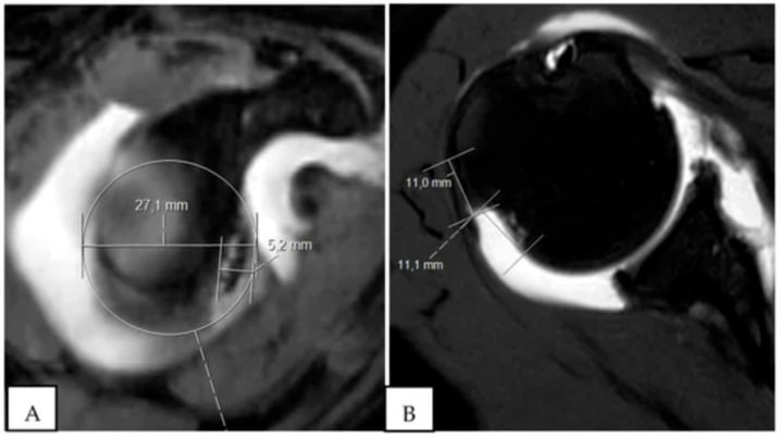
Clinical Assessment
History. Establish the mechanism (an abduction-external rotation position or a direct blow), whether the shoulder reduced spontaneously or needed reduction, the number of dislocations and their pattern, the level of sport (contact, overhead, competitive), and the age at first dislocation, which predicts recurrence.
Examination. The apprehension-relocation test is the most specific clinical test for anterior instability. With the patient supine, abduct the arm to 90° and externally rotate: the test is positive when the patient is apprehensive, not merely in pain. Then apply a posterior force to the humeral head; relief of the apprehension is a positive relocation test. Complete the examination with:
- Anterior load and shift, which grades translation
- Sulcus sign for inferior laxity, positive if greater than 2cm
- Beighton score for generalised laxity
The acute dislocation. The arm is held in external rotation and abduction, the deltoid contour is lost, and the humeral head is palpable anteriorly. After reduction, assess for neurovascular injury (especially the axillary nerve), rotator cuff integrity in patients over 40, and range of motion.
The axillary nerve is the commonest nerve injured in anterior dislocation. It is tethered as it passes around the surgical neck of the humerus and through the quadrilateral space, so it is stretched when the head dislocates anteroinferiorly. Test and document it before and after every reduction: sensation over the "regimental badge" area (lateral/upper deltoid) is the practical screen, since deltoid motor testing is unreliable in an acutely painful shoulder. Most injuries are a neurapraxia with a high rate of spontaneous recovery over weeks to a few months; a deficit persisting beyond about 3 months warrants nerve conduction studies/EMG and consideration of exploration. Documenting the pre-reduction status is medicolegally essential: a deficit found only afterwards must not be wrongly attributed to the reduction.
The older patient. In patients over about 40, a dislocation is more likely to combine dislocation, rotator cuff tear and neurological (axillary nerve or brachial plexus) injury, the so-called "terrible triad" of the shoulder. Persistent weakness after reduction in an older patient should not be assumed to be nerve injury alone: actively exclude a rotator cuff tear with ultrasound or MRI as well.
Investigations
Radiographs. The standard views, and what each one shows:
- True AP (Grashey view): glenohumeral alignment
- Axillary lateral: the relationship of the glenoid and humeral head; essential
- Scapular Y: confirms the direction of dislocation
- West Point view: the anteroinferior glenoid (Bankart)
- Stryker notch view: the Hill-Sachs lesion
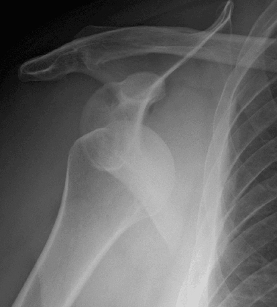
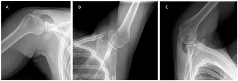
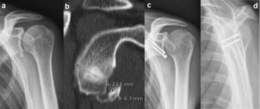
What the plain films tell you. A Hill-Sachs visible on the AP indicates a significant lesion, one that is engaged in external rotation, and glenoid loss visible on the AP suggests bone loss greater than 20%. Either finding prompts CT quantification.
CT with 3D reconstruction is the gold standard for quantifying bone loss and essential for surgical planning. Glenoid loss is measured by the best-fit circle method on the en face view, as described under Classification. The Hill-Sachs is measured for width, depth and location, and glenoid version is checked to ensure the glenoid is not retroverted.
MRI shows the soft-tissue lesions: the Bankart, ALPSA and HAGL lesions described above, GLAD (glenoid labrum articular disruption), and the rotator cuff, which matters especially in patients over 40. MR arthrography is superior for labral pathology.
Acute Reduction Techniques
The acute dislocation must be reduced before any instability work-up, and naming and contrasting the reduction manoeuvres, with the safe principles around them, is a clinical and OSCE favourite. Provide adequate analgesia first, either intravenous sedation and analgesia or intra-articular local anaesthetic, which is effective and avoids the risks of sedation. Document neurovascular status, the axillary nerve in particular, before and after, and obtain a post-reduction radiograph to confirm reduction and exclude an iatrogenic fracture.
- Principle
- Longitudinal traction on the arm against countertraction from a sheet around the chest
- Notes
- Effective and widely used; needs an assistant
- Principle
- Patient prone, arm hanging over the edge with a hanging weight; gravity fatigues spasm over 15-20 min
- Notes
- Gentle, minimal force; slow
- Principle
- Rotate the inferior scapular tip medially (often with the patient prone/seated) to reposition the glenoid
- Notes
- High success, low complication; can combine with gentle traction
- Principle
- Seated, arm adducted on the shoulder; analgesic massage of biceps/deltoid/trapezius with scapular positioning - no traction
- Notes
- Best for cooperative, atraumatic/recurrent cases
- Principle
- Abduct and externally rotate the arm overhead, then push the humeral head back with the thumb
- Notes
- Low force, low complication
- Principle
- Longitudinal traction with the arm at the side plus gentle vertical oscillations while abducting and then externally rotating
- Notes
- Single operator, quick, well tolerated
- Principle
- Lever technique: traction, external rotation, adduction, then internal rotation
- Notes
- Historically associated with humeral neck fracture and nerve/vascular injury if forced - use gently or prefer atraumatic methods
Forceful leverage, classically the original Kocher technique, risks iatrogenic proximal humeral fracture, axillary nerve injury and even vascular injury, especially in older osteopenic patients. Prefer low-force traction, scapular or gravity methods, and ensure muscle relaxation.
Differential Diagnosis
The painful or unstable-feeling shoulder in a young patient has several mimics. The exam trap is labelling everything "anterior instability" without distinguishing the direction and the driver, traumatic structural against atraumatic or volitional.
- Direction / Pattern
- Unidirectional anterior
- Discriminating Features
- Discrete injury in abduction-external rotation, positive apprehension-relocation, Bankart on imaging
- Key Investigation
- MRA plus CT for bone loss
- Direction / Pattern
- Unidirectional posterior
- Discriminating Features
- Seizure, electrocution or posteriorly-directed load; pain on flexion-adduction-IR; jerk test positive
- Key Investigation
- Axillary view, CT/MRI (reverse Bankart)
- Direction / Pattern
- Atraumatic, two or more directions
- Discriminating Features
- Generalised laxity (Beighton), bilateral, positive sulcus sign, no discrete trauma
- Key Investigation
- Clinical; MRI shows capsular redundancy
- Direction / Pattern
- Often muscle-patterning
- Discriminating Features
- Voluntary or positional, abnormal muscle recruitment, psychosocial factors
- Key Investigation
- Dynamic EMG; clinical observation
- Direction / Pattern
- Stable joint
- Discriminating Features
- Deep pain, catching with overhead loading, positive O'Brien/dynamic labral shear, no true apprehension
- Key Investigation
- MRA
- Direction / Pattern
- Stable joint
- Discriminating Features
- Weakness and night pain after dislocation in older patients; pseudo-instability
- Key Investigation
- MRI / ultrasound
The Stanmore (polar-type) triangle frames instability as Type I (traumatic structural), Type II (atraumatic structural) and Type III (muscle-patterning, non-structural). It is high-yield because surgery helps Type I and selected Type II but typically harms Type III: operating on a muscle-patterning shoulder makes it worse.
Management Algorithm
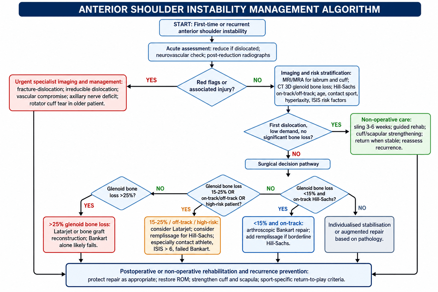
The first dislocation. After closed reduction under sedation, post-reduction radiographs and assessment of the axillary nerve, immobilise in a sling. MRI assesses the labrum and rotator cuff; CT with 3D reconstruction quantifies bone loss and is essential for surgical planning.
Conservative treatment. A sling for 3-6 weeks, then progressive range of motion with rotator cuff and periscapular strengthening. This may be appropriate for the patient over 40, the low-demand patient, the shoulder with no bone loss, and the non-contact athlete.
Early surgery. Consider it in the patient under 20, the contact athlete, the shoulder with significant bone loss and the in-season athlete. Stabilisation reduces recurrence from 67% to less than 10%.
Recurrent instability. Quantify the bone loss first: CT with 3D reconstruction, glenoid loss by the best-fit circle, and the Hill-Sachs classified on-track or off-track. The procedure then follows the bone loss and the risk profile. The table uses 15% and 25% as its working cut-offs; the reasoning behind them, the 13.5% threshold for function and the 20-25% threshold for redislocation, is set out under Classification.
- Bone Loss
- Minimal
- Recommended Procedure
- Conservative
- Key Consideration
- Rehab, reassess if recurrent
- Bone Loss
- Less than 15%
- Recommended Procedure
- Arthroscopic Bankart
- Key Consideration
- Higher recurrence in contact sports
- Bone Loss
- 15-25%
- Recommended Procedure
- Consider Latarjet
- Key Consideration
- Especially if engaging Hill-Sachs
- Bone Loss
- Greater than 25%
- Recommended Procedure
- Latarjet
- Key Consideration
- Bankart will fail
- Bone Loss
- Any
- Recommended Procedure
- Latarjet or remplissage
- Key Consideration
- Address engaging defect
Arthroscopic Bankart suits glenoid bone loss under 15% with an on-track Hill-Sachs, a first stabilisation attempt and non-contact sport. Remplissage may be added if the Hill-Sachs is borderline.
Latarjet is mandatory above 25% glenoid bone loss, where the alternative is an iliac crest bone graft (Bristow-Eden-Hybinette technique). It is also the procedure for 15-25% loss with an engaging Hill-Sachs, for contact sport with an ISIS greater than 6, and after a failed Bankart, and many dislocations are a further reason to consider it. Combining Latarjet with remplissage is rarely needed and is reserved for an off-track Hill-Sachs with glenoid loss.
A bony Bankart (glenoid fracture with labral avulsion) carries a higher recurrence after arthroscopic repair than a soft-tissue Bankart. Consider Latarjet when the bony fragment is significant or glenoid bone loss is greater than 15%.
Surgical Technique
Arthroscopic Bankart Repair
Surgical Steps
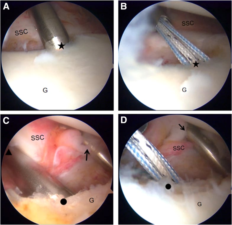
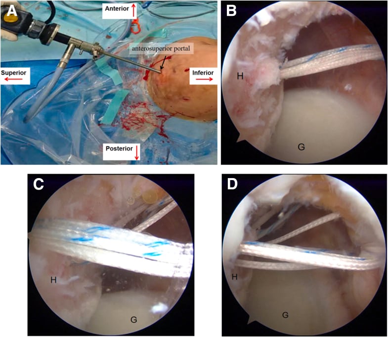
Beach chair or lateral decubitus. Standard posterior viewing portal. Anterior portals for anchor placement.
Elevate the labrum from the glenoid neck. Decorticate the glenoid rim to bleeding bone. Ensure adequate mobilisation for a tension-free repair.
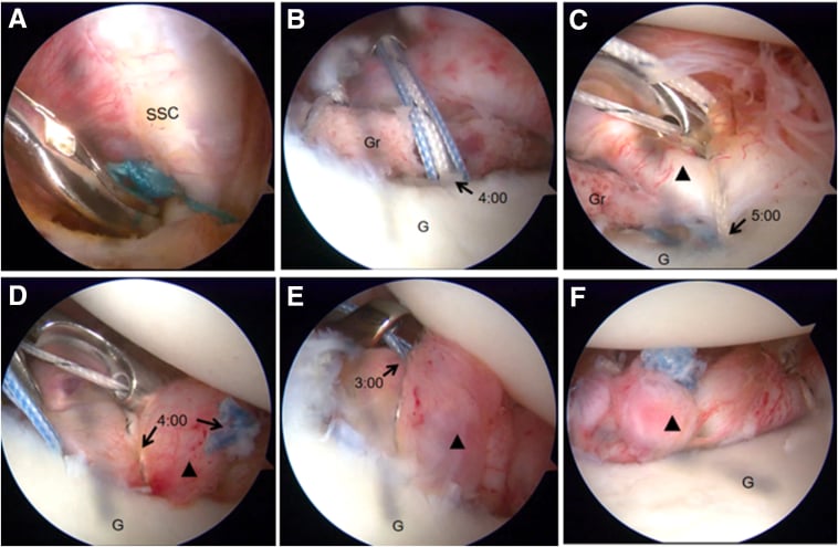
3-4 anchors along the anteroinferior glenoid rim (5 to 3 o'clock for a right shoulder). Suture anchors or knotless.
Mattress or simple sutures through the labrum. Restore the labral bumper. Capsular plication if redundant.
Anchors go ON the glenoid rim at the articular margin, not on the neck, and there should be at least 3. The 5:30 position (6:30 in a left shoulder) is critical because it addresses the IGHL attachment. Superior anchors must avoid the suprascapular nerve.
Complications
- Procedure
- Bankart
- Incidence
- 10-20%
- Prevention/Management
- Proper patient selection, technique
- Procedure
- Latarjet
- Incidence
- 0-5%
- Prevention/Management
- Rare if technique correct
- Procedure
- Latarjet
- Incidence
- 5-7%
- Prevention/Management
- Proper screw placement, length
- Procedure
- Latarjet
- Incidence
- 5%
- Prevention/Management
- Avoid over-medialisation, good contact
- Procedure
- Latarjet
- Incidence
- Variable
- Prevention/Management
- Split (not tenotomy) preferred
- Procedure
- Both
- Incidence
- Variable
- Prevention/Management
- Early ROM, appropriate capsular tension
- Procedure
- Latarjet
- Incidence
- Rare
- Prevention/Management
- Protect musculocutaneous and axillary
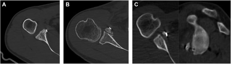
The most common causes of Latarjet failure are graft malposition (too lateral or too medial), graft non-union, screw pullout and a missed HAGL lesion. Meticulous technique and preoperative planning are essential.
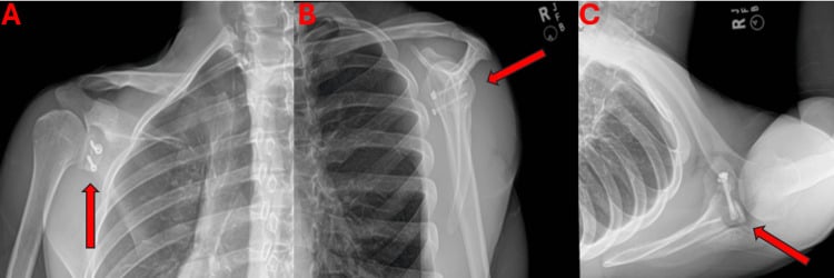
Postoperative Care
Rehabilitation Protocol
Sling immobilisation. Elbow and hand exercises. Pendulum exercises only. No external rotation past neutral.
Wean the sling. Gentle active range of motion. Limit external rotation (based on surgeon preference). Scapular stabilisation exercises.
Full range of motion by 12 weeks. Rotator cuff strengthening. Avoid apprehension positions.
Progressive resistance. Sport-specific training started.
After a Bankart, return to contact sport at 6-9 months. After a Latarjet, some allow an earlier return at 4-6 months because of the bony stability; confirm graft healing on CT before high-risk activities.
Outcomes and Prognosis
Arthroscopic Bankart succeeds in 80-90% of appropriately selected patients. Recurrence is higher in young contact athletes and with bone loss or an engaging Hill-Sachs.
Latarjet gives 95-98% stability, with low recurrence even in high-risk groups, at the cost of some risk of osteoarthritis in the long term.
Prognosis. Older age, non-contact sport, minimal bone loss, a first stabilisation and compliant rehabilitation predict a good result. Age under 20, contact sport, significant bone loss, an engaging Hill-Sachs, hyperlaxity and failed previous surgery predict a poor one.
Guidelines, Registries & Global Practice
Global Epidemiology
- Glenohumeral dislocation is the most common large-joint dislocation; population incidence is reported around 23-24 per 100,000 person-years, with anterior dislocations making up roughly 95% of cases.
- Bimodal distribution: a large peak in young males (15-25 years, sport and high-energy trauma) and a smaller peak in older patients (often with concomitant rotator cuff tears or greater tuberosity fractures).
- Recurrence is driven overwhelmingly by age and bone loss, a pattern consistent across cohorts worldwide (Robinson, Edinburgh; Hovelius, Sweden).
Society Guidance Side by Side
- Emphasis
- Evidence appraisal
- Practical Position
- Notes limited high-level evidence; supports surgical stabilisation reducing recurrence in young active patients; individualised first-dislocation decisions
- Emphasis
- Pathway and patient selection
- Practical Position
- Stanmore polar-type framework; reserve surgery for structural (Type I/II) instability; avoid operating on muscle-patterning shoulders
- Emphasis
- Bony injury and fixation
- Practical Position
- Structured assessment and fixation principles for bony Bankart and glenoid reconstruction
- Emphasis
- Bone-loss-driven algorithm
- Practical Position
- Glenoid track and bipolar bone loss central to choosing soft-tissue vs bony procedure
Registry and Outcome Signals
- Large series and registry-type data consistently show Latarjet with lower recurrence than isolated Bankart, balanced against a higher-acuity complication profile (An et al. meta-analysis).
- Revision and complication tracking (e.g. coracoid graft non-union, hardware removal) is increasingly captured in national shoulder-instability and arthroplasty-adjacent datasets to benchmark Latarjet safety.
High- vs Limited-Resource Practice Variation
- Routine 3D CT volumetric bone-loss quantification and MRA
- Access to arthroscopic Bankart, remplissage and arthroscopic or open Latarjet
- Structured return-to-sport testing before clearance
- Greater reliance on plain radiographs (AP in ER, axillary, Stryker/West Point) and clinical assessment
- Open Latarjet/Bristow favoured where arthroscopy or advanced imaging is scarce
- Iliac crest autograft is a low-cost bone-block option without implant dependence
Anterior shoulder instability is a common viva topic in fellowship examinations worldwide. Be prepared to: quantify glenoid and bipolar bone loss, explain on-track vs off-track, articulate the Bankart vs Latarjet decision algorithm, describe both surgical techniques and their complications, and apply the ISIS and Stanmore frameworks.
Controversies and Areas of Uncertainty
How to measure bone loss. No single method is universal. The best-fit circle on 3D CT is the most used, but linear, area and surface-based methods give different percentages for the same defect, and the subcritical zone increasingly prompts augmentation below the classic threshold.
Surgery after the first dislocation. Whether to stabilise every young first-time dislocator is debated. The Robinson and Hovelius data show high but not universal recurrence, so shared decision-making that weighs sport, age, bone loss and patient preference is favoured over a blanket policy.
Arthroscopic versus open Latarjet. The arthroscopic Latarjet offers lower morbidity in expert hands but has a steep learning curve and graft-positioning and hardware concerns. Open Latarjet remains the reference standard, and no high-level evidence proves one superior for stability.
Free bone block alternatives. Iliac crest autograft and distal tibial allograft (anatomic glenoid reconstruction) avoid the conjoint sling but lose its dynamic effect. Their role versus Latarjet, and bone-block versus screw-versus-suture fixation, is unsettled.
Latarjet lowers recurrence (An et al. meta-analysis) but carries a distinct, sometimes serious complication profile: neurovascular injury, graft non-union or resorption, hardware problems and later arthritis. The honest answer is patient- and pathology-specific selection, not "Latarjet is always better."
MCQ Practice Points
Q: Above what percentage of glenoid bone loss is Latarjet/bony procedure required? A: Greater than 25%. At this level, Bankart repair will fail. 15-25% is the gray zone where engaging Hill-Sachs tips toward Latarjet.
Q: What is the recurrence rate after first dislocation in patients under 20? A: 67%. Young age is the strongest predictor of recurrence. This justifies early surgical consideration in young athletes.
Q: What are the three stabilizing effects of the Latarjet procedure? A: 1) Bone block augmenting glenoid, 2) Sling effect of conjoint tendon, 3) Capsular repair to the bone block.
Q: Which ligament is the primary static restraint to anterior translation? A: Inferior glenohumeral ligament (IGHL) - specifically the anterior band when the arm is abducted and externally rotated.
Q: What percentage of traumatic anterior dislocations have a Bankart lesion? A: Greater than 90%. The anteroinferior labrum avulses with the IGHL attachment in almost all traumatic dislocations.
Q: What does an off-track Hill-Sachs lesion indicate? A: The Hill-Sachs engages the glenoid rim during movement, causing instability. Requires Latarjet or remplissage rather than simple Bankart.
Exam Viva Scenarios
Practise clinical reasoning and management decisions out loud
“An 18-year-old rugby player presents after his first anterior shoulder dislocation during a tackle. He required reduction in the emergency department. Radiographs show no fracture. How would you manage this?”
“A 24-year-old AFL player has had 5 anterior dislocations over 3 years. CT shows 22% glenoid bone loss and an engaging Hill-Sachs lesion. What would you recommend?”
“A 22-year-old had arthroscopic Bankart repair 2 years ago but has had 3 further dislocations. CT shows the anchors are in place but there is now 18% glenoid bone loss. What is your approach?”
Pathoanatomy
- Bankart lesion in greater than 90% of dislocations
- Hill-Sachs in 65-70% of first dislocations
- IGHL is primary static restraint
- Subscapularis is key dynamic stabilizer
- Bony Bankart reduces glenoid surface area
Bone Loss Thresholds
- Less than 15%: Arthroscopic Bankart likely sufficient
- 15-25%: Gray zone - consider Latarjet if engaging HS
- Greater than 25%: Latarjet or bone graft required
- Off-track HS: Latarjet or remplissage
- Critical bone loss dramatically increases recurrence
Age and Recurrence
- Under 20 years: 67% recurrence
- 20-40 years: 30-40% recurrence
- Over 40 years: 10-15% (but cuff tears)
- Young age = strongest predictor
- Male gender increases risk further
Latarjet Triple Effect (BSC)
- Bone block: Augments glenoid
- Sling effect: Conjoint tendon dynamics
- Capsular repair: Capsule sutured to block
- Benefits: Addresses bone loss and prevents engagement
- Ideal for high-risk contact athletes
ISIS Score greater than 6 = Consider Latarjet
- Age 20 or younger at surgery: 2 points
- Competitive sport: 2 points
- Contact or forced-overhead sport: 1 point
- Shoulder hyperlaxity (anterior or inferior): 1 point
- Hill-Sachs visible on AP in external rotation: 2 points
- Loss of inferior glenoid contour on AP: 2 points
Outcomes
- Bankart: 10-20% recurrence overall
- Latarjet: 0-5% recurrence
- Latarjet superior in high-risk groups
- Both have high patient satisfaction
- Open Latarjet has higher complication rate than Bankart
Evidence Base and Key Studies
Recurrence Risk After Primary Dislocation in the Young
- Prospective cohort of 252 patients aged 15-35 years, nonoperative treatment
- Recurrent instability in 55.7% within 2 years, rising to 66.8% by 5 years
- Younger males at highest risk; females much lower risk
- 86.7% of those who recurred did so within the first 2 years
25-Year Outcome of Nonoperative Treatment
- Prospective multicentre study, 255 patients (257 shoulders), age 12-40, 25-year follow-up
- Roughly half of dislocations in patients aged 12-25 had not recurred or had stabilised over time
- 62 shoulders (27%) ultimately underwent surgical stabilisation
- Immobilisation after primary dislocation did not change the prognosis
Critical Glenoid Bone Loss After Bankart Repair
- Cadaveric study of sequential simulated glenoid defects after Bankart repair
- A defect at least 21% of glenoid LENGTH caused instability and restricted external rotation
- Average loss of 25 degrees external rotation per cm of defect
- Defined the biomechanical basis for bone-loss thresholds
Inverted-Pear Glenoid and Engaging Hill-Sachs
- 194 consecutive arthroscopic Bankart repairs (suture anchor)
- No significant bone defect: 4% recurrence
- Significant bone defect (inverted-pear or engaging Hill-Sachs): 67% recurrence
- Contact athletes with bone defect: 89% recurrence
Glenoid Track Concept
- Nine fresh-frozen cadaveric shoulders, contact mapping in abduction and external rotation
- Defined the glenoid track - the zone of glenoid-humeral contact
- Medial margin of track at 84% (plus or minus 14%) of glenoid width from the cuff footprint
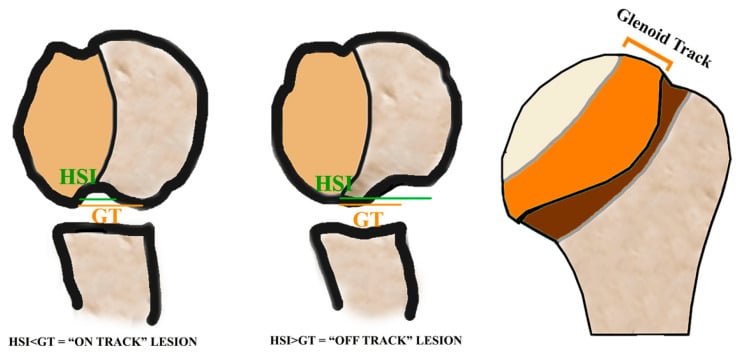
- A Hill-Sachs lesion extending medial to the track risks engagement
On-Track / Off-Track Treatment Paradigm
- Translated engaging/non-engaging into the quantitative on-track/off-track concept
- Integrates bipolar (glenoid plus humeral) bone loss into one framework
- Glenoid track narrows as glenoid bone loss increases, making a fixed Hill-Sachs more likely off-track
- Provides surgical criteria for Bankart, remplissage and bony procedures
Instability Severity Index Score (ISIS)
- Prospective case-control of 131 patients after arthroscopic Bankart, mean 31 months
- 10-point preoperative score (age, sport, hyperlaxity, Hill-Sachs and glenoid contour on AP)
- Score over 6 points: 70% recurrence after arthroscopic Bankart
- Authors suggest a Bristow-Latarjet procedure instead for high scorers
Latarjet vs Bankart - Systematic Review
- Systematic review and meta-analysis, 8 comparative studies, 795 shoulders (Bankart 416, Latarjet 379)
- Latarjet conferred significantly lower risk of recurrence and redislocation
- No significant difference in reoperation rate between procedures
- Higher Rowe scores and less external-rotation loss with Latarjet