Degenerative Cascade | Radiculopathy | Myelopathy
- Degenerative cascade: Disc desiccation leads to Osteophytes leads to Facet hypertrophy leads to Ligamentum flavum hypertrophy
- C5-6 level: Most mobile segment, thus most prone to degeneration
- Radiculopathy vs Myelopathy: Differentiate clinically (LMN vs UMN signs)
- Natural history: at 10 years 79% had LESS pain but only 43% were pain-FREE and 32% still had moderate or severe pain (PMID 3576350) - 'improves' is not 'resolves'; myelopathy is the exception and tends to progress
- Surgical indications: Progressive myelopathy, refractory radiculopathy
- “Do not attribute all neck pain to radiographic spondylosis - it is ubiquitous in elderly
- “T2 cord signal change (myelomalacia) is a poor prognostic factor for recovery
- “Spurling test has high specificity (93%) for radiculopathy
- “Uncovertebral joints (Luschka) are unique to cervical spine and cause foraminal stenosis
Overview and Epidemiology
What it is. Cervical spondylosis is the age-related degenerative cascade of the cervical spine, involving the intervertebral discs, the vertebral bodies (osteophytes), the facet joints and the ligaments. It spans everything from an asymptomatic radiographic finding to debilitating myelopathy, and the same set of changes produces all three clinical syndromes: axial pain, radiculopathy and myelopathy.
Who has it. Prevalence increases linearly with age: 25% at age 40 and over 85% at age 60. The segments that give way are C5-6, the most mobile and the most commonly affected, and C6-7.
Risk factors. Age is the primary one, and a family history of early degeneration is the other that cannot be changed. The modifiable list is worth taking a history for:
- Smoking - accelerates disc desiccation
- Heavy labour - axial loading
- Vibration exposure - jackhammering
- Contact sports
Previous whiplash or cervical injury often initiates the cascade early.
Natural history. Generally benign, but be precise about what "benign" means. In the 10-year follow-up of 205 patients with neck pain (Gore, Spine 1987, PMID 3576350), 79% had a decrease in pain - yet only 43% became pain-free, and 32% still had moderate or severe residual pain. So the honest counselling line is that most people get better but a third do not get better enough, and that is a different sentence from "it resolves".
The same study found pain severity was not related to the degree of degenerative change, canal diameter or lordosis, which is why treating the radiograph rather than the patient goes wrong here. Myelopathy is the exception: it deteriorates in a stepwise fashion rather than improving spontaneously (Nurick, Brain 1972, PMID 5023079).
Pathophysiology and Mechanisms
It begins in the disc. Disc desiccation - loss of water content - is the first event, and the rest of the cascade follows from it.
- Loss of disc height reduces the tension on the annular fibres and the ligaments
- Segmental instability follows, the vertebral bodies moving more than they should
- Osteophytes are the body's answer to that instability, increasing surface area to restabilise the joint
- Uncinate hypertrophy develops as the uncovertebral joints carry more load with the disc collapsed, and those osteophytes project into the foramen - radiculopathy
- Facet hypertrophy follows in the posterior joints, causing pain and encroaching on the canal
- Ligamentum flavum buckling completes it: with height lost the ligament is redundant and buckles inwards, compressing the cord from behind - myelopathy
The Kirkaldy-Willis cascade divides the same degeneration into three stages by age.
- Dysfunction, age 15-45. Annular tears, endplate microfractures and facet synovitis, with the potential for disc herniation. Episodic neck pain.
- Instability, age 35-70. Disc height loss and a dark, desiccated disc, lax facet capsules, the beginning of osteophyte formation, and segmental instability.
- Stabilisation, age over 60. Advanced osteophytes bridging levels and facet hypertrophy leave a stiff, restabilised spine in which stenosis - foraminal or central - is the main problem.
OPLL. Ossification of the posterior longitudinal ligament is a distinct but related pathology, often co-existing, and particularly common in Asian populations (Japanese and Korean). It causes severe, hard compression of the anterior cord.
Applied Anatomy
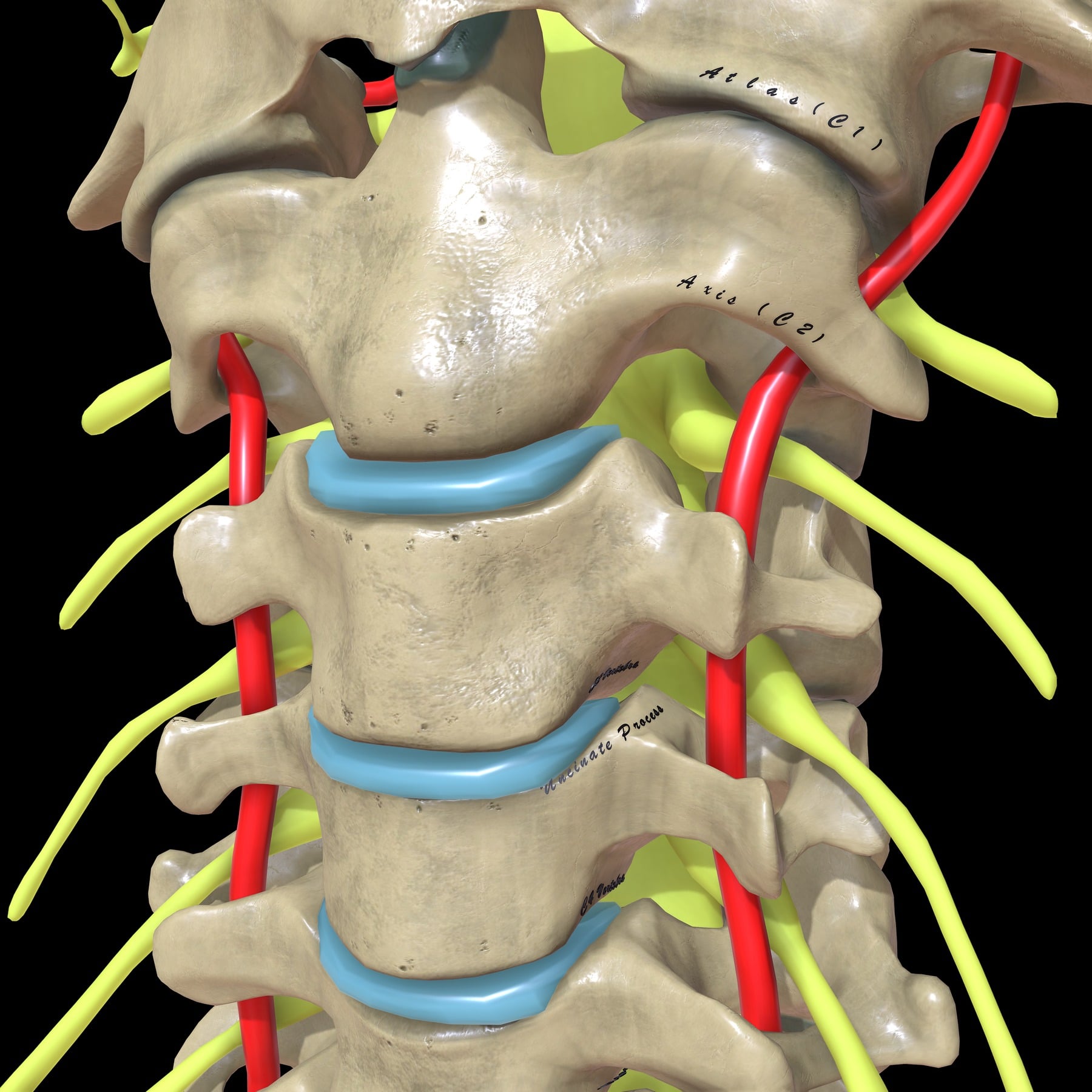
Unique to C3-C7, the uncinate processes form the anterior border of the neural foramen, so hypertrophy and osteophytes here cause foraminal stenosis and radiculopathy. This is distinct from the lumbar spine, where disc and facet pathology dominates and the facets sit posteriorly. They develop by age 10-14 and are considered adventitious joints resulting from fissuring of the annulus.
The ligaments. The anterior longitudinal ligament is a strong broad band that prevents hyperextension and is often the site of large anterior osteophytes (DISH). The posterior longitudinal ligament is a narrower band behind the bodies that prevents hyperflexion, and is the ligament that ossifies in OPLL. The ligamentum flavum connects the laminae and is elastic and yellow; it compresses the cord from behind when it hypertrophies or buckles in extension.
The neural foramen. Its boundaries and contents:
- Anterior: uncovertebral joint, disc and the posterior aspect of the vertebral body
- Posterior: facet joint (the superior articular process of the lower vertebra) and ligamentum flavum
- Superior and inferior: the pedicles of the levels above and below
- Contents: the exiting nerve root, with the dorsal root ganglion usually inside the foramen, the radicular artery and a venous plexus
So an uncinate osteophyte compresses the root from in front, and facet hypertrophy compresses it from behind.
Canal dimensions. The numbers worth carrying:
- Normal: 17-18mm
- Relative stenosis: under 13mm - watch for symptoms
- Absolute stenosis: under 10mm - high risk of cord injury
- Cord compression: under 8mm effective diameter
The Torg ratio, canal diameter divided by vertebral body diameter, suggests stenosis below 0.8, but it has a high false positive rate and an absolute measurement on MRI is preferred.
Blood supply of the cord. The anterior spinal artery supplies the anterior two-thirds - the motor tracts and the spinothalamic tracts - and is prone to compression from anterior osteophytes and disc herniation. The paired posterior spinal arteries supply the posterior columns and therefore proprioception. The grey matter is highly vascular, but the watershed zone leaves the anterior cord susceptible to ischaemia from compression by osteophytes, and that ischaemia contributes to the pathophysiology of cervical spondylotic myelopathy. Radicular arteries enter through the foramen; the largest is the artery of Adamkiewicz, usually T9-L1 but it can be higher.
The vertebral artery. It ascends in the transverse foramen from C6 to C1, usually skipping the C7 transverse foramen, and is described in four segments: V1 pre-foraminal, V2 foraminal from C6 to C2, V3 the extradural loop at the atlas, and V4 intradural. It is vulnerable during lateral dissection and during lateral mass or pedicle screw insertion. The left is often larger than the right, and occlusion of a dominant artery can be fatal (Wallenberg syndrome, stroke).
Classification Systems
Compression is named for the structure causing it, and the distinction between a soft disc and a hard osteophyte is critical for surgical planning.
- Structure
- Soft Disc Protrusion
- Syndrome
- Radiculopathy (Acute)
- Structure
- Hard Osteophyte
- Syndrome
- Radiculopathy (Chronic)
- Structure
- Luschka Joint Spur
- Syndrome
- Foraminal Stenosis
- Structure
- Buckled Lig Flavum
- Syndrome
- Central Stenosis (Myelopathy)
Clinical Assessment
History. The first task is to work out which of the three syndromes is in front of you.
- Axial Pain
- Neck/trapezius pain
- Radiculopathy
- Arm pain greater than Neck pain
- Myelopathy
- Gait disturbance, hand clumsiness
- Axial Pain
- Deep ache, stiffness
- Radiculopathy
- Shooting, electric, burning
- Myelopathy
- Often painless or vague ache
- Axial Pain
- Upright posture, extension
- Radiculopathy
- Extension, arm dependency
- Myelopathy
- Extension (canal narrowing)
- Axial Pain
- Intact
- Radiculopathy
- LMN signs (root level)
- Myelopathy
- UMN signs (long tract)
Red flags. These take the patient out of the degenerative pathway and into urgent MRI.
PINSRed Flags (PINS)
Hook:PINS requiring urgent MRI
Differential Diagnosis
- Discriminating Features
- Distal, focal nerve distribution; can co-exist (double-crush)
- Key Test / Clue
- Tinel/Phalen, nerve conduction studies
- Discriminating Features
- Pain with active ROM, normal sensation, no neck reproduction
- Key Test / Clue
- Impingement signs, subacromial LA test
- Discriminating Features
- Younger, visual symptoms, multifocal CNS signs
- Key Test / Clue
- Brain/cord MRI, oligoclonal bands
- Discriminating Features
- Progressive painless weakness, fasciculations, NO sensory loss
- Key Test / Clue
- EMG, absence of sensory signs
- Discriminating Features
- Night pain, fever, weight loss, constitutional symptoms
- Key Test / Clue
- Red flags then urgent MRI
- Discriminating Features
- Viral prodrome, severe acute pain then patchy weakness/wasting
- Key Test / Clue
- Clinical course, EMG
The double crush. Concurrent proximal compression at the cervical root and distal compression such as a carpal tunnel can produce symptoms greater than either lesion alone, so examine the whole limb before settling on one level.
Which page you need. Spondylosis is the substrate; what it does is what has its own page. If the picture is root - dermatomal arm pain, Spurling - go to cervical radiculopathy. If it is cord - gait, hands, hyperreflexia - go to cervical myelopathy, where the grading and the urgency live and where delay costs recovery. The disc itself and its operative thresholds are cervical disc disease, and when the operation is anterior it is ACDF. Note the asymmetry the Boden study (PMID 2398088) forces on this whole page: degenerative change is near-universal on MRI in asymptomatic people, so the imaging cannot make the diagnosis - the syndrome does.
Physical Examination
Provocative tests. These are the bedside tests that separate a compressed root from a compressed cord.
- Spurling test - extension, lateral rotation and axial load. Reproduction of radicular pain is positive, and specificity is 93%, so a positive test rules radiculopathy in.
- Shoulder abduction (Bakody) test - relief of arm pain with the hand on the head is C5/C6 radiculopathy, the position relieving tension on the root.
- Lhermitte's sign - a shock sensation down the spine on flexion, indicating myelopathy or cord compression.
- Finger escape sign - the patient cannot hold the fingers extended and adducted (C8/T1 weakness in myelopathy).
- Grip and release test - 20 times in 10 seconds is normal; the myelopathic hand is slower.
- Inverted radial reflex - tapping the supinator produces finger flexion instead of the wrist or supinator response, indicating C5/6 cord compression.
- Valsalva manoeuvre - raises intrathecal pressure and aggravates radicular pain.
The myelopathic patient. The story is hand clumsiness, weak grip and wasting of the intrinsics in the first web space, together with a broad-based, spastic, unsteady gait. The long-tract signs to elicit are Hoffman, Babinski, hyperreflexia and clonus.
Root levels. The motor, sensory and reflex findings that localise the root:
- C5: deltoid, lateral arm, biceps reflex
- C6: wrist extension, thumb and index finger, brachioradialis reflex
- C7: triceps, middle finger, triceps reflex
- C8: finger flexion, little finger
- T1: interossei, medial forearm
Investigations
Diagnostic Workup
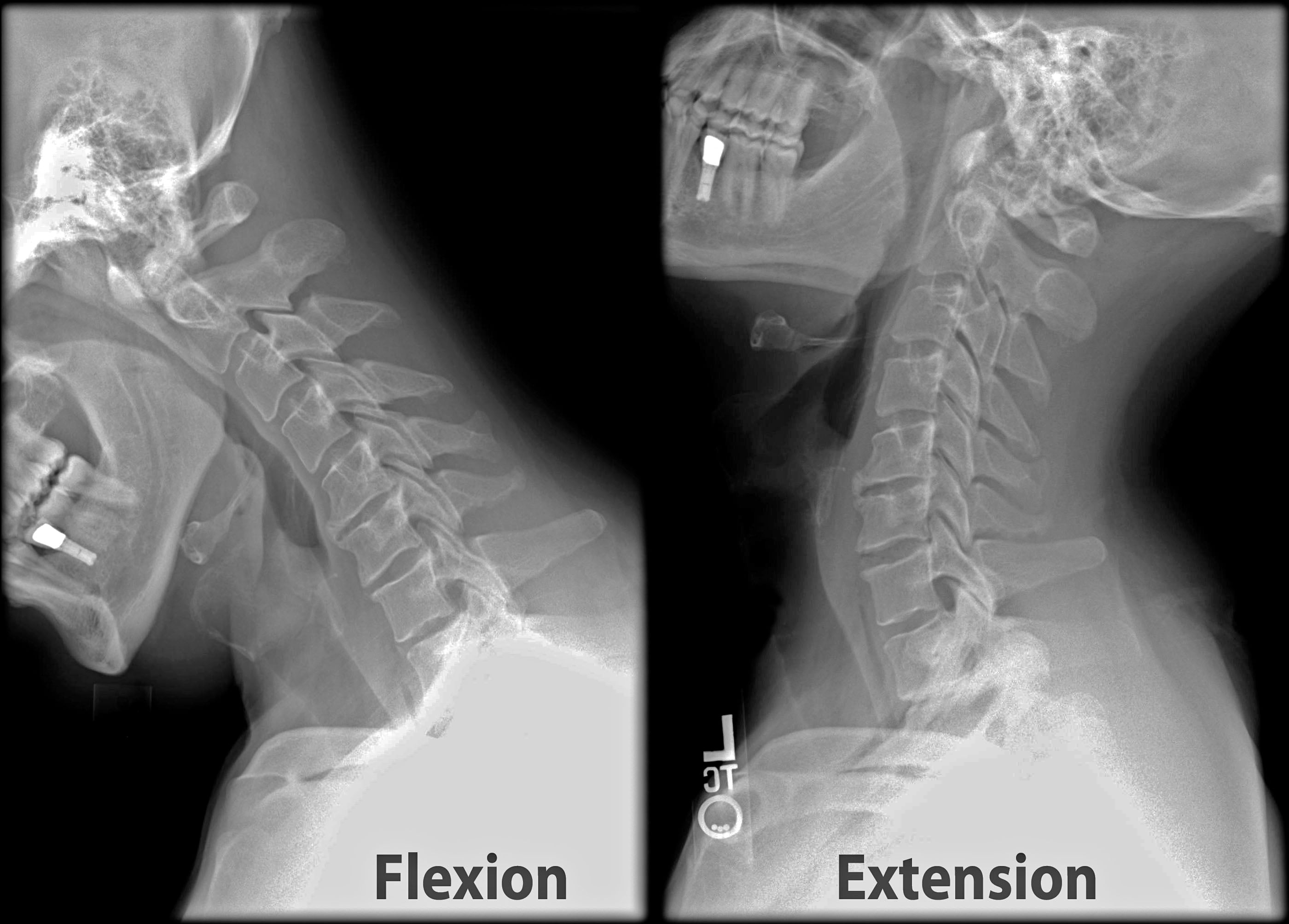
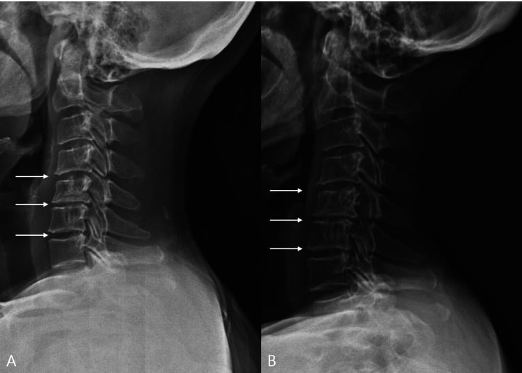
Start with AP, lateral, open-mouth peg, oblique views for the foramina and flexion/extension views for instability. Look for disc height loss, osteophytes, spondylolisthesis, sagittal alignment and signs of OPLL.
Instability on the dynamic views means translation greater than 3.5mm or a difference in angulation greater than 11 degrees between flexion and extension.
Indicated for radiculopathy lasting over 6 weeks, a progressive deficit, myelopathy, or red flags. MRI is the gold standard for soft tissue and neural compression.
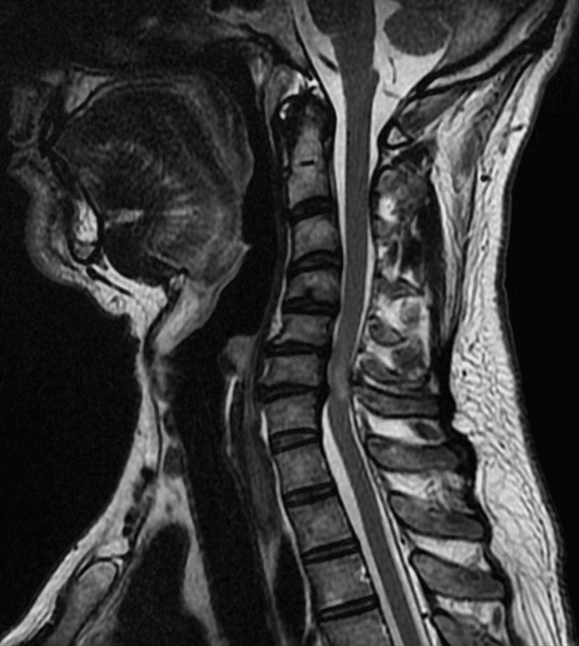
Assess disc hydration (dark on T2), foraminal nerve root compression, central cord compression and cord signal change (myelomalacia). T2 sagittal is best for screening and T2 axial for level-specific compression.
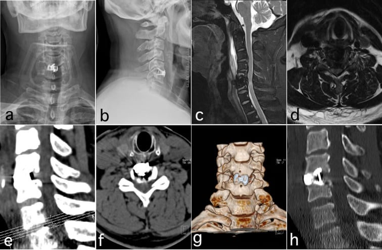
Indicated for surgical planning, suspected OPLL, or when MRI is contraindicated. It shows the bony anatomy, separates osteophyte from soft disc and demonstrates ossification of the PLL, and a high quality CT is crucial preoperatively to assess bone quality for fusion.
Indicated when the diagnosis is equivocal or a double crush is suspected (carpal tunnel versus radiculopathy). Sensitivity is variable, and the value is in ruling out a peripheral neuropathy.
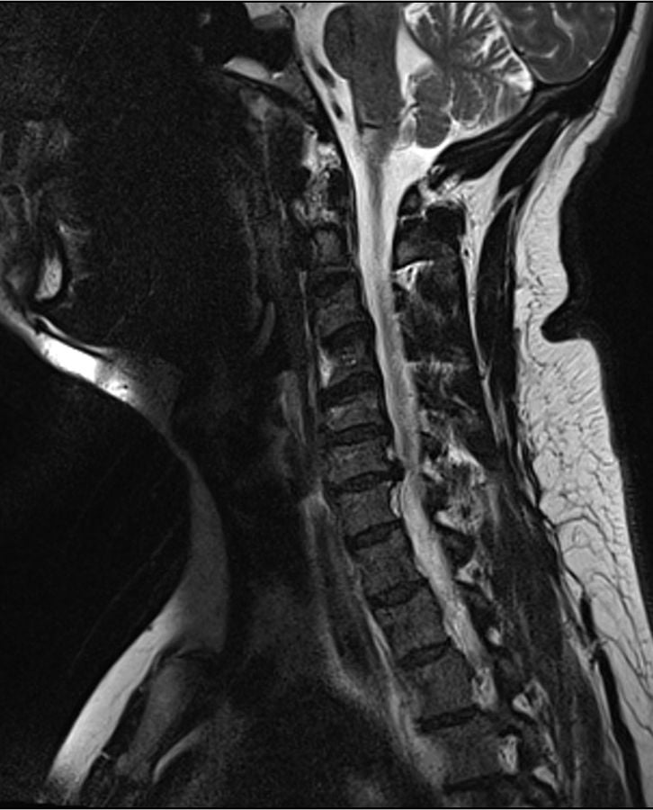
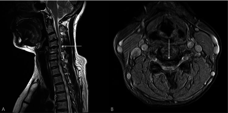
High T2 signal in the cord is myelomalacia - oedema, gliosis or ischaemia - and it predicts poorer surgical recovery, with complete resolution of symptoms less likely. T1 hypointensity, a dark cord, is worse still: cystic necrosis and atrophy, indicating irreversible damage.
Management Algorithm
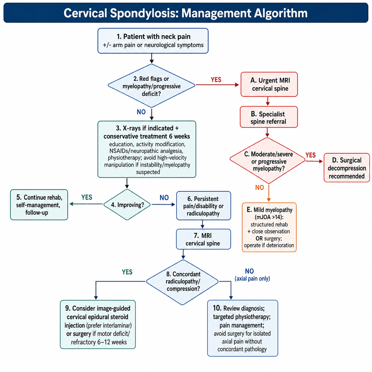
Conservative treatment succeeds in 75-90% of radiculopathy and axial pain.
Medication. NSAIDs are first line as a short course, and gabapentin or pregabalin are the choice for radicular pain. Muscle relaxants are for acute spasm only, under a week, and a tricyclic such as amitriptyline can help chronic pain. Avoid opioids: poor efficacy, high risk.
Physiotherapy. Strengthen the deep neck flexors and the scapular stabilisers, trapezius and rhomboids, and get an ergonomic assessment including monitor height. Manual therapy means mobilisation, and high-velocity manipulation is avoided for the risk of stroke and dissection. Traction gives short-term relief in radiculopathy, and home devices are available.
Cervical epidural steroid injection has moderate evidence for short-term relief of radiculopathy. The transforaminal approach risks catastrophic injury from cord infarct, so the interlaminar approach is preferred and safer in the cervical spine. Fluoroscopic guidance is mandatory.
Surgical Technique
- Anterior (ACDF/Corpectomy)
- Anterior (Disc/Osteophyte)
- Posterior (Laminectomy/Fusion/Plasty)
- Posterior (Lig flavum/shingles)
- Anterior (ACDF/Corpectomy)
- Kyphosis or Straight (Need to restore lordosis)
- Posterior (Laminectomy/Fusion/Plasty)
- Maintained Lordosis (Required for drift)
- Anterior (ACDF/Corpectomy)
- 1-3 Levels
- Posterior (Laminectomy/Fusion/Plasty)
- 3+ Levels (Multilevel)
- Anterior (ACDF/Corpectomy)
- Better relief (stabilises segment)
- Posterior (Laminectomy/Fusion/Plasty)
- Can worsen (muscle stripping)
- Anterior (ACDF/Corpectomy)
- Dysphagia, RLN palsy, Horner's
- Posterior (Laminectomy/Fusion/Plasty)
- C5 palsy, wound healing, muscle atrophy
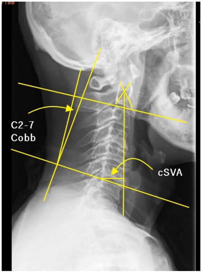
Alignment. The choice depends heavily on alignment. A kyphotic spine cannot be decompressed from behind, because the cord bowstrings forward against the osteophytes instead of drifting away from them, and it is the anterior approach that allows lordosis to be corrected.
OPLL: Classification and the K-line
For ossification of the posterior longitudinal ligament, two extra tools guide the approach:
OPLL radiographic classification (Japanese): continuous (bridges multiple bodies), segmental (behind individual bodies, discontinuous), mixed (both), and localised/circumscribed (single disc level).
The K-line (a high-yield surgical-decision rule): on a lateral radiograph/CT, draw a line connecting the midpoints of the spinal canal at C2 and C7.
- K-line positive — the OPLL does not cross the line → the cord can drift backward after a posterior decompression (laminoplasty/laminectomy), which works well.
- K-line negative — the OPLL crosses/exceeds the line (large, kyphotic) → posterior decompression alone is insufficient (the cord cannot drift back far enough) → favour an anterior decompression (corpectomy) or a combined anterior-posterior approach.
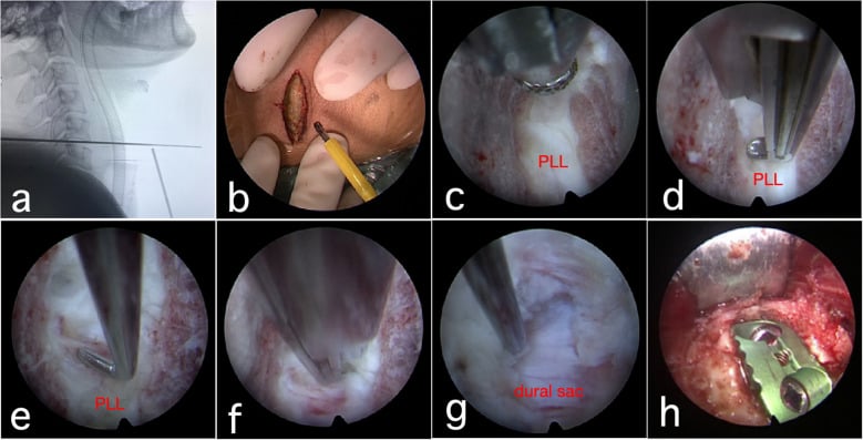
The other reason to go behind is the dura. Ossified ligament is hard to remove anteriorly and the dural tear risk is high, because the ossification can merge with the dura, so the presence of OPLL often dictates a posterior approach (laminoplasty) or a corpectomy. This, together with overall alignment, is why large anterior OPLL with kyphosis is not treated by laminoplasty alone.
Complications
The airway. After an anterior approach the airway is the first concern: a postoperative haematoma is an emergency, and oedema adds to it.
Dysphagia. The commonest complaint after an anterior approach, transient in up to 70%. Set that beside the measured trajectory in the Bazaz series below - 50.2% at one month falling to 12.5% at a year - and the two figures say the same thing: the early rate is high, most of it settles, and chronic dysphagia is rare.
Preventing it. Minimise retractor pressure, relax the retractors intermittently, and deflate the endotracheal tube cuff during retraction.
Managing it. Reassurance, since most resolve, with speech pathology review, a nasogastric tube if the aspiration risk is high, and intraoperative dexamethasone, which may help the oedema.
Adjacent segment disease. The rate is about 2.9% per year for symptomatic disease, reaching 25.6% by ten years (Hilibrand), of whom more than two-thirds needed a further operation. Radiographic change is far commoner: 92% of 180 patients followed beyond five years had additional degeneration at an adjacent level (Goffin 2004). Keep the two numbers apart, because one is a new radiculopathy or myelopathy and the other is an appearance on a film.
The cause is genuinely two competing mechanisms, and the evidence does not cleanly favour the biomechanical one. Increased stress at the levels above and below a fusion is the intuitive explanation, but Hilibrand found adjacent-segment disease was less common after multilevel than single-level fusion - the opposite of what stress transfer predicts - and Goffin found the same rate of progression in younger trauma patients as in older degenerative ones, concluding that fusion biomechanics and the natural progression of pre-existing spondylosis both contribute. Manage conservatively at first and extend the fusion if symptoms are concordant, and counsel before the index operation, because this is the commonest reason a fused patient returns years later.
C5 palsy. The incidence tracks the operation: 3.3% after ACDF, 5.1% after laminoplasty alone, 7.5% after anterior corpectomy and fusion and 11.0% after laminectomy with fusion, pooling to 5.3% across 13,621 patients.
Posterior drift of the cord tethering the shorter, more direct C5 root is the classic explanation, but it is contested. Pooled across 107 studies, anterior decompression for myelopathy carries a similar rate to posterior (4% versus 7%, p=0.999) despite causing no posterior drift, while anterior surgery for pure radiculopathy causes essentially none - so the presence of a compressed cord, not the direction of approach, is what predicts it. Reperfusion injury of the cord is the better-supported mechanism, with tethering at most contributory.
The prognosis splits by approach, and the usual line that most resolve in 6-12 months needs qualifying. Pooled recovery at one year is 100% after ACDF but only 52.9% after laminoplasty and 50% after posterior decompression and fusion, so after the posterior operations roughly half are still not fully recovered at a year.
Other major risks. The remaining items on the consent list:
- Cord injury and carotid injury
- Recurrent laryngeal nerve palsy - hoarseness in 1-2%, more common on the right where the nerve is variable. ENT scope if it persists
- Oesophageal injury - rare but catastrophic and can lead to mediastinitis. Early recognition is key; immediate repair or a flap
- Vertebral artery injury - rare, during drilling or screw insertion. Tamponade, or sacrifice if the contralateral vessel is patent
- Horner's syndrome - sympathetic chain injury, with ptosis, miosis and anhidrosis
- Dural tear - CSF leak, repaired primarily or with a patch or sealant, with bed rest or a drain
- Pseudarthrosis and malposition of the fusion - the late construct failures, alongside adjacent segment disease
Postoperative Care
Support and diet. A collar is usually not required because the plate provides the stability, and a soft collar for comfort for 1-2 weeks is enough. A soft diet for two weeks manages the swallowing, with steroids for severe oedema.
What to check. An erect radiograph on day 1 confirms hardware position and alignment, and the first 24 hours are spent watching closely for haematoma and the airway. Wounds are usually closed with absorbable sutures and strip tapes or glue, kept dry for 10 days.
Getting back. Most patients are discharged on day 1 or 2. Desk work at two weeks, manual work at three months or once fusion is confirmed, and avoid lifting more than 5kg for six weeks.
Outcomes and Prognosis
Surgical Decompression for CSM — AOSpine North America
- Prospective multicentre study, 278 patients with CSM across 12 North American centres
- Significant improvement at 1 year in mJOA, Nurick grade, NDI and SF-36v2 (all p less than 0.05)
- Benefit seen across mild, moderate and severe baseline severity groups
- Overall treatment-related complication rate 18.7%
- Established surgery as effective across the full myelopathy severity spectrum
Anterior versus Posterior Approach for CSM — AOSpine NA
- 264 of 278 prospective patients analysed by approach (169 anterior, 95 posterior)
- Anterior cases were younger with less severe, more focal disease
- Raw mJOA gain was lower anteriorly (+2.47) than posteriorly (+3.62)
- After adjusting for baseline differences, anterior and posterior approaches had equivalent efficacy
- NDI and SF-36 improvements did not differ between approaches
Natural History of Cervical Neck Pain — 10-Year Follow-up
- 205 patients with neck pain followed clinically and radiographically for at least 10 years
- 79% had a decrease in pain and 43% became pain-free over time
- 32% had persistent moderate or severe residual pain
- Pain severity was NOT related to degree of degenerative change, canal diameter or lordosis
- Patients injured with initially severe pain had the worst outcomes
Natural History of Cervical Spondylotic Myelopathy & Nurick Grade
- Classic study of the natural history and surgical results of spondylotic cord disorder
- Introduced the Nurick grading system (0-5) based on gait and ambulation
- Disability frequently progressed in a stepwise fashion rather than improving spontaneously
- Older patients and longer symptom duration carried worse prognosis
Abnormal MRI of the Cervical Spine in Asymptomatic Subjects
- MRI of 63 asymptomatic volunteers, blinded among symptomatic scans for 3 neuroradiologists
- 19% of asymptomatic subjects had an abnormality on MRI
- Under age 40: 14% abnormal (10% disc herniation, 4% foraminal stenosis)
- Over age 40: 28% abnormal (20% foraminal stenosis), disc degeneration in ~60%
Incidence of Dysphagia after Anterior Cervical Spine Surgery
- Prospective longitudinal study of 249 consecutive anterior cervical surgery patients
- Dysphagia incidence 50.2% at 1 month, 32.2% at 2 months, 17.8% at 6 months, 12.5% at 12 months
- Only 4.8% had moderate or severe dysphagia at 6 months
- Female gender and multilevel surgery were significant risk factors
- Vocal cord paresis identified in 1.3% at 12 months
Cervical Disc Arthroplasty vs ACDF — PRESTIGE ST RCT
- Prospective randomised multicentre IDE trial, 541 patients with single-level cervical disease and radiculopathy
- 276 arthroplasty (PRESTIGE ST) vs 265 ACDF; 2-year follow-up
- Arthroplasty maintained segmental motion (mean greater than 7 degrees) and gave a 2-point greater NDI improvement
- Higher neurological success and lower rate of secondary/revision surgery with arthroplasty
- Lower rate of adjacent-segment reoperation in the arthroplasty group
Adjacent-Segment Disease after Anterior Cervical Arthrodesis
- 374 patients / 409 anterior cervical arthrodeses followed up to 21 years
- Symptomatic adjacent-segment disease occurred at a constant ~2.9% per year
- Kaplan-Meier prediction: 25.6% develop adjacent-level disease within 10 years
- Highest risk at C5-6 and C6-7; risk was lower after multilevel than single-level fusion
- Over two-thirds of affected patients failed nonoperative care and needed further surgery
Radiographic versus symptomatic adjacent-segment change - where the 92% comes from
- 180 patients followed for more than 60 months after anterior cervical interbody fusion, examined clinically and radiologically by INDEPENDENT investigators
- Additional radiologic degeneration at an adjacent disc level was found in 92% of cases - the source of the figure usually quoted
- Severity of that additional degeneration correlated with the time elapsed since surgery
- Progression was SIMILAR in younger trauma patients and older non-trauma patients, which is the paper's key inference: both the biomechanical effect of the fusion AND the natural progression of pre-existing degenerative disease act as triggers
AOSpine / CSRS Clinical Practice Guideline for DCM
- Multidisciplinary, GRADE-based guideline built on five systematic reviews
- Recommends surgical intervention for moderate and severe DCM
- For mild DCM, suggests surgery OR a supervised trial of structured rehabilitation, with surgery if deterioration occurs
- Suggests NOT offering prophylactic surgery to non-myelopathic cord compression without radiculopathy — counsel and follow
- Non-myelopathic cord compression WITH radiculopathy carries higher risk of developing myelopathy
Guidelines, Registries & Global Practice
Global Epidemiology
Degenerative cervical myelopathy (DCM) is the most common cause of non-traumatic spinal cord dysfunction in adults worldwide, a point emphasised by both the AOSpine/CSRS clinical practice guideline (Fehlings et al, 2017) and the AOSpine North America cohort. Radiographic spondylosis rises steeply with age and is near-universal in older adults, yet only a minority become symptomatic. Ossification of the posterior longitudinal ligament (OPLL) is a major contributor to cervical stenosis in East Asian populations (notably Japan and Korea) and shifts surgical decision-making toward posterior or corpectomy strategies.
Side-by-Side Guidance
- Core Position
- Surgery for moderate/severe DCM; surgery or structured rehab for mild; no prophylactic surgery for asymptomatic cord compression without radiculopathy
- Evidence Basis
- GRADE-based, 5 systematic reviews
- Core Position
- Stepped care: conservative management first for axial pain and radiculopathy; urgent specialist referral and MRI for suspected myelopathy or progressive deficit
- Evidence Basis
- Guideline + consensus
- Core Position
- MRI is the imaging gold standard for neural compression; surgery reserved for refractory radiculopathy or myelopathy with concordant imaging
- Evidence Basis
- Appropriate-use criteria
- Core Position
- Emphasise early recognition of myelopathy and approach selection driven by alignment and pathology level
- Evidence Basis
- Expert consensus
Registry & Practice Variation
Spine procedures are tracked in national datasets such as the British Spine Registry and various European spine registries, which consistently show that ACDF and posterior decompression are the dominant constructs, with cervical disc arthroplasty used selectively for single-level disease in younger patients. Practice varies internationally: a higher prevalence of OPLL drives greater use of laminoplasty in Japan and Korea, whereas anterior approaches predominate for focal one- to two-level pathology elsewhere. Across systems, progressive myelopathy is prioritised for earlier surgery over isolated axial pain.
Medication & Rehabilitation Notes
Neuropathic agents (gabapentin, pregabalin) are used for radicular pain but show modest benefit and carry sedation and misuse risk; access may be restricted under national subsidy schemes. Opioids are discouraged for chronic non-cancer neck pain in favour of multidisciplinary, exercise-based rehabilitation. High-velocity cervical manipulation should be avoided where myelopathy or instability is suspected.
Return to Driving
Patients should not drive in a rigid collar and should resume driving only when able to comfortably rotate the head to check blind spots and perform an emergency stop — typically a few weeks postoperatively, guided by local licensing authority advice.
Viva Scenarios
Clinical Decision Scenarios
Practise clinical reasoning and management decisions out loud
“A 55-year-old female presents with neck pain after a minor MVA. X-rays show moderate C5/6 spondylosis. Neurologically intact.”
“A 68-year-old male presents with worsening balance and difficulty buttoning his shirts. He has hyperreflexia and a positive Hoffman's sign.”
“A 50-year-old plasterer presents with right arm pain and weakness in shoulder abduction. He has a history of rotator cuff tendonitis.”
“You have just performed a C5/6 ACDF. In the recovery room, the nurse calls you because the patient has stridor and neck swelling.”
MCQ Practice Points
Q: Which nerve root is affected by a C5-6 posterolateral disc herniation? A: C6 nerve root. In the cervical spine, nerve roots exit ABOVE their corresponding pedicle (e.g., C6 nerve exits above C6 pedicle), so C5-6 disc affects the exiting C6 root. Note: A C4-5 disc affects C5.
Q: What is the clinical significance of the Joints of Luschka (Uncovertebral joints)? A: Foraminal Stenosis. These joints form the anterior border of the neural foramen. Osteophytes here compress the nerve root, causing radiculopathy.
Q: What is the mechanism and utility of the Spurling test? A: Foraminal Compression. Extension and rotation to the affected side narrows the foramen. Reproduction of radicular pain is positive. It has High Specificity (93%) but low sensitivity.
Q: Which of the following is an Upper Motor Neuron sign seen in cervical myelopathy? A: Hoffman's Sign. Also Babinski response, Hyperreflexia, and Clonus. Muscle atrophy in the hands is a Lower Motor Neuron sign (segmental cord damage) often seen at the level of compression.
Q: What does high T2 signal in the spinal cord signify? A: Myelomalacia. It represents edema, gliosis, or ischemia. It is a predictor of poorer surgical recovery.
Definitions
- Spondylosis: Degenerative OA of spine (Disc + facets)
- Radiculopathy: Nerve root compression (LMN signs)
- Myelopathy: Cord compression (UMN signs)
- OPLL: Ossification of PLL (Asian population, hard compression)
- Disc Desiccation: Early fluid loss in nucleus
Key Anatomy
- C5-6: Most common level involved
- Uncovertebral Joints: unique to C-spine, cause foraminal stenosis
- Cord Signal (T2): Myelomalacia, poor prognosis
- Vertebral Artery: V2 segment in transverse foramen
- PLL: Behind body, can ossify (OPLL)
Management
- Axial/Radicular: 75% improve with conservative Rx
- Myelopathy: Surgery indicated (stop progression)
- ACDF: Anterior approach, 1-2 levels, restores lordosis
- Laminectomy: Posterior approach, more than 3 levels, requires lordosis
- CDR: Disc Replacement for soft disc in young
Complications
- Dysphagia (Anterior approach)
- C5 Palsy (Posterior decompression)
- Adjacent Segment Disease (2-3% per year)
- Vertebral Artery Injury: Rare but fatal
- Dural Tear: CSF leak risk