Hyperextension Deformity | Neonatal Presentation | Quadriceps Contracture
- CDK = knee hyperextension or anterior dislocation present at birth
- Tibia displaced anterior to femur; quadriceps and anterior capsule contracted
- Strong association with DDH (screen hips), clubfoot, arthrogryposis, Larsen syndrome
- Grade I and II usually respond to serial casting within 6-8 weeks
- Grade III or resistant cases require quadricepsplasty (V-Y lengthening or Curtis-Fisher)
- “Always screen hips in CDK: DDH coexists in up to 40-100 percent of cases
- “Hyperextension at birth is CDK until proven otherwise
- “Serial casting reduces the knee in stages: hyperextension to neutral to flexion
- “Failed closed treatment by 3 months strongly indicates surgical intervention
Overview and Epidemiology
Congenital dislocation of the knee (CDK) is a knee that presents at birth in hyperextension or frank anterior dislocation: the tibia is displaced anterior to the femur, and the quadriceps mechanism and anterior joint capsule are contracted. It sits on a spectrum from simple genu recurvatum through subluxation to irreducible anterior dislocation, and in severe cases the knee cannot be flexed passively beyond a right angle.
Why it matters. Early recognition is critical: outcomes are excellent with prompt serial casting, but delayed or inadequate treatment leads to progressive deformity, growth disturbance and the need for complex surgery. CDK is rarely isolated. The high association with developmental dysplasia of the hip means every CDK baby must have hip screening, and the association with clubfoot, arthrogryposis and Larsen syndrome means the knee is often the presenting sign of something wider, so every newborn with CDK gets a full musculoskeletal examination.
Who. Approximately 1 per 100,000 live births, with a female predominance of roughly 2:1 to 3:1. It is bilateral in roughly 40-60 percent of cases. Most cases are sporadic; familial forms are reported.
What it costs untreated. Untreated CDK causes severe gait disturbance, and knee instability limits standing, walking and sitting. Chronic anterior displacement can distort the distal femoral physis. The cosmetic deformity and functional limitation carry their own psychosocial cost through childhood.
Pathophysiology
Two theories. The exact aetiology remains debated. The mechanical theory attributes CDK to intrauterine positioning, a breech presentation with the feet trapped under the chin or against the uterine wall, leading to sustained knee hyperextension. The muscular theory emphasises primary quadriceps fibrosis and contracture as the driving force, analogous to the contracture seen in arthrogryposis.
The case for each. The association with breech presentation and with oligohydramnios, which restricts movement and maintains hyperextension, supports mechanical compression; a first-born's tighter uterus leaves less room for movement. Against pure positioning, the absence of the cruciates suggests developmental arrest, and familial CDK and the syndromic forms (Larsen) support an inherent tissue abnormality. CDK may represent a continuum from positional deformity to true congenital absence of joint formation.
Pathoanatomy. In severe (Grade III) cases the quadriceps tendon, patellar ligament and anterior joint capsule are all contracted and shortened. The tibia is displaced anteriorly and laterally relative to the femur, and the hamstrings are attenuated and displaced anteriorly. The cruciate ligaments may be absent, attenuated or abnormally positioned; the table sets out how each structure changes across the grades.
- Grade I (Recurvatum)
- Mild contracture
- Grade II (Subluxation)
- Moderate contracture
- Grade III (Dislocation)
- Severe fibrosis and shortening
- Grade I (Recurvatum)
- Lax or minimally tight
- Grade II (Subluxation)
- Contracted
- Grade III (Dislocation)
- Severely contracted, suprapatellar pouch obliterated
- Grade I (Recurvatum)
- Normal position
- Grade II (Subluxation)
- Mild anterior displacement
- Grade III (Dislocation)
- Displaced anteriorly, become knee extensors
- Grade I (Recurvatum)
- Usually present
- Grade II (Subluxation)
- May be attenuated
- Grade III (Dislocation)
- Often absent or severely abnormal
- Grade I (Recurvatum)
- Congruent, hyperextends
- Grade II (Subluxation)
- Subluxed anteriorly
- Grade III (Dislocation)
- Completely dislocated anteriorly
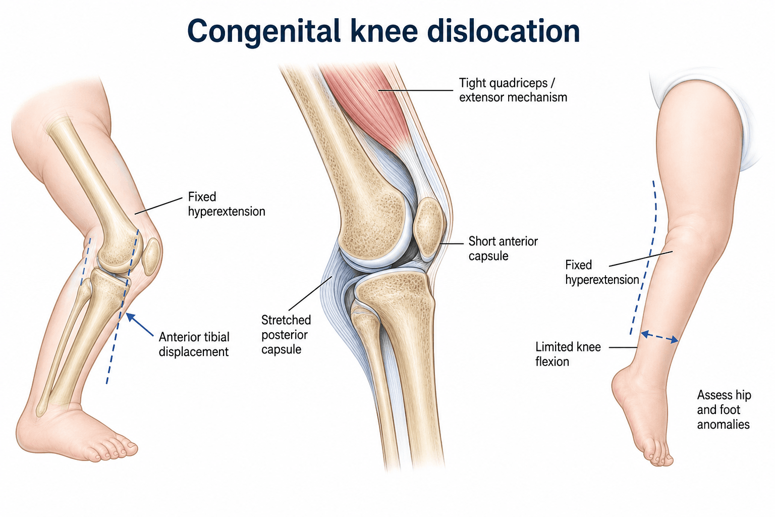
The patella. The patella is frequently abnormal in CDK and is a source of later morbidity that is easy to overlook in the neonate. The neonatal patella is unossified, but the underlying cartilaginous patella is often small (hypoplastic), laterally positioned, or even absent, and the extensor mechanism may be laterally subluxed, features that are especially pronounced in Larsen syndrome. After the tibiofemoral dislocation is reduced, persistent patellar maltracking or recurrent lateral patellar instability may emerge as the child grows and the patella ossifies, sometimes requiring later extensor-mechanism realignment; see paediatric patellar instability for those options (lateral release, medial reefing, or distal realignment). Examine and document patellar position at presentation and follow it through growth, because a reduced tibiofemoral joint does not guarantee a stable patellofemoral joint.
Classification and Types
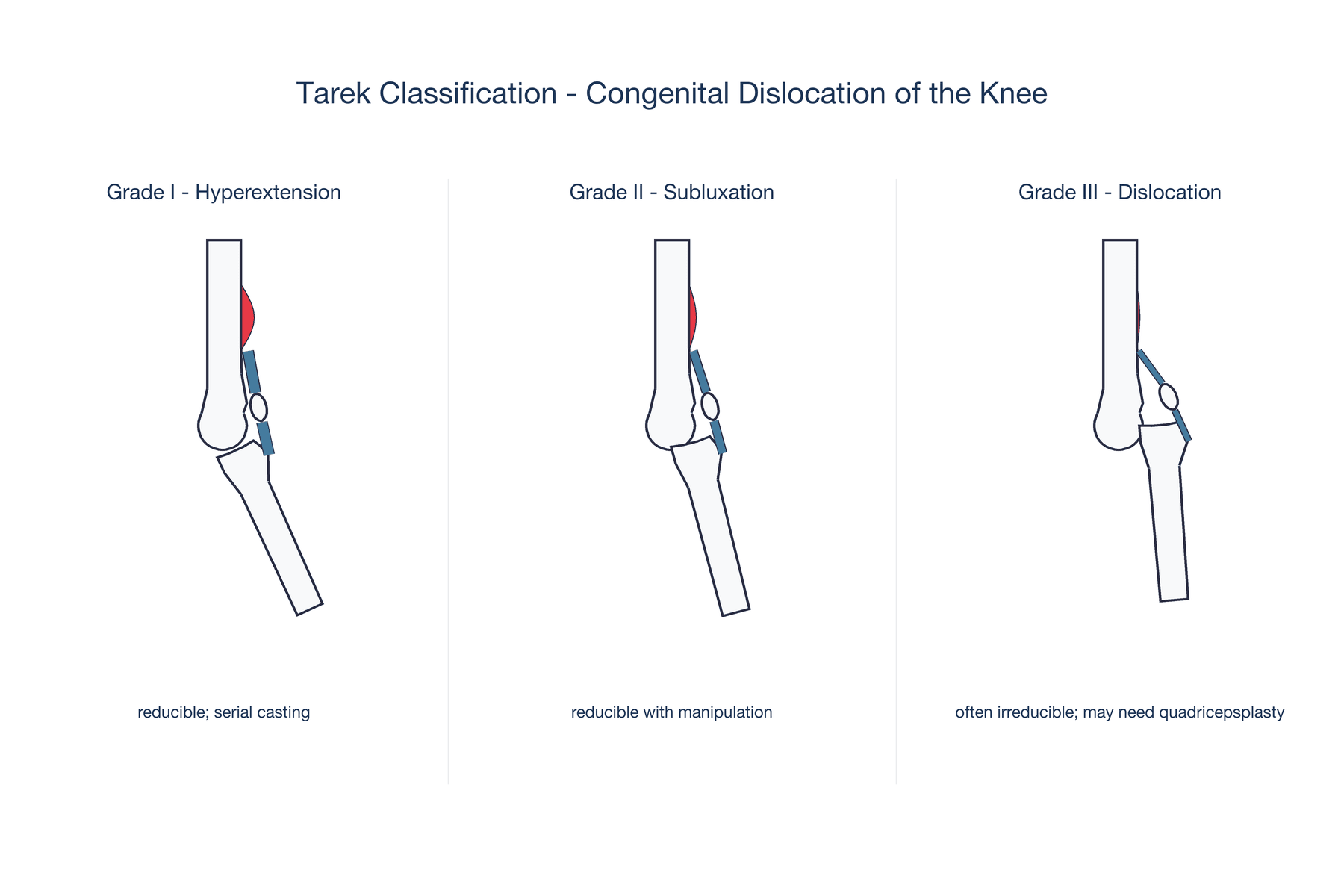
Know the ladder, not the surname. Several systems grade CDK: Tarek's, and the older descriptive sequence Curtis and Fisher used in the very title of their 1969 paper, "congenital hyperextension with anterior subluxation of the knee". They all climb the same three anatomical steps, hyperextension with the joint still congruent, then anterior subluxation, then frank anterior dislocation. What differs between sources is the surname attached and whether the cut-offs are stated as a passive flexion arc. A candidate who describes the step and its flexion arc is right whichever eponym the examiner prefers; a candidate who has memorised only a name is stuck when a different one is used. Tarek's grading is set out here because it states the flexion arc explicitly, which is what actually drives treatment.
- Clinical Finding
- Genu recurvatum: severe hyperextension without displacement, joint congruent
- Passive Flexion
- Beyond 90 degrees
- Reduction
- Passively correctable to flexion
- Treatment
- Serial casting, excellent prognosis
- Clinical Finding
- Anterior subluxation of tibia on femur
- Passive Flexion
- 30 to 90 degrees
- Reduction
- Reducible under gentle manipulation or under anaesthesia
- Treatment
- Serial casting, majority resolve
- Clinical Finding
- Complete anterior dislocation
- Passive Flexion
- Less than 30 degrees
- Reduction
- Irreducible or requires significant force
- Treatment
- Casting trial, most need quadricepsplasty
The grade guides both prognosis and treatment selection. Grade I has the best prognosis and is nearly always treated successfully; Grade III carries a high surgical rate and is the grade in which to check for Larsen syndrome.
Clinical Assessment
History. Ask about the pregnancy and the delivery: breech presentation, oligohydramnios, any prenatal ultrasound findings, and whether the hyperextended knee was noted at delivery. Ask about consanguinity and familial joint disorders. Then ask about the other joints: a foot deformity, a hip click or limited abduction.
The knee. Inspect for hyperextension or anterior dislocation; the skin folds may be abnormal. Palpate the patella, which may be displaced or poorly palpable, and the tibiofemoral relationship. Assess passive flexion, because the arc is what grades the knee.
The rest of the baby. Every newborn with CDK must have:
- Both knees examined, since the condition is so often bilateral
- Both hips screened by clinical examination (Ortolani, Barlow, abduction) and by hip ultrasound
- Both feet examined for clubfoot
- Full musculoskeletal survey of elbows, shoulders and spine for other contractures or dislocations
- Neurological examination to exclude myelodysplasia
- Dysmorphology assessment for Larsen syndrome features: prominent forehead, hypertelorism, depressed nasal bridge
Failing to screen for DDH in a baby with CDK is a significant clinical error.
A note on "40-100 percent", because it is quoted throughout this page and it is not really a figure. A range whose upper bound is 100 percent and whose lower bound is 40 percent cannot be used to estimate anything for an individual baby; it is the spread across small series that differ in whether they studied isolated or syndromic CDK, and in whether "DDH" meant frank dislocation or any sonographic dysplasia. Do not quote it as a prevalence in a viva. The only inference that survives the spread is the one that changes behaviour: the association is high enough that every CDK baby gets hip imaging irrespective of how normal the hip examination feels. The hip examination is itself less reliable here, because a stiff hyperextended knee makes Ortolani and Barlow testing difficult to perform properly, which is why the ultrasound is mandatory rather than confirmatory.
- Key Feature
- Hyperextension or anterior dislocation at birth
- Discriminating Finding
- Anterior tibial displacement, tight quadriceps
- Management
- Serial casting, possible quadricepsplasty
- Key Feature
- Hyperextends but flexes fully and easily
- Discriminating Finding
- No fixed deformity, reduces with gentle flexion
- Management
- Observation, usually resolves spontaneously
- Key Feature
- Limited flexion without frank dislocation
- Discriminating Finding
- Quadriceps tight, joint not subluxed
- Management
- Stretching programme, rarely surgery
- Key Feature
- Stiff knee with multiple other joint contractures
- Discriminating Finding
- Multiple rigid joints, absent skin creases
- Management
- Multidisciplinary, early casting, often surgical
- Key Feature
- Multiple large joint dislocations
- Discriminating Finding
- Bilateral knee dislocations, hip dislocations, characteristic facies
- Management
- Genetic workup, staged surgical reduction
Positional or true? A positional hyperextended knee is soft and passively correctable to full flexion without resistance, and the tibiofemoral joint is congruent on radiographs. True CDK has a firm endpoint to passive flexion, the tibia is anteriorly displaced, and radiographs show anterior subluxation or dislocation. If you can flex the knee to 90 degrees easily with no firm resistance, it is likely positional and will resolve without casting.
Investigations
Radiographs. AP and lateral views of both knees at diagnosis. On the lateral, look for anterior displacement of the tibia relative to the femur, the position of the patella and the physeal status. Grade I may appear near-normal with hyperextension only; in Grade III the tibia is obviously displaced anterior to the femoral condyles. The diagnosis is primarily clinical: radiographs confirm the degree of tibial displacement and guide grading, and casting is not delayed while waiting for advanced imaging.
Hip ultrasound. At 6 weeks, by the Graf technique or dynamic ultrasound, in every CDK baby regardless of the clinical hip examination. This is the most important investigation, because DDH coexists so frequently and has its own treatment window.
MRI. For Grade III or syndromic cases before quadricepsplasty. It shows cruciate ligament status, meniscal position and cartilage integrity, and identifies the absent or abnormal structures that affect the surgical approach.
Skeletal survey and genetics. For bilateral CDK, multiple joint dislocations or dysmorphic features. Look for spinal anomalies, other joint dislocations and features of skeletal dysplasia; request FLNB gene testing for Larsen syndrome, and chromosome analysis if indicated.
Management Algorithm
The principle. Gradual reduction by serial casting, first line for every grade; in Grade I, passive stretching accompanies it and casting runs about 4-6 weeks. Casts are changed weekly with progressively more knee flexion, and the goal is a congruent reduction with at least 90 degrees of flexion. Grade I and II usually reduce within 6-8 weeks; failure of closed treatment by 3 months of age indicates surgery.
The first cast. Begin within the first few days of life. A well-padded long-leg plaster holds the knee in the maximum obtainable flexion, even if that is still hyperextended, moulded to hold the tibia posteriorly; the aim of the first cast is to correct hyperextension toward neutral.
Weekly changes. Each cast increases flexion by 10-15 degrees, with a posterior moulding force on the tibia to maintain the reduction as flexion increases. At every change check the skin, the neurovascular status and the hip position. A Grade I knee typically reaches neutral to 30 degrees of flexion by week 2-3.
Reduction. The target is at least 90 degrees of knee flexion with a stable reduction. A Grade I or II knee is usually reduced and stable by 6-8 weeks, and once stable, casting gives way to bracing. A Grade III knee may still have anterior tibial displacement at this point; if it is not progressing, plan surgery.
Maintenance, weeks 8-12. A night-time knee flexion splint for a further 4-6 weeks, with gentle passive and active range-of-motion exercises. Watch for relapse into hyperextension, and follow hip stability and gait development. This is the stage at which a dysplastic hip is treated, not the neonatal one; the sequencing is set out below.
Never force the knee into flexion in a single cast change: the risks are physeal injury (distal femoral or proximal tibial growth plate), peroneal nerve palsy, femoral fracture and neurovascular compromise. If reduction is not progressing after 6-8 weeks of proper serial casting, surgery is indicated rather than continuing futile casting.
Knee before hip. When CDK and DDH coexist, as they frequently do, the knee is reduced first. The reason is mechanical: the Pavlik harness and the hip spica hold the hip in flexion and abduction, which requires a flexed knee. A knee fixed in hyperextension or anterior dislocation cannot be positioned in either device, so treating the hip first is futile and may even displace it. Serial casting, or surgery, is therefore directed at restoring knee flexion first; once the knee flexes past roughly 90 degrees a Pavlik harness can be applied, and one device may then treat a reducible knee and a dysplastic hip together. "Concurrent" describes that stage, not the neonatal one. In bilateral Grade III disease the same sequencing, knees then hips, dictates the order of any staged surgery. The hip side of the plan is set out in DDH treatment options; where a neurological cause is suspected, see myelomeningocele.
Complications
- Incidence
- 10-20 percent after casting alone
- Risk Factors
- Grade III, syndromic, delayed treatment
- Management
- Night splinting, extension osteotomy if severe in older child
- Incidence
- Rare but reported with forced flexion
- Risk Factors
- Aggressive casting, intraoperative hyperflexion
- Management
- Immediate release of flexion position, nerve exploration if no recovery
- Incidence
- Rare with proper technique
- Risk Factors
- Forced manipulation during casting or surgery
- Management
- Monitor growth with serial radiographs; reconstructive osteotomy if deformity develops
- Incidence
- 15-25 percent in Grade III
- Risk Factors
- Absent cruciate ligaments, inadequate capsular release
- Management
- Cruciate reconstruction in older child, brace management
- Incidence
- Common after quadricepsplasty
- Risk Factors
- Scarring, arthrogryposis
- Management
- Physiotherapy, gentle manipulation under anaesthesia
- Incidence
- Expect a minor lag even when power returns to Grade 5
- Risk Factors
- Inherent to V-Y lengthening of the extensor mechanism
- Management
- Warn at consent; quadriceps strengthening; usually minor and compatible with community walking
- Incidence
- Demonstrated on 3D gait analysis in operated knees
- Risk Factors
- Any operation on the extensor mechanism; type-3 disease
- Management
- Gait analysis once ambulatory; counsel that scores are near-normal but high-demand activity is limited
- Incidence
- 2 of 16 knees in one casting-and-tenotomy series
- Risk Factors
- Force applied during physiotherapy, not only during casting
- Management
- Resolved with time in both; extend the never-force principle to the therapist, not just the cast room
- Incidence
- Significant if hips not screened
- Risk Factors
- Failure to perform hip ultrasound
- Management
- Late DDH treatment is more complex; always screen hips early
Prevention. The most important preventable complication is missed DDH, which is why every child with CDK has hip screening by clinical examination at birth and ultrasound at 6 weeks. The second is physeal injury from forced manipulation: cast changes progress gradually, never forcing the knee beyond what it will comfortably accept. Finally, peroneal nerve palsy is prevented by avoiding excessive flexion in a single cast and by monitoring neurovascular status after every cast change.
Outcomes and Prognosis
- Functional Outcome
- Excellent; near-normal ROM
- Residual Deformity
- Minimal or none
- Long-term Function
- Normal gait and activities
- Functional Outcome
- Good to excellent
- Residual Deformity
- Mild residual hyperextension possible
- Long-term Function
- Functional gait, may have mild cosmetic concern
- Functional Outcome
- Good functional result
- Residual Deformity
- Variable; residual laxity or stiffness common
- Long-term Function
- Ambulatory, may have limitations in high-demand activities
- Functional Outcome
- Guarded
- Residual Deformity
- Residual stiffness and deformity common
- Long-term Function
- Ambulation achievable but with limitations
- Functional Outcome
- Variable
- Residual Deformity
- Multiple joint issues persist
- Long-term Function
- Requires lifelong multidisciplinary care
How to read the table. The gradient is right, but "good functional result" for an operated Grade III understates what the long-term data show and overstates what you should promise. In Honcharuk's series the operated knees had less flexion in swing, less overall motion, greater coronal instability and slower gait than controls, and the authors concluded that surgery for type-3 CDK will not likely restore normal knee function because the joint is teratologic (PMID 38666580): the knee was never normally formed, so reduction is achievable but normality is not.
The reassurance, from the same unit. In Oetgen's series these children walked without braces, averaged 112 degrees of arc and scored comparably to normal controls, with limitations only in high-demand activity, and 7 of 9 knees showed instability on examination that none of them felt (PMID 20357585). Clinically detectable instability is not the same as functional instability, and both facts belong in the same conversation with a parent. Both are true: reduction and a functional knee are realistic; a normal knee is not. The diagnosis matters more than the operation, since idiopathic CDK had the most control-like gait, then Larsen syndrome, then arthrogryposis.
Best prognosis
- Grade I or II
- Isolated CDK, with no syndrome
- Treatment started within the first week of life
- Compliant serial casting
Poor prognosis
- Grade III
- Bilateral involvement
- Underlying arthrogryposis or Larsen syndrome
- Delayed treatment initiation beyond 3 months
Guidelines, Registries & Global Practice
- CDK incidence approximately 1 per 100,000 live births worldwide
- Female predominance (2:1 to 3:1) consistently reported across populations
- Breech association reported globally but may vary with local breech delivery rates
- Syndromic forms (arthrogryposis, Larsen) appear to have similar prevalence across ethnic groups
- High-resource: neonatal identification, immediate serial casting with paediatric orthopaedic follow-up, MRI pre-operatively
- Limited-resource: diagnosis may be delayed; plaster casting is low-cost and effective; surgery in specialist paediatric centres
- Universal principle: serial casting is the first-line treatment globally, regardless of resources
- Key message: the cost of treatment is in the expertise and follow-up, not the materials
- Diagnosis
- Clinical examination + lateral radiograph; classify by Tarek
- Conservative Treatment
- Serial casting from birth, weekly changes, 6-8 week target
- Surgical Approach
- Curtis-Fisher quadricepsplasty at 3-6 months if casting fails
- Diagnosis
- Same clinical-radiographic approach; mandatory hip screening
- Conservative Treatment
- Same serial casting principles; some advocate earlier surgical referral for Grade III
- Surgical Approach
- V-Y quadricepsplasty; some centres use mini-open tenotomy first, or femoral shortening as an alternative
- Diagnosis
- Clinical grading + AP/lateral knee radiographs + hip ultrasound
- Conservative Treatment
- Immediate serial casting; 3-month threshold for surgical decision
- Surgical Approach
- Curtis-Fisher or modifications; cruciate reconstruction considered in older children
There is no dedicated registry for congenital knee dislocation. The evidence base consists entirely of retrospective case series and expert opinion. No randomised controlled trials exist comparing casting protocols or surgical techniques. Management is therefore guided by clinical grading (Tarek), institutional experience, and the consistent principle that early, gradual serial casting gives the best outcomes for Grades I and II.
Record in every newborn with CDK:
- Tarek grade (I, II, or III) with clinical description
- Serial casting start date and weekly flexion progression
- Hip examination findings and hip ultrasound arranged
- Foot examination findings
- Full musculoskeletal survey for other contractures or dislocations
- Genetic or syndromic referral if bilateral or Grade III
- Parent counselling: treatment plan, timeline, and prognosis
Missed DDH in a child with CDK is a recurring and preventable error. Always document hip screening in the CDK chart.
MCQ Practice Points
Q: What is the Tarek classification of congenital knee dislocation? A: Grade I = genu recurvatum (hyperextension without displacement, reducible). Grade II = anterior subluxation of tibia on femur (reducible with manipulation). Grade III = complete anterior dislocation (irreducible or requires significant force). Grade correlates with treatment: Grades I and II usually respond to casting; Grade III often requires quadricepsplasty.
Q: What is the most common association with congenital dislocation of the knee? A: Developmental dysplasia of the hip (DDH), reported in 40-100 percent of CDK cases. Every newborn with CDK must have clinical hip examination (Ortolani, Barlow) and hip ultrasound at 6 weeks. Other common associations include clubfoot (30-50 percent), arthrogryposis, and Larsen syndrome.
Q: What is the first-line treatment for congenital dislocation of the knee? A: Serial long-leg casting, beginning immediately after birth. Casts are changed weekly with progressive increase in knee flexion of 10-15 degrees per week. The goal is congruent reduction with at least 90 degrees of flexion within 6-8 weeks. If casting fails by 3 months, surgical quadricepsplasty (Curtis-Fisher procedure) is indicated.
Q: Describe the Curtis-Fisher quadricepsplasty for resistant CDK. A: V-Y lengthening of the quadriceps tendon combined with anterior capsular release. Through an anterior longitudinal incision, the contracted anterior capsule is released medially and laterally, the quadriceps tendon is divided in a V pattern and repaired in a lengthened Y configuration, and tibiofemoral reduction is confirmed. Post-operative casting at 60-90 degrees flexion for 4-6 weeks.
Q: What syndrome should you suspect in a newborn with bilateral knee dislocations, bilateral hip dislocations, and characteristic facies? A: Larsen syndrome, an autosomal dominant or recessive condition caused by FLNB gene mutations. Features include multiple large joint dislocations, prominent forehead, hypertelorism, depressed nasal bridge, and spinal anomalies (scoliosis, cervical kyphosis). Management requires staged surgical treatment and genetic counselling.
Exam Viva Scenarios
Practise clinical reasoning and management decisions out loud
“A newborn girl is noted at delivery to have a hyperextended left knee. The knee rests at 30 degrees of hyperextension. It can be flexed passively to 60 degrees but with firm resistance. The right knee is normal. The baby was born in breech position. What is your diagnosis, classification, and management plan?”
“A 4-month-old infant with bilateral congenital knee dislocations has undergone 12 weeks of serial casting without achieving reduction. The knees remain in anterior dislocation with fixed hyperextension. The child also has bilateral hip dislocations. No dysmorphic features are noted. What is your next step?”
Key Facts
- CDK = hyperextension or anterior dislocation of the knee present at birth
- Incidence approximately 1 per 100,000 live births; female predominance 2-3:1
- Tarek grading: Grade I (recurvatum), Grade II (subluxation), Grade III (complete dislocation)
- Quadriceps fibrosis and anterior capsule contracture are the primary pathoanatomy
Associations (Must Screen)
- DDH: 40-100 percent association; clinical exam + hip ultrasound mandatory
- Clubfoot: 30-50 percent; examine both feet at birth
- Arthrogryposis: multiple joint contractures suggest underlying myopathic or neuropathic cause
- Larsen syndrome: multiple joint dislocations with flat facies; FLNB gene
Treatment Ladder
- First-line: serial casting from birth, weekly changes, progressive flexion 10-15 degrees per week
- Target: congruent reduction with at least 90 degrees flexion within 6-8 weeks
- If casting fails by 3 months: Curtis-Fisher quadricepsplasty (V-Y lengthening + anterior capsular release)
- Syndromic CDK: higher surgical rate; staged procedures; realistic expectations
Surgical Details
- Curtis-Fisher: anterior approach, V-Y quadriceps lengthening, anterior capsule release
- Intraoperatively assess cruciate ligaments (often absent in Grade III)
- Post-op: long-leg cast at 60-90 degrees flexion for 4-6 weeks
- Address DDH after knee stability is achieved (staged approach)
Complications
- Residual hyperextension: night splinting, osteotomy if severe in older child
- Peroneal nerve palsy: avoid forced flexion; immediate release if occurs
- Physeal injury: never force manipulation; monitor growth serially
- Missed DDH: the most preventable error; always screen hips in CDK
Evidence Base and Key Trials
Quadricepsplasty for congenital dislocation of the knee and congenital quadriceps contracture
- The authors wrote this paper because they encountered THREE specific problems with the original Curtis and Fisher technique: wound dehiscence, insufficient lengthening of the quadriceps, and instability of the knee
- Modified technique in 20 children (33 knees) using a LATERAL incision, raising a long tongue of rectus femoris and mobilising the vasti without dividing the lateral retinaculae as far as the collateral ligaments
- Primary wound healing in 32 of 33 knees; lengthening adequate to allow flexion to 90 degrees; mean follow-up 63 months
- In non-syndromic CDK quadriceps power recovered to Grade 5, but MINOR DEGREES OF EXTENSOR LAG were noted, and minor joint instability was present in a proportion
- Results were better in non-syndromic CDK than in arthrogryposis, though not all differences were significant; the majority of children were community walkers
Congenital hyperextension with anterior subluxation of the knee: surgical treatment and long-term observations
- Described the V-Y quadriceps lengthening with anterior capsular release for resistant CDK
- Reported successful reduction in the majority of cases unresponsive to casting
- Emphasised the importance of anterior capsular release in addition to quadriceps lengthening
- Recommended casting for at least 6 weeks before considering surgical intervention
A minimally invasive treatment protocol for the congenital dislocation of the knee
- 8 consecutive patients with 16 congenitally dislocated knees, treated by a single surgeon; mean age at presentation 5.3 weeks, mean follow-up 33 months
- Protocol was serial casting followed by a MINI-OPEN quadriceps tenotomy (not a percutaneous tenotomy)
- Serial casting ALONE achieved correction in only 3 of the 16 knees; the other 13 had an average of 7 casts (range 5 to 9) before surgery
- 10 knees had mini-open tenotomy alone and 3 needed an additional anterior capsulotomy; 2 knees recurred and needed further surgery
- 2 knees sustained plastic deformation of the proximal tibia during physiotherapy, which resolved with time
- Final outcome excellent in 11 knees (69%), good in 3 (19%), fair in 2 (12%)
Functional results after surgical treatment for congenital knee dislocation
- 7 patients (9 knees) treated surgically, reviewed at a mean 12 plus or minus 6 years with examination, Lysholm, PODCI and 3D gait analysis
- This is a head-to-head comparison of QUADRICEPSPLASTY versus FEMORAL SHORTENING - the two surgical approaches used for CDK
- Total knee arc averaged 112 degrees, with 8 of 9 knees flexing beyond 90 degrees
- 7 of the 9 knees had some instability on examination, yet NO patient used a brace to walk
- Functional scores were good and comparable to normal controls, with limitations seen only in higher-demand activities
- Gait analysis showed a STIFF-KNEE GAIT pattern versus controls; there were no clinical or functional differences between the two operations
Long-term outcomes of surgically treated congenital dislocation of the knee
- 12 patients operated 1985-2015 (mean age 19 months) and studied 9 to 30 YEARS later with gait analysis and patient-reported outcomes: 3 idiopathic, 5 Larsen syndrome, 4 arthrogryposis
- Compared the two operations for type-3 dislocation: 11 knees had QUADRICEPSPLASTY (QP) and 7 had FEMORAL DIAPHYSEAL SHORTENING (FS)
- Versus controls, operated knees had less flexion in swing, less overall motion, greater coronal instability and slower gait (all p less than 0.05)
- QP knees had MORE instability in midstance (p=0.03) and less flexion during gait than FS knees, and trended toward lower KOOS and Lysholm scores - in this small series FEMORAL SHORTENING produced better gait mechanics and patient-reported outcomes than quadricepsplasty
- The authors conclude surgery for type-3 CDK will not likely restore normal knee function, suggesting a TERATOLOGIC joint abnormality rather than a purely positional one
- Idiopathic CDK had the most control-like gait, then Larsen syndrome, then arthrogryposis; idiopathic had a better UCLA Activity Score than arthrogryposis (p=0.03)