Unilateral vs Bilateral | MRI Before Reduction Debate | Urgent Decompression for SCI
- Bilateral dislocation = translation more than 50% = complete SCI in 50%
- Unilateral dislocation = translation approximately 25% = root injury common
- MRI before reduction is for the patient who CANNOT BE EXAMINED - not a clock, and not 'neurologically intact' (PMID 12431286)
- Closed reduction safe with prior MRI or awake monitored reduction
- Posterior approach preferred for reduction, anterior for discectomy if needed
- “25% translation suggests unilateral, 50%+ suggests bilateral
- “Awake closed reduction allows real-time neurological monitoring
- “Disc herniation is common and rises with severity: Rizzolo found it in 42% of all cervical trauma but 80% of BILATERAL facet dislocations - this is the MRI debate crux
- “Posterior approach allows direct facet manipulation and reduction
Overview and Epidemiology
A facet dislocation is a spectrum rather than a single lesion, running from a unilateral perched facet to bilateral locked facets with a complete cord injury. All of it is high-energy: road traffic collisions, diving and falls.
Mechanism. Flexion-distraction is the primary mechanism. Add a rotational component and one facet fails, which gives a unilateral dislocation; pure flexion-distraction fails both and gives a bilateral one.
On the lateral radiograph, count vertebral body widths of displacement. 25% anterior translation of the vertebral body suggests a unilateral dislocation; more than 50% indicates bilateral. It is a quick screening tool before CT.
Anatomy and Biomechanics
The facet joints. The subaxial facets sit at roughly 45 degrees to the horizontal, the superior articular process facing posterolaterally and the inferior process facing anteromedially. The capsule is the primary restraint to flexion, and it is the structure a flexion force overpowers.
How a facet locks. With the capsule torn, the superior facets slide superiorly and anteriorly; carried past the inferior facet they come to rest in front of it and are locked. The same capsular rupture is what allows the abnormal translation seen on the lateral film.
Stability. These are three-column injuries. The posterior tension band is destroyed and the disco-ligamentous complex is always disrupted, so they are unstable and require surgical stabilisation.
- Unilateral
- Approximately 25%
- Bilateral
- More than 50%
- Unilateral
- Present
- Bilateral
- Minimal
- Unilateral
- 30%
- Bilateral
- 50-75%
- Unilateral
- Common (same level)
- Bilateral
- Less common
- Unilateral
- Moderate
- Bilateral
- High
- Unilateral
- Flexion + rotation
- Bilateral
- Pure flexion-distraction
What the cord and roots do. A unilateral dislocation compresses the root at the level of the injury, so a C6-7 dislocation compresses the C7 root, and those root injuries generally recover well. A bilateral dislocation compresses the cord, with a complete injury in up to 50%; an incomplete injury carries a better prognosis than a complete one.
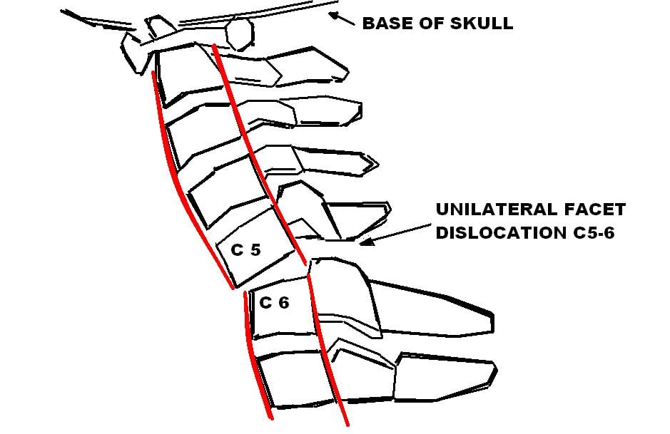
Classification
Perched or locked. Perched facets sit tip-to-tip, balanced on one another. A locked facet has jumped past its partner completely and lies in front of it. Perched facets may reduce more easily; locked facets often require open reduction.
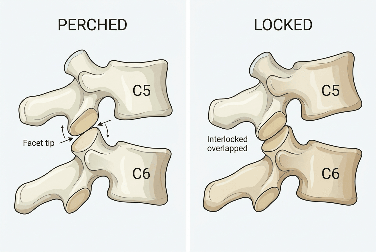
Practical Classification
The practical description is how many facets are out, whether they are perched or locked, and whether a facet is broken.
- Definition
- Facet tip-to-tip
- Key Features
- Rotation, approximately 25% translation
- Definition
- Facet jumped completely
- Key Features
- More rotation, root symptoms common
- Definition
- Both facets subluxed
- Key Features
- Less than 50% translation, unstable
- Definition
- Both facets jumped
- Key Features
- More than 50% translation, high SCI rate
- Definition
- Facet fracture-dislocation
- Key Features
- May affect reduction strategy
Clinical Assessment
History. The mechanism (collision, dive, fall), any transient neurological symptoms at the scene, where the neck hurts, and what the patient can feel and move now.
Inspection. Swelling, bruising and deformity of the neck, and torticollis, which is classic for a unilateral dislocation: the chin is rotated away from the side of the dislocated facet. Log-roll and examine the whole spine.
Neurological examination. Motor testing myotome by myotome, dermatomal sensation including the perianal area, deep tendon reflexes and Babinski. Look specifically for sacral sparing, because it separates ASIA A from ASIA B and so decides the prognosis.
What each pattern looks like. A unilateral dislocation gives torticollis, cervical tenderness and possibly a radiculopathy. A bilateral dislocation gives severe pain, the patient frequently holding the head with both hands, with quadriparesis or quadriplegia if the cord is injured.
Grade the patient before you intervene, and write it down.
- A - complete: no motor or sensory function below the level
- B - sensory incomplete: sensation present, no motor function
- C - motor incomplete: fewer than half the muscles grade 3 or better
- D - motor incomplete: at least half the muscles grade 3 or better
- E - normal
Differential Diagnosis
The painful, deformed, post-traumatic cervical spine has several mimics. Distinguishing them changes urgency and approach.
- Distinguishing Features
- Approx 25% translation, rotation, torticollis, same-level radiculopathy
- Key Discriminator
- Rotation with translation under 50%
- Distinguishing Features
- More than 50% translation, high SCI rate, severe instability
- Key Discriminator
- Translation more than 50% without rotation
- Distinguishing Features
- Fractured superior/inferior articular process, may be reducible without lock
- Key Discriminator
- Fracture line on CT through the facet
- Distinguishing Features
- Widened interspinous distance, focal kyphosis, no jumped facet
- Key Discriminator
- Facets reduced/perched, ligamentous only
- Distinguishing Features
- Anterior column comminution, retropulsion, axial-flexion mechanism
- Key Discriminator
- Vertebral body fracture dominates, facets congruent
- Distinguishing Features
- Upper cervical, fixed torticollis, paediatric/atraumatic
- Key Discriminator
- C1-C2 level, abnormal odontoid relationship
- Distinguishing Features
- Fused spine, trivial trauma, highly unstable transverse fracture
- Key Discriminator
- Bamboo spine, fracture through ankylosed segment
Investigations
Imaging answers three questions: what the bones are doing, what the disc and cord are doing, and whether the vertebral artery survived.
Imaging Algorithm
First-line imaging. Shows facet relationship (perched, locked), associated fractures, translation percentage. 3D reconstructions helpful for surgical planning.
Critical for disc assessment. Shows disc herniation, cord contusion and ligamentous injury, and so influences reduction strategy. Whether to obtain it before reduction is the debate below.
Vertebral artery assessment. Injury in 20-40% of facet dislocations. Particularly important C1-C3.
Confirm reduction. Assess for iatrogenic fracture, hardware position if immediate fixation.
What to record from the CT. The report that is useful to the surgeon who has to reduce the injury says five things:
- Facet relationship - normal, perched or locked
- Translation, as a percentage of vertebral body width
- Rotation, seen as asymmetry of the spinous processes and facets
- Associated fractures of the facet, vertebral body or lamina
- Canal compromise, as percentage occlusion
On CT axial images, the naked facet sign shows an "empty" facet joint with the superior articular process displaced anteriorly. This confirms jumped facet.
Blunt vertebral artery injury (BVAI) accompanies cervical facet dislocations frequently because the displaced lateral mass and transverse foramen shear the artery - subaxial dislocations, fractures through the foramen transversarium, upper-cervical (C1-C3) fractures and rotational patterns all raise the risk.
Screen with CT angiography using the (modified) Denver criteria - any facet dislocation or subluxation, a fracture involving the transverse foramen, a C1-C3 fracture, or a displaced or rotational cervical injury should trigger CTA.
Grade with the Biffl/Denver scale:
- I - intimal irregularity
- II - dissection or intramural haematoma with luminal narrowing
- III - pseudoaneurysm
- IV - complete occlusion
- V - transection with active extravasation
Why it matters. Most are unilateral and clinically silent, because contralateral and collateral flow compensate; the real danger is a delayed posterior-circulation (brainstem or cerebellar) stroke.
Treatment is antithrombotic - antiplatelet (aspirin) or therapeutic anticoagulation (heparin) for grades I-IV, balanced against the bleeding risk of the polytrauma or cord-injured patient and the timing of spinal surgery. A grade III pseudoaneurysm or a symptomatic lesion may need endovascular treatment, and grade V needs urgent intervention.
Operative relevance. If one artery is already occluded, iatrogenic injury to the patent contralateral artery during lateral mass or pedicle screw placement can be catastrophic. Know the CTA before you instrument.
Management
The clock. An incomplete cord injury is the emergency: reduce urgently, with decompression inside 24 hours. A complete injury is less urgent, but it is still reduced. A root injury is painful and distressing and allows time for a full workup.
Pre-Reduction MRI: The Controversy
The question. Should MRI be obtained before an attempt at closed reduction? Reduction can push herniated disc material into the canal and onto the cord, and disc herniation is common in these injuries.
How common. Rizzolo found acute disc herniation in 42% of all cervical trauma imaged within 72 hours, but in 80% of bilateral facet dislocations and 100% of anterior cord syndromes. That gradient is the crux of the argument: the bilateral dislocation is both the pattern most likely to have a disc in the way and the pattern most likely to have an injured cord.
The case for imaging first. Beyond the disc, MRI shows cord injury for prognostication and allows the whole operation to be planned in one sitting.
The case against waiting. Time is cord, and STASCIS showed that early decompression improves outcomes. An awake reduction with continuous monitoring is safe, and reduction under anaesthesia is safe once the MRI has been seen.
Where this sits among cervical injuries. A facet dislocation is one pattern within the wider problem set of cervical spine fracture, and the reason it is treated as its own emergency is the cord: the deficit, its grading and the decompression evidence belong to spinal cord injury. If the translation is degenerative rather than traumatic, the reader wants cervical spondylolisthesis instead - the same slip on a lateral film with an entirely different clock and no urgency. When the operation is anterior, it is the ACDF exposure.
The discriminator is not the neurology and it is not a clock - it is whether the patient can be examined during the reduction. That is how the AANS/CNS guideline frames it (Hadley, Neurosurgery 2002, PMID 12431286), and it is the version that survives a viva.
- Awake and examinable - early closed reduction with craniocervical traction is recommended, examining the patient continuously as you go. Pre-reduction MRI will show a disrupted or herniated disc in one third to one half of these patients, but the guideline states those findings "do not seem to significantly influence outcome after closed reduction in awake patients", so the usefulness of pre-reduction MRI here is explicitly uncertain. An awake reduction is itself the monitor.
- Cannot be examined - intubated, obtunded, head-injured, intoxicated - MRI before any attempted reduction. This is the group the rule exists for, because you have lost the only real-time neurological monitor you had.
- Failed closed reduction - MRI is recommended before proceeding.
- A significant disc herniation on pre-reduction MRI is a relative indication for ventral decompression before reduction.
- An additional rostral (more cranial) injury is a contraindication to closed traction reduction.
Do not quote a fixed number of hours for "MRI if available within X". No such threshold is established, and practice is genuinely variable - a Spine Trauma Study Group survey of 25 fellowship-trained surgeons found interrater reliability of the treatment decision was very poor, and got worse once an MRI was available in complete cord injury (PMID 19342930). What you can defend is the examinability rule above.
- MRI before reduction?
- Usefulness explicitly uncertain
- Reduction method
- Early awake closed traction reduction
- Key pearl
- The examination during reduction is the monitor
- MRI before reduction?
- Yes - before any attempt
- Reduction method
- Reduction once the imaging has been seen
- Key pearl
- This is the group the rule exists for
- MRI before reduction?
- Not if it delays decompression
- Reduction method
- Urgent reduction (closed or open)
- Key pearl
- STASCIS: less than 24h decompression improves outcomes
- MRI before reduction?
- Not mandatory before reduction
- Reduction method
- Closed or open reduction
- Key pearl
- Prognosis already poor - prioritise reduction
- MRI before reduction?
- Yes - allows full workup
- Reduction method
- Posterior open or closed awake
- Key pearl
- Not as urgent - allows complete planning
- MRI before reduction?
- Recommended before proceeding
- Reduction method
- Open reduction
- Key pearl
- Usually truly locked facets or interposed disc or bone
If the MRI shows a disc herniation. Many surgeons prefer anterior discectomy first, removing the disc that reduction could otherwise push into the canal, and then reducing and fusing posteriorly - or doing both in one anterior-posterior procedure.
A facet dislocation that presents late - a missed diagnosis, or a patient reaching definitive care days to weeks after injury - behaves very differently from the acute injury and is a favourite viva curveball. Once the facets are fixed by early fibrosis and callus, closed traction reduction usually fails, and a forceful attempt is both futile and dangerous.
Re-characterise the deformity and the cord and canal status with CT and MRI before any intervention, then plan an open reduction that almost always requires direct facetectomy, resecting the locked articular processes to disengage the bony block. Frequently this must be combined with an anterior release or discectomy when an organised interposed disc or anterior tethering prevents reduction, which makes it a planned anterior-posterior procedure.
Reduction of a long-standing deformity carries a higher neurological risk because the cord has accommodated to the malalignment. Run neuromonitoring, and be prepared to accept an in-situ fusion if the signals deteriorate. A patient with an established complete injury and a stable, painless, fused malunion may be best served by in-situ stabilisation rather than a risky realignment. Counsel the patient that neurological and pain outcomes after late reduction are less predictable than after acute reduction.
Surgical Technique
Consent. Name neurological worsening, which is higher during the reduction itself, failure of reduction and the need for a different or second approach if instability persists, hardware failure, non-union, and adjacent segment disease in the long term. The figures are in the complications table below.
Equipment. Neuromonitoring with SSEPs and MEPs, a Mayfield head holder for prone positioning, lateral mass screws in a range of sizes, levering instruments (Cobb elevator, Penfield, lamina spreader) and fluoroscopy to confirm both reduction and hardware.
Complications
- Incidence
- 1-5%
- Prevention/Management
- Neuromonitoring, careful reduction, pre-reduction MRI
- Incidence
- 5-10%
- Prevention/Management
- May need open or combined approach
- Incidence
- 5-10%
- Prevention/Management
- Adequate fixation length, consider combined approach
- Incidence
- 5-10%
- Prevention/Management
- Bone graft, smoking cessation, stable fixation
- Incidence
- Less than 1%
- Prevention/Management
- Pre-op CTA, careful screw placement
- Incidence
- 10-20% at 10 years
- Prevention/Management
- Limit fusion levels
- Incidence
- 5%
- Prevention/Management
- Adequate fixation, compliance with collar
Neurological worsening is the complication that is feared above the others. It follows a disc herniation that was not addressed, a forced reduction or over-distraction, which is why pre-reduction MRI, neuromonitoring and gentle technique are the prevention.
Failed closed reduction occurs in 10-30% of closed attempts, usually because the facets are severely locked or because disc or bone is interposed. The answer is open reduction, not more weight.
Postoperative Care
Rehabilitation Timeline
ICU for a cord injury, neurological checks every 4 hours, drain out at 24-48 hours if one was used, and DVT prophylaxis.
Mobilise in the collar, radiograph to confirm alignment, wound check, and start cord-injury rehabilitation where it applies.
Collar continues, mobilisation progresses, physiotherapy for conditioning, radiograph at 6 weeks.
CT to assess the fusion, collar removed if it has fused, activity increased, and return to work planned.
The collar. A hard collar - Miami J or similar - for 6-12 weeks after surgery. Wean earlier if the fusion is evidently solid and the construct stable, and keep it on longer in osteoporosis or where there is concern about the fixation.
Outcomes and Prognosis
Neurological recovery follows the presenting grade. A complete injury (ASIA A) has a poor neurological prognosis and the effort turns to stability and rehabilitation. An incomplete injury has significant potential to improve, particularly with early decompression. A root injury generally recovers well over 6-12 months.
What else predicts the result. The ASIA grade at presentation, the time to reduction and decompression, the quality of the reduction, associated head and chest injuries, and patient factors such as age and comorbidities.
Sacral sparing (any perianal sensation or voluntary anal contraction) converts ASIA A to B and dramatically improves prognosis. Always check carefully.
Guidelines, Registries & Global Practice
Global epidemiology. Cervical facet dislocations are high-energy injuries of the subaxial spine, most often at C5-6 and C6-7, typically from road traffic collisions, falls and diving. They sit within the broader burden of traumatic spinal cord injury: in the STASCIS cohort the majority of subaxial dislocation/translation injuries presented with complete or incomplete cord injury, and early decompression (less than 24h) more than doubled the odds of a ≥2-grade AIS improvement at 6 months (Fehlings 2012, PMID 22384132). Bilateral locked facets carry the highest cord-injury risk; unilateral injuries more often produce a same-level radiculopathy.
- Recommendation
- Offer decompression ≤24h for adult acute SCI regardless of level; consider early surgery in central cord syndrome (Fehlings 2017, PMID 29164024)
- Evidence Level
- GRADE: conditional, low quality
- Recommendation
- Early closed reduction recommended for awake, examinable patients with cervical fracture-dislocation; reduction safe without prior MRI in this group (Grant 1999, PMID 10413120)
- Evidence Level
- Level III option
- Recommendation
- Subaxial injuries scoring 4 or more should be considered operative; facet dislocations score in the operative range (Vaccaro 2007, PMID 17906580)
- Evidence Level
- Validated classification
- Recommendation
- Major trauma networks: immobilise, transfer to a spinal-capable centre, MRI before reduction in the neurologically intact, urgent surgery for evolving deficit
- Evidence Level
- Consensus / guideline
Practice variation. The pre-reduction MRI question is the main genuine divergence. North American practice (AANS/CNS, supported by Grant 1999) favours immediate awake closed reduction in alert, examinable patients with significant deficit, accepting that reduction can displace disc material but rarely worsens neurology. Many UK/European units prefer MRI before reduction in the neurologically intact, citing the rise in disc herniation after reduction (Vaccaro 1999, PMID 10382247). The shared ground: in incomplete SCI, do not let MRI delay decompression beyond the ≤24h window (Badhiwala 2021, PMID 33357514). In limited-resource settings without rapid MRI or neuromonitoring, awake closed traction reduction with serial neurological checks remains the pragmatic standard.
Registry note. There is no implant joint registry for cervical trauma equivalent to the arthroplasty registries (NJR, AJRR, AOANJRR); evidence is driven by prospective cohorts and pooled analyses such as STASCIS and the Badhiwala 2021 individual-patient pooled analysis rather than registry data.
- Major trauma centres with 24/7 spinal surgery and neuromonitoring
- Early transfer of SCI to a specialised spinal unit
- Coordinated pre-hospital retrieval and spinal immobilisation
- MAP support (target 85-90 mmHg) to limit secondary cord injury
- Pre-reduction MRI in the intact: routine (UK/Europe) vs selective (North America)
- Awake closed reduction availability depends on MRI/monitoring access
- ≤24h decompression target is universal for incomplete SCI
- Anterior-first when disc herniation present is widely accepted
- Baseline neurological exam before any intervention
- ASIA grade at presentation
- Imaging interpretation and timing of MRI
- Decision-making rationale for reduction approach
- Informed consent including neurological worsening risk
- Failure to document baseline neurology
- Delayed recognition of facet dislocation
- Neurological deterioration during reduction without documented monitoring
MCQ Practice Points
Q: On lateral cervical X-ray, what percentage translation suggests bilateral facet dislocation? A: More than 50% translation indicates bilateral dislocation. Approximately 25% suggests unilateral.
Q: What percentage of facet dislocations have associated disc herniation on MRI? A: There is no single number, and the honest answer is stratified by severity. Rizzolo found acute disc herniation in 42% of all cervical trauma within 72 hours, but in 80% of bilateral facet dislocations and 100% of anterior cord syndromes. Grant's closed-reduction series found post-reduction herniation in 22% and disruption in 24%. So quote it as common and severity-dependent - highest in bilateral facet dislocation - rather than as a single figure, and give the clinical point: it is the main argument for pre-reduction MRI in the neurologically intact patient.
Q: What is the maximum traction weight for a C5-6 facet dislocation? A: Approximately 10 lbs per level above injury. For C5-6: 5 levels x 10 = 50 lbs (some allow up to 70-80 briefly).
Q: When reducing a locked facet operatively, should you flex or extend the neck? A: Flex first to unlock the facets (opens the joint), then extend to complete reduction.
Q: A C6-7 facet dislocation will compress which nerve root? A: C7 root - cervical roots exit above their numbered vertebra, so the C7 root exits at C6-7.
Q: What differentiates ASIA A from ASIA B? A: Sacral sparing - any perianal sensation or voluntary anal contraction converts complete (A) to sensory incomplete (B).
Exam Viva Scenarios
Practise clinical reasoning and management decisions out loud
“A 32-year-old man presents after a diving accident. He has bilateral C5-6 locked facet dislocation on CT with ASIA C incomplete quadriplegia. How do you manage this patient?”
“A 45-year-old woman has a unilateral C6-7 locked facet dislocation after a car accident. She has C7 radiculopathy with weakness of her triceps and finger extensors. She is neurologically otherwise intact. Walk me through your management.”
“You have attempted awake closed reduction for bilateral C5-6 locked facets. Despite 70 lbs of traction, the facets remain locked. The patient has incomplete SCI (ASIA C). What do you do now?”
Classification
- Unilateral: approximately 25% translation, single locked facet
- Bilateral: more than 50% translation, both facets locked
- Perched: tip-to-tip, may reduce easier
- Locked: jumped completely, often needs open reduction
MRI Decision
- Disc herniation common (42% all trauma, 80% bilateral facet) - key reason for MRI
- Awake and examinable: reduce early - pre-reduction MRI usefulness uncertain
- Cannot be examined: MRI before any reduction attempt
- Complete SCI: don't delay reduction for MRI
Reduction Methods
- Awake closed: safe with continuous neuro monitoring
- Open posterior: direct facet access, preferred for failed closed
- Weight max: approximately 10 lbs per level above injury
- Flex to unlock, extend to reduce
Approach Selection
- Posterior: direct facet reduction, lateral mass screws
- Anterior: if disc herniation needs removal first
- Combined: disc herniation + facet dislocation
- Posterior first if facet dislocation + disc (then anterior)
Complications
- Neurological worsening: 1-5%
- Failed reduction: 10-30% of closed attempts
- Hardware failure: 5-10%
- Always use neuromonitoring during reduction
Evidence Base
STASCIS: Surgical Timing in Acute Spinal Cord Injury
- Prospective multicentre cohort of 313 adults with acute cervical SCI (182 early, 131 late)
- 19.8% of early-surgery patients improved ≥2 AIS grades at 6 months vs 8.8% in the late group (OR 2.57, 95% CI 1.11–5.97)
- Adjusted odds of ≥2 grade AIS improvement 2.8x higher with early surgery
- Complication rates similar (24.2% early vs 30.5% late, p=0.21) - early decompression is safe and feasible
SLIC: Subaxial Cervical Spine Injury Classification and Severity Scale
- Scores three domains: injury morphology, disco-ligamentous complex (DLC), and neurological status
- Distraction morphology = 3 points; rotation/translation = 4 points; DLC disrupted = 2 points
- Facet dislocation is a distraction/translation injury that almost always scores in the operative range
- Score of 4 or more favours surgery; 3 or less favours non-operative care; raters agreed with the algorithm in 93.3% of cases