Glenoid Track in Shoulder Instability
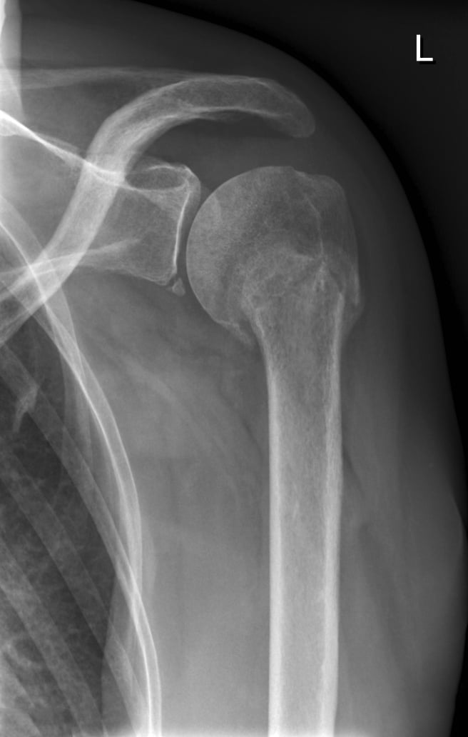
- Bone loss in anterior shoulder instability is BIPOLAR: it involves both the anterior GLENOID (a bony Bankart fragment or attritional/erosive loss) and the posterosuperior humeral head (a HILL-SACHS impaction lesion), and because the two interact during instability, both must be assessed TOGETHER rather than in isolation.
- GLENOID bone loss is quantified on an EN-FACE (sagittal-oblique) reconstruction using a best-fit circle of the inferior glenoid (the inferior glenoid is normally a near-perfect circle), expressing the defect as a percentage of the circle; 3D CT is the GOLD STANDARD for this static assessment, and 3D MRI has been validated as a reliable alternative (with the advantage of also assessing soft tissues).
- Recognised THRESHOLDS guide management: 'critical' glenoid bone loss is classically around 20-25% (above which an isolated soft-tissue Bankart repair has unacceptably high failure), and there is now emphasis on 'SUBCRITICAL' bone loss (around 13.5-20%), where outcomes of isolated Bankart repair are already compromised and augmentation should be considered.
- The GLENOID TRACK concept (Yamamoto/Di Giacomo) is the key dynamic, bipolar tool: the glenoid track is the zone of contact of the glenoid on the humeral head in the provocative abduction-external rotation position (its width is approximately 0.83 times the glenoid diameter minus the glenoid bone-loss width); if the Hill-Sachs lesion extends MEDIAL to the medial margin of the track it is 'OFF-TRACK' and will ENGAGE the glenoid rim (high recurrence risk), whereas if it stays within the track it is 'ON-TRACK'.
- These measurements DRIVE the surgical choice: an ON-TRACK lesion with minimal glenoid bone loss is suitable for an isolated arthroscopic BANKART repair; an OFF-TRACK Hill-Sachs (or significant bipolar loss) needs the Hill-Sachs addressed - typically a Bankart plus REMPLISSAGE (filling the Hill-Sachs with posterior capsule/infraspinatus) - and significant GLENOID bone loss (around 20% or more, or subcritical loss with an off-track lesion or other risk factors) needs a BONY procedure such as a LATARJET (coracoid transfer) or bone block.
- Accurate, reproducible assessment matters because under-estimating bone loss leads to recurrent instability after an inadequate soft-tissue repair: 3D CT (or validated 3D MRI) should be used to measure glenoid bone loss and the Hill-Sachs and to compute the glenoid track / on-off-track status as part of preoperative planning, integrated with clinical risk factors (age, sport, hyperlaxity) and instability scores.
- “Bone loss is BIPOLAR: anterior GLENOID (bony Bankart) + posterosuperior humeral head (HILL-SACHS) - assess BOTH together.
- “Quantify glenoid loss on EN-FACE 3D CT (gold standard; 3D MRI validated) with a best-fit inferior-glenoid circle; subcritical ~13.5-20%, critical ~20-25%.
- “GLENOID TRACK: track width ~0.83 x glenoid diameter - glenoid bone-loss width; Hill-Sachs medial to the track = OFF-TRACK (engages). Drives Bankart (on-track) vs +remplissage (off-track) vs Latarjet (significant glenoid loss).
Bone loss is bipolar (glenoid + Hill-Sachs). Measure glenoid loss on en-face 3D CT (gold standard) with a best-fit inferior-glenoid circle; subcritical ~13.5-20%, critical ~20-25%.
Compute the glenoid track: a Hill-Sachs medial to the track margin = OFF-TRACK (engages, high recurrence). This drives Bankart vs remplissage vs Latarjet.
Bipolar Bone Loss, Quantification & the Glenoid Track
Bone loss in anterior instability is bipolar - both the anterior glenoid (bony Bankart/attritional loss) and the posterosuperior humeral head (Hill-Sachs) - and the two interact, so both are assessed together. Glenoid bone loss is quantified on an en-face (sagittal-oblique) 3D CT with a best-fit circle of the inferior glenoid (3D CT is the gold standard; 3D MRI is a validated alternative); thresholds are subcritical (~13.5-20%) and critical (~20-25%). The glenoid track concept is the key dynamic tool: the track is the glenoid's contact zone on the humeral head in abduction-external rotation (width approximately 0.83 x glenoid diameter minus the glenoid bone-loss width); a Hill-Sachs lesion medial to the medial margin of the track is OFF-TRACK (it engages the rim, high recurrence) whereas one within the track is ON-TRACK. These measurements drive the surgical choice.
| Scenario | Bone status | Typical procedure |
|---|---|---|
| Minimal loss, on-track | Glenoid loss minimal; Hill-Sachs on-track | Arthroscopic Bankart repair |
| Off-track Hill-Sachs (limited glenoid loss) | Hill-Sachs engages; glenoid loss subcritical | Bankart + remplissage |
| Significant glenoid loss | Glenoid loss ~20%+ (or subcritical + off-track / high risk) | Bony procedure (Latarjet / bone block) |
| Severe bipolar loss | Large glenoid + large Hill-Sachs | Latarjet (+/- humeral-side procedure) |
How the Numbers Drive Surgery
- On-track, minimal glenoid loss: isolated arthroscopic Bankart repair.
- Off-track Hill-Sachs (with subcritical glenoid loss): address the Hill-Sachs - Bankart + remplissage (fill the Hill-Sachs with posterior capsule/infraspinatus) to make it functionally on-track.
- Significant glenoid bone loss (~20% or more, or subcritical loss with an off-track lesion / high-risk patient): a bony procedure - Latarjet (coracoid transfer, restoring glenoid arc + the sling effect) or a glenoid bone block.
- Integrate clinical risk: age, sport (contact/collision), hyperlaxity and instability scores modify the thresholds.
- Measure accurately: use 3D CT (or validated 3D MRI) for glenoid loss, the Hill-Sachs and the glenoid track preoperatively - under-estimating bone loss is a key cause of recurrence."
The central error in shoulder instability surgery is performing an isolated soft-tissue Bankart repair while under-estimating or ignoring bone loss, because an off-track Hill-Sachs or significant glenoid bone loss will cause the repair to fail with recurrent instability. The bone loss is bipolar, so both the glenoid and the Hill-Sachs must be measured and combined in the glenoid-track assessment: a Hill-Sachs that is off-track, or glenoid bone loss in the subcritical-to-critical range, signals that the soft-tissue repair needs augmentation - remplissage for the engaging Hill-Sachs, and a bony procedure such as a Latarjet for significant glenoid loss. Use 3D CT (or validated 3D MRI) to quantify bone loss and compute on/off-track status preoperatively, and integrate clinical risk factors, rather than deciding from a plain film alone.
Evidence & Key Studies
3D MRI vs 3D CT for static bone loss and dynamic bipolar (glenoid track) assessment in instability
- 3D CT has been the gold standard for evaluating static glenoid bone loss; 3D MRI was validated as a reliable alternative for both static bone loss and dynamic morphological variables.
- Measured variables included glenoid and humeral bone loss, Hill-Sachs occupancy, glenoid track zones and distance to dislocation; both modalities accurately identified on-track/off-track lesions.
- Inter- and intra-rater reliability was good to excellent, supporting either modality for preoperative bipolar bone-loss and glenoid-track assessment.
Bone-loss-guided treatment: dynamic anterior stabilization with remplissage for off-track/bipolar loss
- Soft-tissue stabilization (dynamic anterior stabilization using the biceps) is indicated for anterior instability with anterior glenoid bone loss of up to about 20%.
- In patients with bipolar bone loss who play contact/collision sports, augmenting with Hill-Sachs remplissage further increases stability and reduces recurrence.
- Treatment selection is explicitly driven by the amount of glenoid bone loss and whether the Hill-Sachs is engaging, supporting the bone-loss/glenoid-track framework.
According to PubMed, 3D CT being the gold standard for static glenoid bone loss (with 3D MRI a validated alternative) and the measurement of glenoid/humeral bone loss, Hill-Sachs occupancy and on/off-track glenoid track zones come from the cited Twomey-Kozak study; the bone-loss-driven treatment thresholds (soft-tissue repair up to ~20% glenoid loss, augmenting with remplissage for bipolar/off-track loss in contact athletes) from the cited de Campos Azevedo technical note. The bipolar-bone-loss concept, the en-face best-fit-circle quantification, the subcritical/critical thresholds, the glenoid-track formula and the Latarjet-for-significant- glenoid-loss principle are standard, well-established teaching. (See also our Anterior Shoulder Instability and Latarjet Procedure topics.)
Clinical Decision Scenarios
Practise clinical reasoning and management decisions out loud
“How do you assess bone loss in a patient with recurrent anterior shoulder instability?”
“How does bone-loss assessment change your surgical choice?”
Mnemonics & Memory Aids
TRACK
Hook:TRACK: Track width formula, Reconstruct on 3D CT, Assess both bones (bipolar), Critical 20-25%, Know procedure (Bankart/remplissage/Latarjet).
Concept
- Bone loss is bipolar: anterior glenoid (bony Bankart) + posterosuperior humeral Hill-Sachs
- Assess both together (they interact)
- Under-estimating bone loss -> recurrence after isolated soft-tissue repair
Quantify glenoid loss
- En-face (sagittal-oblique) best-fit circle of the inferior glenoid
- 3D CT gold standard; 3D MRI validated alternative
- Subcritical ~13.5-20%; critical ~20-25%
Glenoid track
- Track width ~0.83 x glenoid diameter - glenoid bone-loss width
- Hill-Sachs medial to the track margin = OFF-TRACK (engages, high recurrence)
- Within track = ON-TRACK
Procedure selection
- On-track + minimal glenoid loss -> arthroscopic Bankart
- Off-track (subcritical glenoid loss) -> Bankart + remplissage
- Significant glenoid loss (~20%+ / subcritical + off-track / high-risk) -> Latarjet / bone block