Schatzker Classification Explained Type by Type, With CT Signs and Fixation Strategy
The Schatzker classification type by type: mechanism, CT signs and fixation for each tibial plateau fracture, where the system fails, and a model exam answer.
By OrthoVellum Editorial TeamPublished 13 min read
Educational content for clinicians, not medical advice. Editorial policy
Key points
- Schatzker I–III are usually lower-energy lateral plateau injuries (split, split-depression, pure depression); IV–VI are the higher-energy medial, bicondylar and metaphyseal-diaphyseal patterns.
- Type IV, the medial plateau, is a knee-dislocation-equivalent: pulses, ABPI, peroneal nerve and compartments come before any talk of plates.
- Schatzker is drawn on the AP radiograph. A posteromedial fragment was found in 59% of 111 bicondylar fractures on CT, and lateral locking screws may not reliably capture it.
- CT changed the Schatzker type in only 6% of cases in one blinded study; its value is the plan: depression depth, coronal fragments, approach and implant.
- Interobserver agreement for Schatzker on plain films is usually fair to moderate (kappa about 0.41–0.47 in most series), and CT improves it.
On this page8 sections
A tibial plateau fracture gets a Schatzker number on every trauma list, and the number is meant to carry the energy of the injury and the operation it needs. The Schatzker classification does that well for the lateral plateau and for the line between one condyle and two. It is drawn on the AP radiograph, though, and it does not describe the posterior fragments that most often decide the approach. The answer in one sentence: use Schatzker for the pattern and the energy, then plan the incisions from the CT, column by column. The full clinical picture is on the tibial plateau fractures topic page.
What are the six Schatzker types?
Schatzker, McBroom and Bruce published the system in 1979 from the Toronto experience of 1968–1975. It describes six patterns on plain radiographs, and the number broadly tracks energy: types I–III are lower-energy injuries of the lateral plateau, types IV–VI the medial, bicondylar and dissociated patterns of higher-energy trauma. The lateral plateau is involved in 55–70% of plateau fractures, and type II is the most common type.
| Type | Pattern | Mechanism | What CT has to answer | Typical fixation |
|---|---|---|---|---|
| I | Lateral split (wedge), no depression | Low-energy valgus; younger, dense bone | Is there hidden depression? How big is the fragment? | Percutaneous lag screws, with or without a lateral buttress plate |
| II | Lateral split with articular depression | Low-energy valgus | Depth and site of the depression; does the split run posteriorly? | Anterolateral approach: open the split, lift the depression, fill the void, raft screws, lateral buttress or locking plate |
| III | Pure lateral depression, cortex intact | Low-energy axial; older, osteoporotic bone | Depression depth; is the rim really intact? | Elevation through a cortical window (open or arthroscopic-assisted), raft screws, void filler |
| IV | Medial plateau | High-energy varus; knee-dislocation-equivalent | Is the medial fragment a posteromedial coronal shear fragment? | Medial or posteromedial buttress (antiglide) plate |
| V | Bicondylar, metaphysis still attached to the shaft | High-energy axial | How many columns? Is there a posteromedial fragment? | Staged: spanning frame, then dual plating through two incisions |
| VI | Metaphyseal-diaphyseal dissociation | High-energy | Articular comminution and the metaphyseal fracture, ideally scanned after a spanning frame | Staged: dual plating, or limited internal fixation plus a ring fixator |
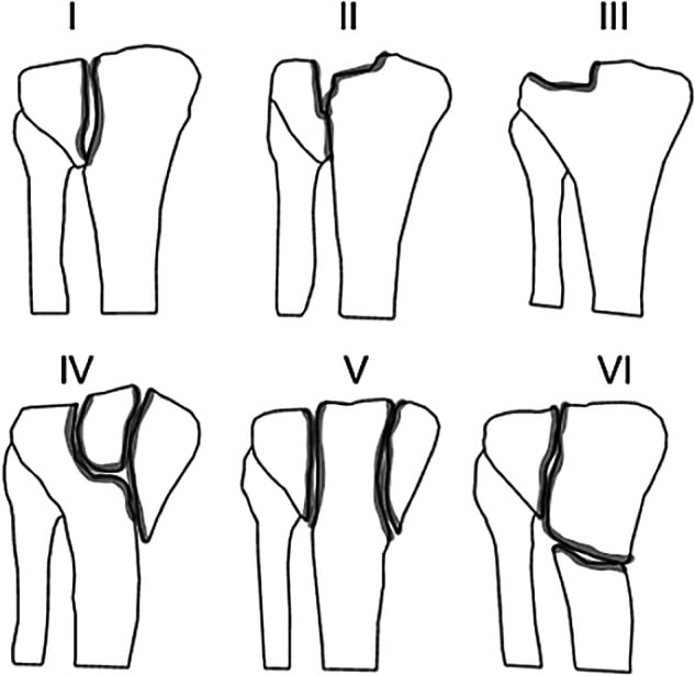
Two boundaries carry most of the weight. Between III and IV the injury moves from the lateral plateau to the medial, which is treated as high-energy until proven otherwise. Between IV and V it moves from one condyle to two, and with it from a single approach to staged surgery and two plates. In AO/OTA terms the bicondylar fractures are complete articular, segment 41-C; the AO/OTA classification page sets out the lettering.
Types I to III: the lateral plateau
The lateral plateau is convex, sits about 3 mm higher than the medial and is the side prone to depression. Three patterns follow from that.
Type I: the pure split. A wedge of lateral condyle is split off by a low-energy valgus force, with no articular depression, in younger and denser bone. Fixation is often percutaneous lag screws, with or without a lateral buttress plate. Plain films underestimate depression, so the CT is there to confirm that no depressed segment is hiding behind the split, and to size the fragment for the screws.
Type II: split-depression. The most common type and the common operative pattern: a lateral split with a depressed articular segment beside it. Through an anterolateral approach with a submeniscal arthrotomy, the split fragment is opened like a book to expose the depression, which is lifted with a tamp. The metaphyseal void is filled, raft screws go immediately beneath the subchondral bone, the split is closed over the restored joint surface, and a lateral buttress or locking plate holds it. Calcium phosphate cement has the higher compressive strength and lower rates of subsidence than calcium sulfate, which is why it is the favoured filler under a reduced surface.
The meniscus is part of the type II injury. Gardner and colleagues obtained pre-operative MRI in 62 consecutive Schatzker II fractures. With depression over 6 mm and condylar widening over 5 mm on the AP radiograph, lateral meniscal injury was present in 83%, against 50% with less displacement. Lateral collateral and posterior cruciate ligament tears were not seen below 4 mm of displacement but approached 30% as it increased. The lateral meniscus can also be trapped in the fracture; tears are addressed at the time of fixation.
Type III: pure depression. A central depression with the lateral cortex intact, usually a low-energy axial injury in osteoporotic bone. With no split to open, the depressed segment is reached through a metaphyseal cortical window, lifted, and supported with raft screws and a void filler. Arthroscopic-assisted reduction suits types I–III: it shows the step-off directly and lets meniscal and chondral injury be treated. The trap is irrigation fluid leaking through the fracture into the leg compartments, so inflow pressure is kept low and the calf watched.
II or III? The difference is the cortical break. If the cortex is split, the split is the door to the depression; if it is intact, the surgeon has to make a window. Because the choice of exposure turns on it, it is worth confirming on the CT rather than the AP film.
Why is a Schatzker IV fracture different?
The medial plateau is concave, congruent, has the stronger subchondral bone and takes about 60% of the load in neutral alignment. Breaking it usually takes a high-energy varus force. The tibial plateau ORIF operative guide treats a medial plateau fracture as a knee-dislocation-equivalent, with the highest risk of popliteal arterial injury, common peroneal nerve palsy and compartment syndrome of the six types. In one series of 67 knees in spanning frames, compartment syndrome followed 9 of 17 (53%) medial plateau fracture-dislocations against 9 of 50 (18%) type VI fractures (Stark 2009 (opens in a new tab)).
The limb before the bone. Document foot pulses, the ankle-brachial pressure index, common peroneal nerve function and all four compartments before discussing fixation. Any abnormal pulse, or an ABPI under 0.9, means CT angiography and vascular review. When the plateau fracture comes with a knee dislocation, Moore's classification is the one used; the vascular pathway is on the knee dislocation page and fasciotomy on the compartment syndrome of the leg page.
The fixation follows the fragment. A medial fragment that is a posteromedial coronal split displaces posteriorly under axial load, so it needs a buttress plate on the posterior surface; an anteromedial approach cannot place a true posterior buttress. That is the job of the posteromedial approach to the tibial plateau: prone, knee flexed 20–30°, through the interval between the pes anserinus and the medial head of gastrocnemius, with the saphenous nerve found and protected early. High-energy type IV injuries are staged like V and VI when the soft tissues demand it, and types IV–VI stay non-weight-bearing for at least 6–8 weeks.
Types V and VI: bicondylar fractures and staged surgery
Type V splits both condyles, classically as an inverted Y, with the metaphysis still in continuity with the shaft. Type VI adds a fracture that dissociates the metaphysis from the diaphysis. Both are high-energy, and in both the soft tissues set the timing.
Stage the knee. A knee-spanning external fixator goes on the day of injury. The leg is kept raised, the wrinkle test is repeated, and definitive fixation follows at about 7–21 days, once the skin wrinkles and blisters have epithelialised. Both blister types are a split at the dermo-epidermal junction, but under a blood-filled blister the dermis is stripped completely of epidermis, a slightly deeper injury in which incisions heal less well, so incisions are kept away from haemorrhagic blisters (Giordano 1994 (opens in a new tab)). In Egol's prospective protocol of 57 high-energy fractures, 74% of them Schatzker VI, deep infection was 5% overall (PubMed 16056075 (opens in a new tab)). The wider logic is in our post on damage control orthopaedics, and the compartment that can turn up after fixation in our compartment syndrome diagnosis and fasciotomy post.
Two incisions, two plates. The standard is an anterolateral approach for the lateral column and a posteromedial approach for the medial, with a skin bridge of over 7 cm. A single lateral plate leaves the medial column unsupported, and it collapses into varus. Barei and colleagues treated 83 AO/OTA 41-C3 fractures with medial and lateral plates through two exposures. Of the 31 patients with complete radiographs, only 55% had a satisfactory articular reduction (step or gap of 2 mm or less), and after adjusting for age and polytrauma a satisfactory reduction was associated with better function. Residual dysfunction was common.
Or a ring. The Canadian Orthopaedic Trauma Society randomised 83 Schatzker V–VI fractures to dual plating or to limited fixation with a circular fixator. Reduction quality was similar and two-year WOMAC scores did not differ. Plating had more blood loss (544 vs 213 mL), a longer stay (23.4 vs 9.9 days), deep infection in 7 of 40 (18%), and more unplanned reoperations (37 procedures against 16). Periarticular wires are kept roughly 14 mm or more below the joint line to stay extracapsular; frame principles are on the Ilizarov external fixation page.
What does CT add to a Schatzker type?
Less to the label than to the plan. Yacoubian and colleagues had three trauma surgeons read radiographs alone, with CT and with MRI, blinded. Adding CT changed the Schatzker type in only 6% of cases and moved interobserver kappa from 0.68 to 0.73. In an earlier series using the AO system, plain films underestimated 18 of 42 fractures, and CT changed the surgical plan in 13 of 22 (Wicky 2000 (opens in a new tab)). Different studies, different systems, but the direction is consistent: the scan changes the operation more often than the number.
What to read on the CT:
- Depression depth and area, which decide elevation and the volume of void filler
- Coronal-plane fracture lines, above all a posteromedial or posterolateral fragment, which decide the approach
- Fragment size, which decides whether screws will hold
- Comminution, which decides between spanning and length-stable fixation
- The lateral cortex, which separates a type II from a type III
Timing matters too. In a high-energy plateau the CT is best taken after the spanning frame, when ligamentotaxis has pulled the fragments towards the shape you will operate on; a scan of the grossly displaced knee overstates the comminution. The physics and protocols are on the CT imaging principles page.
Where does the Schatzker classification fall short?
Kfuri and Schatzker put it plainly when they revisited the system in 2018: it is a two-dimensional representation of the fracture. The AP film shows which condyle is broken but not whether the main fracture plane runs anterior or posterior, and the posterior fragments are where the fixation fails.
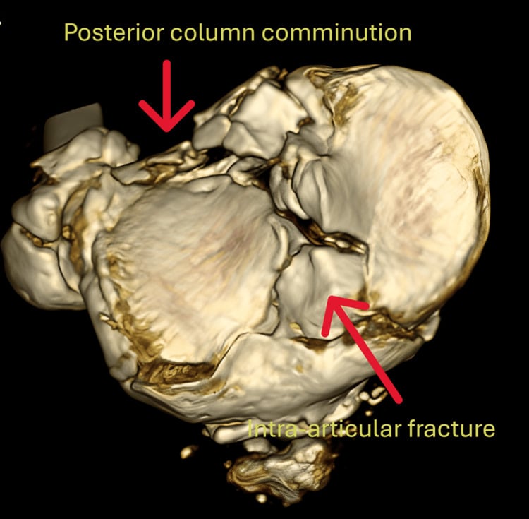
The posteromedial fragment. Higgins and colleagues reviewed CT in 111 bicondylar fractures and found a posteromedial fragment in 65 (59%). It averaged 25% of the joint surface, had over 5 mm of articular displacement in 55%, and ran nearly vertically (mean sagittal angle 73°), the shape of a shear fragment. Barei's group found it in 42 of 57 bicondylar fractures that involved the medial articular surface, nearly a third of the bicondylar fractures they evaluated. In a composite-bone model, every construct failed at the posteromedial fragment first, and the authors suggested lateral locking screws may not reliably reach it (Yoo 2010 (opens in a new tab)).
The three-column concept. Luo and colleagues divided the plateau on axial CT into lateral, medial and posterior columns. A column is involved when it holds a separate articular fragment, and each involved column gets its own approach and fixation. In their 29 three-column (Schatzker V–VI) fractures, fixed through a posterior inverted-L approach combined with an anterolateral one, all but one had a satisfactory reduction and none showed secondary articular depression, in a single-centre cohort without a comparison group.
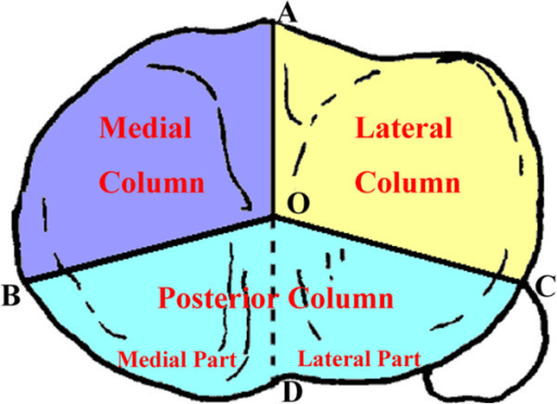
Schatzker's own revision. Kfuri and Schatzker kept the six types but added a coronal "equator" that splits each condyle into anterior and posterior quadrants. Types I–IV take an A or P modifier for the location of the main fracture plane, and V–VI take one for each column. The point is the same as Luo's: positioning, approach and plate position follow the main fracture plane, which only CT shows.
| Schatzker | Three-column (Luo) | Revised Schatzker (2018) | |
|---|---|---|---|
| Imaging | AP radiograph | Axial CT | CT, three-dimensional |
| Describes | Pattern and energy | Which columns hold a separate fragment | Pattern, energy and the main fracture plane |
| Strength | Shared vocabulary; energy and staging | Maps directly to approaches and plates | Keeps the familiar types, adds location |
| Blind spot | Posterior and coronal fragments | Depression, comminution, energy | Newer, less tested |
How reliable is the Schatzker classification?
Usually only moderately on plain films.
- Walton (2003): three observers, 30 fractures; Schatzker interobserver agreement was only fair, and AO was more reliable between observers (PubMed 14574596 (opens in a new tab)).
- Charalambous (2007): six observers, 50 fractures, AP and lateral films; interobserver kappa 0.41 for Schatzker and 0.43 for AO/OTA. Grouping fractures as unicondylar or bicondylar, and as pure split or depression with or without split, improved agreement.
- Maripuri (2008): four observers, 50 fractures; Schatzker interobserver kappa 0.47 and intraobserver 0.91, better than AO (0.36) and Hohl and Moore (0.14).
- Brunner (2010): four observers, 45 fractures; adding CT improved interobserver reliability from moderate to good for Schatzker, AO/OTA and Hohl alike (PubMed 19744652 (opens in a new tab)).
- Millar (2018), systematic review: plain-film reliability was frequently moderate (kappa 0.40–0.60), CT typically improved it, and the simpler three-column system scored higher (interobserver kappa 0.71–0.87) at the cost of less detail on morphology.
The practical reading matches what Charalambous found. Observers agree on the large decisions, one condyle or two, split or depression, and disagree on the finer boundaries. Say what you see in those terms, then give the number.
In the exam
Examiners want the type named, the energy recognised, the CT used for the right reason and the limits of the system acknowledged. For holding six types and their consequences in memory, see how to memorise orthopaedic classifications.
Frequently asked questions
What are the six Schatzker types of tibial plateau fracture?
Type I is a split of the lateral plateau without depression. Type II is a lateral split with articular depression, and the most common type. Type III is a pure depression of the lateral plateau with the cortex intact. Type IV is a fracture of the medial plateau. Type V is bicondylar with the metaphysis still attached to the shaft. Type VI adds a fracture dissociating the metaphysis from the diaphysis.
What is the difference between a Schatzker II and a Schatzker III fracture?
Both have a depressed lateral articular surface. In type II the lateral cortex is also split, so the surgeon can open the split fragment like a book through an anterolateral approach to reach and lift the depression. In type III the cortex is intact, typically in older osteoporotic bone, so the depression is reached through a metaphyseal cortical window, lifted, and held with raft screws and a void filler.
Why is a Schatzker IV tibial plateau fracture dangerous?
A medial plateau fracture usually needs a high-energy varus force and is treated as a knee-dislocation-equivalent, with the highest risk of popliteal artery injury, peroneal nerve palsy and compartment syndrome of the six types. In one series compartment syndrome followed 53% of medial plateau fracture-dislocations against 18% of type VI fractures. Pulses, ABPI, the peroneal nerve and the compartments are documented first; an abnormal pulse or ABPI under 0.9 leads to CT angiography.
Do you need a CT scan if the Schatzker type is clear on X-ray?
Yes, for any displaced fracture being considered for surgery. In one blinded study CT changed the Schatzker type in only 6% of cases, but in another it changed the plain-film surgical plan in 13 of 22 fractures. CT measures the depression, shows coronal-plane posterior fragments, sizes fragments for screws and decides the approach. In high-energy injuries it is most useful after a spanning external fixator has restored length.
What is the three-column classification of tibial plateau fractures?
Luo and colleagues divided the plateau on axial CT into lateral, medial and posterior columns. A column is involved when it contains a separate articular fragment, and each involved column needs its own approach and fixation. It was introduced for complex Schatzker V and VI fractures and is most useful for recognising the posterior fragments that the Schatzker system, drawn on the AP radiograph, does not describe.
References
- Schatzker J, McBroom R, Bruce D. The tibial plateau fracture. The Toronto experience 1968-1975. Clin Orthop Relat Res 1979;(138):94-104. PubMed (opens in a new tab)
- Kfuri M, Schatzker J. Revisiting the Schatzker classification of tibial plateau fractures. Injury 2018;49(12):2252-2263. DOI (opens in a new tab)
- Luo CF, Sun H, Zhang B, Zeng BF. Three-column fixation for complex tibial plateau fractures. J Orthop Trauma 2010;24(11):683-692. DOI (opens in a new tab)
- Higgins TF, Kemper D, Klatt J. Incidence and morphology of the posteromedial fragment in bicondylar tibial plateau fractures. J Orthop Trauma 2009;23(1):45-51. DOI (opens in a new tab)
- Barei DP, O'Mara TJ, Taitsman LA, et al. Frequency and fracture morphology of the posteromedial fragment in bicondylar tibial plateau fracture patterns. J Orthop Trauma 2008;22(3):176-182. DOI (opens in a new tab)
- Yacoubian SV, Nevins RT, Sallis JG, et al. Impact of MRI on treatment plan and fracture classification of tibial plateau fractures. J Orthop Trauma 2002;16(9):632-637. DOI (opens in a new tab)
- Charalambous CP, Tryfonidis M, Alvi F, et al. Inter- and intra-observer variation of the Schatzker and AO/OTA classifications of tibial plateau fractures and a proposal of a new classification system. Ann R Coll Surg Engl 2007;89(4):400-404. DOI (opens in a new tab)
- Maripuri SN, Rao P, Manoj-Thomas A, Mohanty K. The classification systems for tibial plateau fractures: how reliable are they? Injury 2008;39(10):1216-1221. DOI (opens in a new tab)
- Millar SC, Arnold JB, Thewlis D, et al. A systematic literature review of tibial plateau fractures: what classifications are used and how reliable and useful are they? Injury 2018;49(3):473-490. DOI (opens in a new tab)
- Gardner MJ, Yacoubian S, Geller D, et al. Prediction of soft-tissue injuries in Schatzker II tibial plateau fractures based on measurements of plain radiographs. J Trauma 2006;60(2):319-323. DOI (opens in a new tab)
- Barei DP, Nork SE, Mills WJ, et al. Functional outcomes of severe bicondylar tibial plateau fractures treated with dual incisions and medial and lateral plates. J Bone Joint Surg Am 2006;88(8):1713-1721. DOI (opens in a new tab)
- Canadian Orthopaedic Trauma Society. Open reduction and internal fixation compared with circular fixator application for bicondylar tibial plateau fractures. J Bone Joint Surg Am 2006;88(12):2613-2623. DOI (opens in a new tab)
Study the full topic
Included in Pro
Go deeper with Pro
- Operative guide: tibial plateau fracture ORIF
- Operative guide: posteromedial approach to the tibial plateau
Share this article
Cite this article
OrthoVellum Editorial Team. Schatzker Classification Explained Type by Type, With CT Signs and Fixation Strategy [Internet]. OrthoVellum; 2026 Oct 14 [cited 2026 Oct 14]. Available from: https://www.orthovellum.com/blog/schatzker-classification-tibial-plateau-fractures
About this content
Prepared by the OrthoVellum Editorial Team from cited sources, under our editorial policy.
For education and exam preparation; not medical advice or a substitute for clinical judgement and local guidance.
Spotted an error? Tell us; every report is read.
Get new articles by email
New clinical explainers, landmark-trial summaries and exam guidance from the OrthoVellum blog. No account needed; unsubscribe from any email.