Böhler and Gissane Angles
Examiners expect you to draw both angles on a lateral calcaneus radiograph, state normal values, and explain what an abnormal value means. Bohler's angle less than 20 degrees indicates posterior facet depression and a displaced intra-articular fracture. A restored Bohler angle after fixation correlates with better functional outcome. Always compare with the contralateral side — normal anatomy varies, and the uninjured foot is the best control.
Bohler's Angle — Definition and Measurement
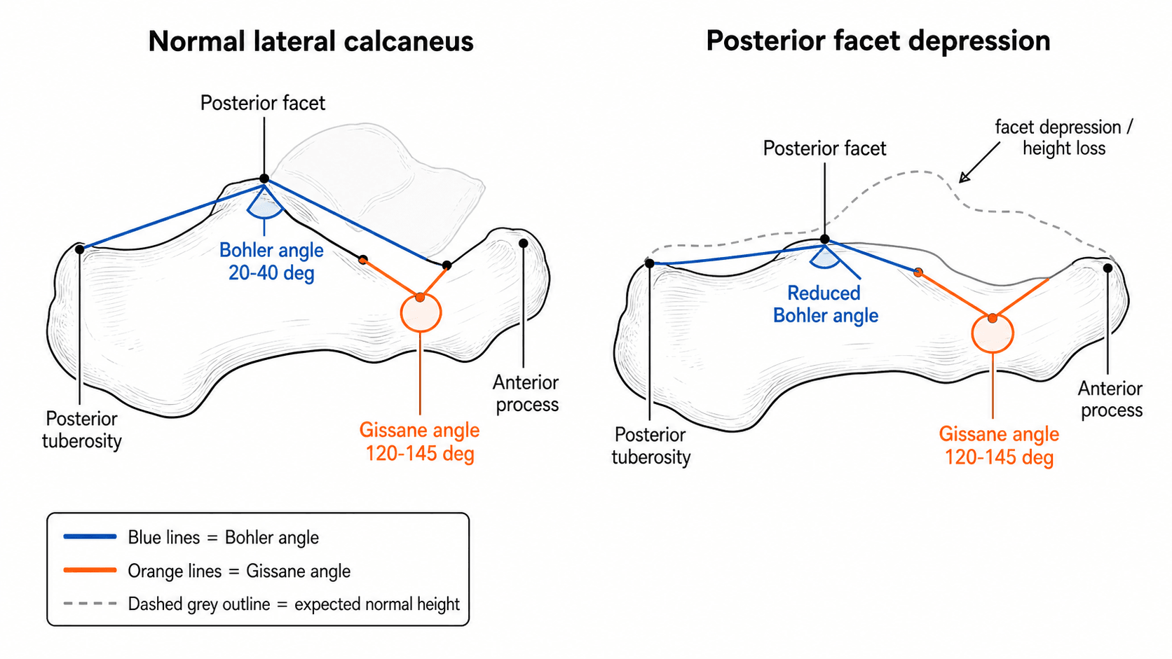
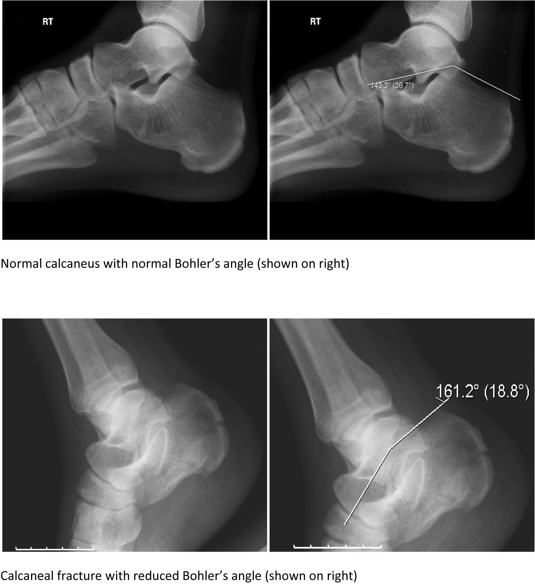
Bohler's angle (also called the tuber joint angle or calcaneal tuber angle) is measured on a true lateral radiograph of the calcaneus. It quantifies the relationship between the posterior tuberosity, the posterior articular facet, and the anterior process — essentially a measure of calcaneal height.
- Description
- Posterior tuberosity to posterior facet
- Anatomical Landmarks
- From the superior-most point of the posterior calcaneal tuberosity to the highest point of the posterior articular facet of the subtalar joint
- Description
- Posterior facet to anterior process
- Anatomical Landmarks
- From the highest point of the posterior articular facet to the superior-most point of the anterior process of the calcaneus
- Description
- Intersection of Line A and Line B
- Anatomical Landmarks
- Normal range: 20 to 40 degrees (average approximately 30 degrees). The angle opens superiorly.
Tuber to Facet, Facet to AnteriorDrawing Bohler's angle — TFA
Hook:Three landmarks, two lines, one angle — all meeting at the posterior facet.
The highest point of the posterior facet is the shared landmark for both lines — it is the apex of Bohler's angle. On a good lateral film, this is the most superior cortex of the posterior subtalar articular surface.
Gissane's (Crucial) Angle
Gissane's angle (the crucial angle) is also measured on a lateral radiograph. It lies at the anterior margin of the posterior facet where the downward slope of the posterior facet meets the upward slope of the anterior process — directly beneath the sinus tarsi.
- Description
- Along the downward (posterior) slope of the posterior facet
- Key Point
- Posterior subtalar articular surface directed inferiorly
- Description
- Along the upward (anterior) slope of the anterior process
- Key Point
- Anterior process directed superiorly toward the talonavicular joint
- Description
- Intersection beneath the sinus tarsi
- Key Point
- Normal: 120 to 145 degrees. Widens (increases) when the posterior facet is depressed.
Crushed Facet equals Gissane WidensGissane's angle — Crushed means it Widens
Hook:Crushed facet equals the crucial angle opens up.
Gissane's angle is less frequently measured in routine practice than Bohler's angle, but examiners may specifically ask about it. An increased Gissane angle indicates disruption at the crucial angle — the cortical break beneath the sinus tarsi — and suggests a joint-depression type injury.
Clinical Application in Calcaneal Fractures
Both angles are used in the assessment and management of intra-articular calcaneal fractures.
- Normal
- 20 to 40 degrees
- Joint-Depression Type
- Decreased (less than 20 degrees) — posterior facet driven into the body
- Tongue-Type
- Decreased but often less severely; the tongue fragment maintains some facet height
- Normal
- 120 to 145 degrees
- Joint-Depression Type
- Increased (greater than 145 degrees) — facet depression widens the angle
- Tongue-Type
- May be near-normal if the tongue fragment remains attached
- Normal
- Baseline
- Joint-Depression Type
- Higher energy, worse prognosis; Bohler less than 0 degrees correlates with poor outcome
- Tongue-Type
- Requires different reduction technique (Essex-Lopresti manoeuvre)
Below Twenty is Badly BrokenBohler in practice — When Low, Watch Out
Hook:Below twenty is badly broken; below zero is worse — and always compare sides.
A Bohler angle less than 0 degrees is associated with significantly worse long-term outcomes regardless of treatment method. This threshold is an important prognostic indicator and should drive discussion about operative versus non-operative management and patient counselling regarding expectations.
Key clinical uses:
- Initial assessment: Reduced Bohler angle and increased Gissane angle on plain radiographs confirm a displaced intra-articular fracture, prompting CT for Sanders classification.
- Surgical planning: The goal of operative fixation is to restore Bohler's angle to within approximately 5 degrees of the uninjured side. Failure to restore the angle correlates with residual subtalar incongruity.
- Post-operative assessment: A restored Bohler angle on intra-operative or post-operative lateral films indicates adequate reduction of the posterior facet. Loss of reduction on follow-up radiographs suggests fixation failure.
- Prognostication: A lower pre-operative Bohler angle (especially less than 0 degrees) and a smaller post-operative Bohler angle both correlate with poorer functional scores.
The angles screen and prognosticate, and Sanders type guides comminution, but the operative-vs-non-operative decision is genuinely contested and examiners want the evidence:
- The UK Heel Fracture Trial (Griffin, BMJ 2014) — a large multicentre RCT — found no significant patient-reported functional benefit of open reduction and internal fixation over non-operative care for displaced intra-articular calcaneal fractures, with more complications (notably wound problems) and no cost-effectiveness in the operative arm. This tempered earlier operative enthusiasm.
- Factors still favouring surgery: a younger, active manual worker, a large Böhler-angle loss / gross displacement, a reconstructable Sanders II-III posterior facet, and a displaced tongue-type at risk of skin compromise.
- Sanders IV (highly comminuted) does poorly with ORIF and is the group most often considered for primary subtalar arthrodesis (± reduction of the body).
- Non-operative management is preferred for the high-risk soft-tissue or medical patient (smoker, diabetic, peripheral vascular disease, poor skin), accepting some loss of height for a much lower wound-complication risk.
Beyond the Two Angles — Completing the Radiographic Assessment
Bohler's and Gissane's angles are measured on the lateral view, but a complete calcaneal assessment uses more than one projection - naming them scores marks:
- Harris (axial) view - shoot tangential to the heel; shows calcaneal width, varus/valgus malalignment of the tuberosity, and medial wall/sustentacular involvement. Widening and varus are reduction targets that the lateral angles cannot show.
- Broden's views - oblique views taken in internal rotation at staged angles (10-40 degrees) that profile the posterior facet - historically used to judge articular congruity before CT became routine.
- Calcaneal pitch (inclination angle) - the angle of the calcaneal inferior cortex to the floor on a weight-bearing lateral; relevant to hindfoot alignment, and reduced when calcaneal height is lost.
- CT (the decision-maker) - axial plus coronal/sagittal reconstructions give the Sanders classification (number of posterior-facet fragments on the coronal slice through the widest part of the facet), which is what actually drives operative planning. The plain-film angles screen and prognosticate; CT decides.
- What the angles contribute
- Reduced Bohler / widened Gissane raises suspicion of a displaced intra-articular fracture
- Next step
- Proceed to CT
- What the angles contribute
- Angles do not classify articular comminution
- Next step
- Sanders type drives operative vs non-operative decision
- What the angles contribute
- Restore Bohler to within 5° of the contralateral side
- Next step
- Also restore height, width and correct varus (axial view)
- What the angles contribute
- Restored, maintained Bohler angle = adequate, durable reduction
- Next step
- Loss of angle suggests fixation failure
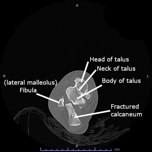
The page keeps sending you to CT "for Sanders" - so be ready to define it. The Sanders classification is read on the coronal CT slice through the widest part of the posterior facet and counts the number of posterior-facet fragments:
- Type I - non-displaced (any number of fracture lines, but no fragment displaced more than 2 mm).
- Type II - two facet fragments (one fracture line); subtyped A, B, C by the line's position from lateral to medial.
- Type III - three facet fragments (two fracture lines), typically with a depressed middle fragment (e.g. IIIAB, IIIAC, IIIBC).
- Type IV - four or more fragments: highly comminuted, the worst prognosis and the group most likely to be considered for primary subtalar fusion.
Higher Sanders type predicts worse articular comminution and outcome - which is why the plain-film angles screen and prognosticate while the Sanders type decides operative versus non-operative management.
A calcaneal fracture is a fall-from-height injury - the radiographic measurement must never distract from the associated-injury sweep. Specifically check the lumbar spine (around 10 percent have an associated spinal fracture from the same axial load), the contralateral calcaneus (bilateral injuries are common), and clinically exclude foot compartment syndrome. Stating this sequence unprompted marks you out as safe.
For calcaneal fractures the soft-tissue envelope governs the operation, and one pattern cannot wait:
- The displaced tongue-type fracture is a surgical emergency. The posterosuperior tongue fragment tents and blanches the posterior heel skin, threatening full-thickness pressure necrosis within hours — it needs urgent reduction (a percutaneous Essex-Lopresti / Gissane-spike manoeuvre) to take the pressure off the skin, ahead of any definitive fixation.
- Timing of definitive ORIF: for the rest, wait for the soft tissues to settle — operate only once swelling subsides and the wrinkle sign returns (typically ~7-14 days); operating through a swollen, fracture-blistered heel courts catastrophic wound breakdown.
- Approach and the wound problem: the traditional extensile lateral (L-shaped) approach has a high wound-complication/infection rate, markedly worse in smokers and diabetics, which has driven the shift to the minimally-invasive sinus tarsi approach and percutaneous/balloon techniques that respect the lateral skin.
So the examiner wants: protect the skin (reduce the tongue-type now), time the definitive surgery to the soft tissues, and choose the approach by the soft-tissue risk.
Limitations and Pitfalls
- Normal variation is wide. Bohler's angle ranges from 20 to 40 degrees in uninjured individuals. Some people naturally sit at 20 degrees, so always compare with the contralateral foot before calling a value abnormal.
- Radiographic technique matters. A poorly positioned lateral radiograph (even slight obliquity) distorts both angles. A true lateral requires the x-ray beam centred on the calcaneus with both malleoli superimposed.
- CT has replaced plain films for operative decision-making. The Sanders CT classification (based on the number of posterior facet fragments on coronal slices) is now the primary tool for surgical planning. Bohler and Gissane angles supplement CT but do not replace it.
- Inter-observer variability in measuring both angles on plain films is moderate. Digital measurement tools reduce this compared with manual goniometry, but even small differences in landmark selection change the result.
- Gissane's angle is less reproducible than Bohler's angle because the cortical lines are shorter and the landmarks are harder to define precisely.
- Neither angle distinguishes tongue-type from joint-depression type fractures reliably — that assessment requires lateral radiograph fracture-line analysis and CT.
- Associated injuries are easily missed. After a fall from height, check the lumbar spine (approximately 10 percent of calcaneal fractures have associated spinal injuries), the contralateral calcaneus (bilateral injuries are common), and assess for foot compartment syndrome.
Exam Viva
Practise clinical reasoning and management decisions out loud
“A 42-year-old labourer falls 4 metres from a ladder onto his feet. He presents with bilateral heel pain, inability to weight-bear, and marked swelling of both heels. A lateral radiograph of the right calcaneus is shown. Describe the radiographic findings and your assessment.”
“A 55-year-old patient underwent open reduction and internal fixation of a displaced intra-articular calcaneal fracture 6 weeks ago. You review the post-operative radiographs. How do you assess the adequacy of reduction and what is the prognostic significance of Bohler's angle in this context?”
Bohler's angle — how to draw it
- Line A: superior posterior tuberosity to highest point of posterior facet
- Line B: highest point of posterior facet to superior anterior process
- Angle at their intersection: normal 20 to 40 degrees (average about 30 degrees)
- Reduced (less than 20 degrees) means posterior facet depression — intra-articular fracture
- Less than 0 degrees: severe displacement, poor prognosis regardless of treatment
Gissane's angle — how to draw it
- Line 1: along the downward slope of the posterior facet
- Line 2: along the upward slope of the anterior process
- Angle at the sinus tarsi: normal 120 to 145 degrees
- Increased (greater than 145 degrees) means posterior facet disruption
- Less reproducible than Bohler's angle — harder to define landmarks precisely
Clinical rules
- Always compare with the contralateral foot — normal anatomy varies widely
- CT (Sanders classification) is the operative planning tool, not plain-film angles alone
- Operative goal: restore Bohler angle to within 5 degrees of the contralateral side
- Bohler angle less than 0 degrees: worst prognosis regardless of treatment method
- After a fall from height: check lumbar spine (about 10 percent have associated spinal injuries)
Evidence Base
Bohler's angle: correlation with outcome in displaced intra-articular calcaneal fractures
- Prospective cohort (95 fractures): a severely depressed Bohler angle predicted a poor 2-year outcome (VAS, SF-36) REGARDLESS of treatment
- Lesser initial displacement (better-preserved angle) gave higher functional scores
- Initial Bohler angle is independently prognostic in calcaneal fractures
Natural Value of Bohler's Angle in Normal Chinese Population
- Normal Bohler angle measured 31.6 +/- 5.2 degrees (range ~20-47) with excellent interobserver reliability
- 30 to 33 degrees recommended as the target for calcaneal fracture reduction
- Not applicable under 10 years of age; values vary between populations