Seatbelt Injury | Flexion-Distraction | High Association with Abdominal Injury
- Classic seatbelt injury = flexion around lap belt fulcrum
- Roughly half have intra-abdominal injury - the published range is wide and the figures measure different things (42% in paediatric Chance fractures, 64% with a seatbelt SIGN). Always CT the abdomen
- Bony Chance may be braced if no translation and intact anterior column
- Ligamentous Chance requires surgery - soft tissue will not heal
- Use compression not distraction during posterior fixation
- “Horizontal fracture through ALL posterior elements is pathognomonic
- “Seatbelt sign on abdomen = high suspicion for Chance AND visceral injury
- “TLICS: Distraction=4, so most are surgical unless purely bony
- “Posterior surgery uses COMPRESSION to close the distraction injury
Overview and Epidemiology
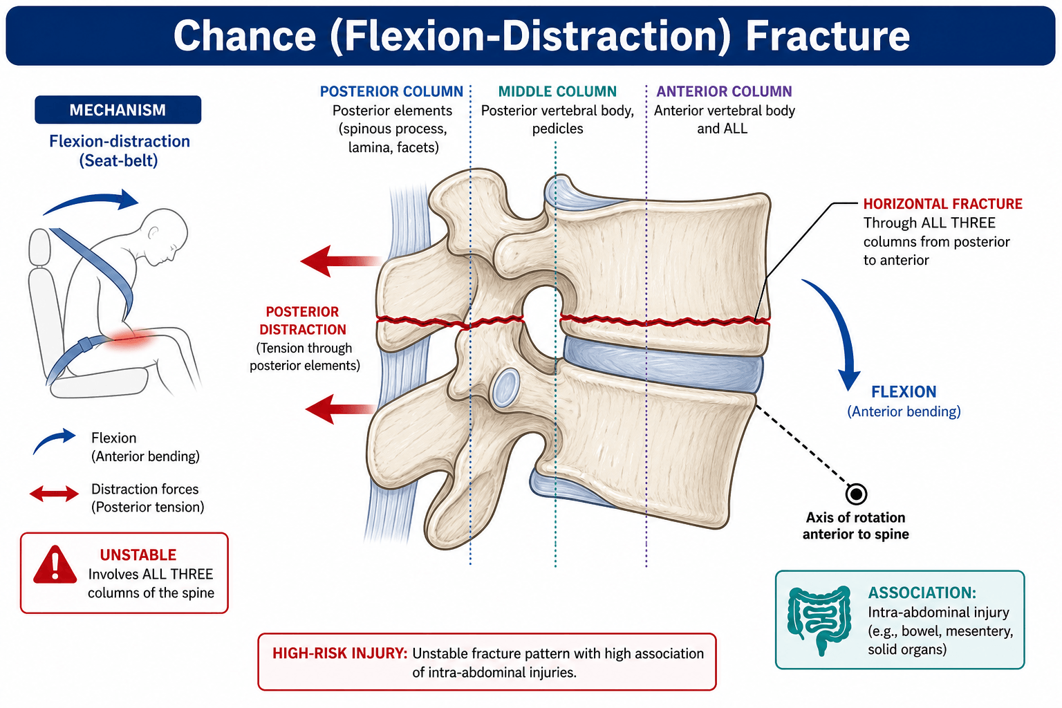
The injury. A Chance fracture is a flexion-distraction injury of the thoracolumbar spine, and the classic patient is a restrained occupant who flexes over a lap seatbelt during a motor vehicle accident. G.Q. Chance described it in 1948 and it was originally called the seatbelt fracture; its hallmark is the classic horizontal fracture line through the posterior elements.
Mechanism. The lap belt acts as a fulcrum. During rapid deceleration the upper body flexes forward while the pelvis stays restrained, and the resulting flexion-distraction force opens the posterior spine while the anterior column pivots over the belt.
Where, and in whom. The peak is L1-L3, at the thoracolumbar junction. A motor vehicle accident with a lap-belt restraint is the commonest mechanism, and a fall with flexion does the same thing. Children are at higher risk because the lap belt rides higher.
In the largest paediatric series, 7 of 26 Chance fractures occurred in children properly restrained by a lap-and-shoulder belt. "She was wearing a proper seatbelt" is not a reason to stop looking.
Where the rest of this sits. A Chance fracture is one pattern in the thoracolumbar fracture family, and the reason it is singled out is that it fails in distraction rather than compression - which is why a burst fracture with a similar-looking loss of height is a different injury with a different operation. And if there is a deficit the grading belongs to spinal cord injury.
Anatomy and Biomechanics
What fails, and in which direction. In Denis's three-column terms the fulcrum is the anterior column, at lap-belt level, so everything behind it is loaded in tension.
- Force Type
- Tension (distraction)
- Result
- Horizontal fracture or ligament rupture
- Force Type
- Tension
- Result
- Fracture through body or disc rupture
- Force Type
- Compression or neutral
- Result
- Usually intact or minimal injury
Tension, not compression. The posterior elements fail in tension. A burst fracture is the opposite: every column fails in compression, and it is fixed in the opposite direction too.
In a pure Chance fracture the anterior column is intact or minimally injured: this is a posterior tension failure pivoting on a column that holds. That is the distinction from a burst fracture.
Stability. The posterior ligamentous complex is disrupted by definition, which is why most Chance fractures are considered unstable.
Classification
The classification that changes management is bony versus ligamentous, because it decides whether the injury can heal in a brace. Denis subdivides by what fails, and TLICS and the AO Spine system turn the findings into the decision to operate.
- Description
- Fracture through vertebral body, pedicles, laminae, spinous process
- Treatment Implication
- Bone heals - may brace if minimal displacement
- Description
- Rupture through disc and posterior ligaments
- Treatment Implication
- Soft tissue does NOT heal - requires surgical fusion
- Description
- Combination of bone and soft tissue failure
- Treatment Implication
- Treat as ligamentous - surgery required
Clinical Assessment
History. The answers that change what happens next:
- The MVA details, especially restraint type: lap belt or three-point
- Mechanism of impact
- Abdominal symptoms: pain, distension, vomiting
- Neurological symptoms
The spine. A widened interspinous space that can be felt is the classic finding, tenderness is focal at the fracture level, and a kyphotic deformity may be visible or palpable.
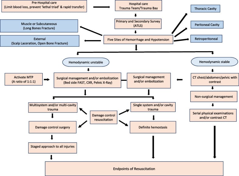
The abdomen. Transverse bruising across the abdomen is the seatbelt sign. This is a two-cavity injury pattern: the bruise raises suspicion for a Chance fracture and for intra-abdominal injury together, so the thoracolumbar spine and the abdomen and pelvis are both imaged, never one alone. Tenderness may be generalised or focal, distension may mean bleeding or an ileus, and guarding and rigidity are the signs of peritonitis.
Neurology. Complete lower limb motor and sensory testing, perianal sensation for sacral sparing, rectal tone, and reflexes, which may be hypo- or hyperreflexic depending on the level.
Associated Injuries
What travels with the fracture. The seatbelt injury spectrum runs through the abdomen as well as the spine, and these are the visceral injuries to look for.
- Incidence
- 15-25%
- Clinical Significance
- Can be delayed presentation - maintain high suspicion
- Incidence
- 10-20%
- Clinical Significance
- Haemorrhage, ischaemia risk
- Incidence
- 5-10%
- Clinical Significance
- Part of seatbelt injury spectrum
- Incidence
- 10-15%
- Clinical Significance
- Peritonitis risk
- Incidence
- Variable
- Clinical Significance
- May be significant
The instruction is not in doubt: every Chance fracture gets a CT abdomen and pelvis with contrast. What is worth being precise about is the figure, because the three studies usually quoted are measuring three different things and a candidate who says "50%" flatly can be asked where it comes from.
- Population
- 26 paediatric Chance fractures - the closest match to the question
- Abdominal injury
- 42% (11/26)
- Population
- 117 blunt-trauma patients, of whom only 14 had a seatbelt SIGN (not necessarily a fracture)
- Abdominal injury
- 64% - but that is 9 of 14 patients, so treat it as "most of a small group"
- Population
- 38 restrained occupants presenting to one hospital - a treated series, not a population
- Abdominal injury
- 84% (32/38); 27 of those 32 had bowel or mesenteric injury
So "roughly half" is a fair summary, and 50% is a round number sitting between them rather than a finding. The clinically important comparison is Chandler's: 64% with a seatbelt sign versus 8.7% without, a seven-fold difference that tells you the abdominal wall bruise, not the vertebral fracture, is the thing driving your suspicion.
A name collision worth knowing, because both papers are from 1983 and both get cited on pages like this one. The Denis of the abdominal-injury series above is R Denis, a general surgeon writing in J Trauma. The Denis of the three-column classification used earlier on this page is Francis Denis, writing in Spine. They are different people and different papers; citing "Denis 1983" without the journal is ambiguous.
Bowel injury in particular can present late, so a normal initial CT does not close the question - serial examination and a low threshold for repeat imaging are what actually prevent the miss.
Investigations
Imaging Algorithm
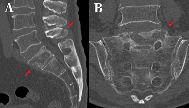
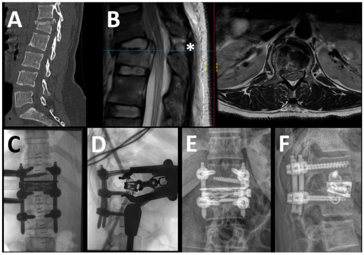
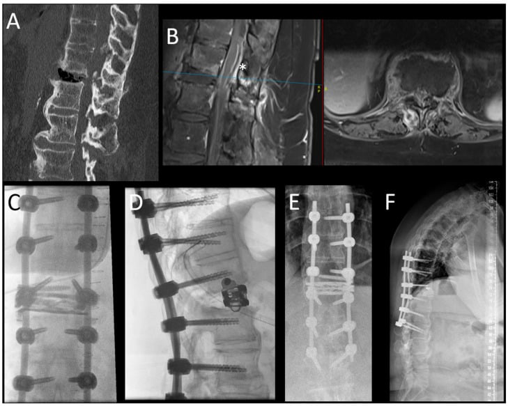
Shows the osseous injury. Look for the horizontal fracture line through the posterior elements; sagittal reconstructions show the flexion-distraction pattern best.
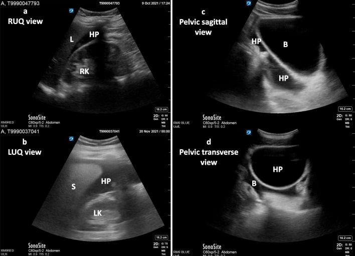
Look for bowel wall thickening, free fluid, mesenteric haematoma and pancreatic injury. Do not wait for symptoms - the injury may declare late.
Determines bony versus ligamentous, and so the treatment. Shows disc status, the extent of PLC injury, and the cord and cauda.
What the CT shows. The classic appearance:
- Horizontal fracture line through spinous process, laminae, transverse processes, pedicles and vertebral body
- Widened interspinous space at the injury level
- Empty facet sign if the facets are subluxed
- Minimal anterior column compression, which distinguishes it from a burst
- Chance Fracture
- Distraction (flexion-distraction)
- Burst Fracture
- Compression (axial)
- Chance Fracture
- Horizontal through posterior elements
- Burst Fracture
- Comminuted vertebral body
- Chance Fracture
- Fractured or widened space
- Burst Fracture
- Usually intact
- Chance Fracture
- Minimal (no retropulsion)
- Burst Fracture
- Common (retropulsed fragment)
- Chance Fracture
- Abdominal in roughly half
- Burst Fracture
- Calcaneus, other spine levels
- Bony: a low-signal fracture line through the vertebral body, with bone marrow oedema
- Ligamentous: high T2 signal in the disc and a disrupted PLC, with no fracture line through the body
- Mixed: features of both
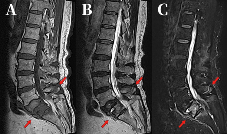
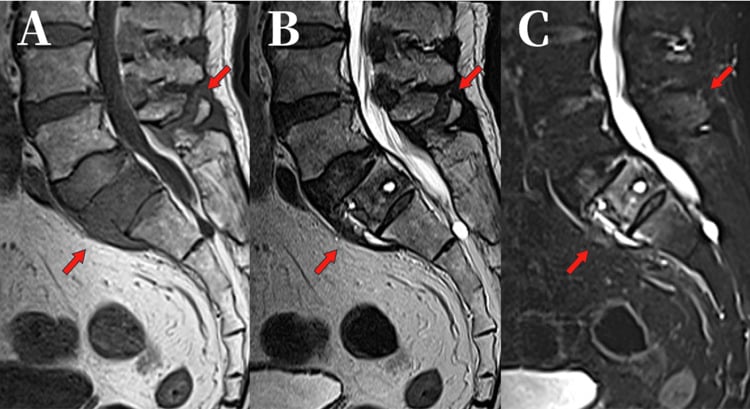
- Mechanism / Key feature
- Posterior tension over lap-belt fulcrum
- Distinguishing feature from Chance
- Horizontal split through posterior elements, intact/minimal anterior column
- Stability / management
- Unstable in tension - compression fixation or brace if purely bony
- Mechanism / Key feature
- Axial compression
- Distinguishing feature from Chance
- Comminuted body with retropulsion and canal compromise; posterior elements often intact
- Stability / management
- Variable - distraction/reconstruction, not compression
- Mechanism / Key feature
- Anterior column compression only
- Distinguishing feature from Chance
- Anterior wedging with intact middle/posterior columns
- Stability / management
- Usually stable - non-operative
- Mechanism / Key feature
- Translation/rotation, all columns
- Distinguishing feature from Chance
- Translation or rotational malalignment, often neurological deficit
- Stability / management
- Highly unstable - surgical stabilisation
- Mechanism / Key feature
- Low-energy, elderly
- Distinguishing feature from Chance
- Marrow oedema without high-energy mechanism; no seatbelt sign
- Stability / management
- Often non-operative or cement augmentation
- Mechanism / Key feature
- Trivial trauma on fused spine
- Distinguishing feature from Chance
- Transverse fracture through ankylosed segment, three-column unstable
- Stability / management
- Highly unstable - long-segment fixation
A transverse, Chance-like fracture in a patient with ankylosing spondylitis or DISH is a different and far more dangerous entity. The rigid, often osteoporotic, fused "bamboo"/flowing-ossification spine behaves like a long bone: a trivial low-energy injury (a simple fall or even hyperextension) produces a highly unstable three-column fracture, frequently through a disc space or ankylosed segment and often an extension rather than flexion pattern.
These fractures are easily missed (the line hides in the fused mass and plain films are unreliable), carry a high rate of epidural haematoma, delayed neurological deterioration and mortality, and mandate whole-spine CT and MRI.
Management differs fundamentally from a typical Chance: long-segment posterior instrumentation (multiple levels above and below, not short-segment) is required because of the long rigid lever arms, and the patient must be handled in their fixed deformity, never forced to neutral.
Management
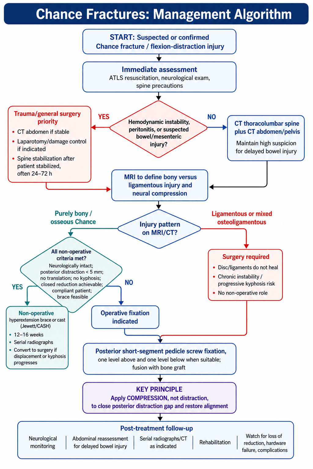
- Displacement
- Less than 5mm, no translation
- Treatment
- Consider hyperextension brace/cast
- Key Pearl
- Must be purely osseous - check MRI
- Displacement
- More than 5mm, kyphosis
- Treatment
- Posterior compression + fusion
- Key Pearl
- Bony heals well with fixation
- Displacement
- Any amount
- Treatment
- Posterior fusion mandatory
- Key Pearl
- Soft tissue will NOT heal
- Displacement
- Variable
- Treatment
- Posterior fusion
- Key Pearl
- Treat as ligamentous
Who can be braced. Non-operative candidates are rare, and every one of these has to hold:
- Purely bony injury, with no soft-tissue component on MRI
- Minimal displacement: less than 5mm posterior distraction, and no translation
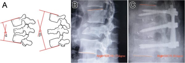
- Kyphosis within accepted limits - under roughly 15 to 20 degrees at the injured segment (published thresholds vary, and there is no trial defining the cut-off)
- Neurologically intact
- Compliant patient
"No kyphosis at all" is a stricter bar than practice uses and would send braceable injuries to theatre.
The brace. A hyperextension brace or cast (Jewett or CASH) for 12-16 weeks, with serial radiographs to watch the alignment and conversion to surgery if the injury progresses.
When a bony Chance is fixed instead. Any of these, and the brace is not the answer:
- Significant displacement, more than 5mm
- Segmental kyphosis beyond that threshold, or kyphosis that progresses on serial upright films
- Closed reduction cannot be achieved
- Neurological deficit
- Associated injuries requiring surgery
- Unable to brace, from body habitus or compliance
The hyperextension brace works because extension closes the posterior distraction - extending the spine approximates the separated posterior elements and lets the bony injury heal in apposition. It is the non-operative equivalent of applying compression across a posterior construct.
Careful with the comparison to compression fractures - the posture is the SAME, not opposite. Both are braced in extension. What differs is what extension is doing: in a Chance injury it closes a posterior gap, whereas in an anterior compression fracture it unloads and helps restore height in the crushed anterior column. Same brace, opposite mechanical target - and that is the distinction examiners are after, not a difference in position.
Surgical Technique
Principles. Short segment, one level above and one below, compressed across the construct and fused with bone graft. Long constructs are avoided unless the injury is multilevel.
Surgical Steps
Prone on a Jackson frame, with neuromonitoring if there is a neurological deficit.
Midline incision and subperiosteal dissection. Note the widened interspinous space at the injured level.
Screws one level above and one level below the injury, through standard entry points and trajectories.
Place the rod and apply compression across the fracture. This is the step that matters: the distracted posterior elements come back together.
Decorticate the facets and transverse processes and lay the graft; the compressed position promotes healing.
Unlike burst fractures, where short segment fixation may fail, a Chance fracture does well with short segment fixation because the anterior column is intact and provides the fulcrum for stability.
Complications
- Incidence
- Initial 5-10%
- Prevention/Management
- High suspicion, repeat imaging if concern
- Incidence
- Less than 5%
- Prevention/Management
- Adequate compression, avoid distraction
- Incidence
- 5%
- Prevention/Management
- Bone graft, smoking cessation
- Incidence
- 5-10%
- Prevention/Management
- Adequate compression, short segment usually sufficient
- Incidence
- Less than 1%
- Prevention/Management
- Careful technique, neuromonitoring
- Incidence
- 10-20%
- Prevention/Management
- Patient education, rehabilitation
A missed abdominal injury is the most dangerous early complication, and bowel injury may present late: the initial CT can be negative, with the bowel injury presenting 24-48 hours after it. Repeat the imaging on any concern, and let these prompt it:
- Seatbelt sign
- Rising inflammatory markers
- Abdominal distension
- Ileus beyond what the spinal surgery explains
Non-union. Rare after a fusion that has been adequately compressed. The risk is higher where a ligamentous injury was treated non-operatively.
Postoperative Care
Rehabilitation Timeline
Monitor for abdominal complications. DVT prophylaxis and pain control. Mobilise when the trauma team clears it.
Progressive mobilisation and a wound check. A TLSO may be used for additional support if wanted.
Continue the brace if one is being used. Serial radiographs, core strengthening, graded activity.
CT to confirm fusion, wean the brace, return to normal activities.
Bracing after surgery. Practice varies. Some use a TLSO for 6-12 weeks as comfort and a reminder, others rely on the instrumentation alone, and short segment fixation is generally stable enough for that.
Long-term. Watch for adjacent segment disease, which is less common than after a burst fracture because the construct is short, and ask about chronic pain.
Outcomes and Prognosis
After surgery. Fusion rates are high when the construct is adequately compressed, hardware failure is low because the anterior column is intact, alignment is restored well, and most patients return to normal function.
By type. A purely bony injury heals excellently, whether braced or fixed, and a ligamentous injury does excellently once fused. A ligamentous injury treated non-operatively does poorly, with chronic instability.
Neurology. Most Chance fractures are neurologically intact. Where there is a deficit the prognosis depends on its severity, and recovery at the cauda equina level is generally good.
The abdomen. An associated abdominal injury increases morbidity and may delay the spine surgery, and needs coordination with the general surgical team.
Guidelines, Registries & Global Practice
Global Epidemiology
Flexion-distraction (Chance) injuries are a minority of thoracolumbar fractures but carry a disproportionate burden of associated visceral and neurological injury. In the original lap-belt era series, 32 of 38 restrained occupants (84%) sustained abdominal injury, with bowel or mesenteric injury in 27 of 32 (Denis et al, 1983). In a prospective cohort, abdominal injury occurred in 64% of patients with a seatbelt sign versus 8.7% without, and small-bowel perforation in 21% of those with the sign (Chandler et al, 1997). In the paediatric population, a two-decade single-centre series found abdominal visceral injury in 42% and neurological injury in 27% of children with lumbar Chance fractures, with injuries occurring even when a three-point belt was correctly worn (Louman-Gardiner et al, 2008). The shift from lap-only to three-point restraints has reduced but not eliminated these injuries, particularly in rear-seat and incorrectly restrained children.
Side-by-Side Guidance & Classification
- Key parameter for Chance injury
- Tension failure of middle and posterior columns
- Surgical threshold
- Instability when 2+ columns disrupted
- Evidence level
- Level 4 (classification)
- Key parameter for Chance injury
- Distraction morphology = 4; PLC disrupted = 3
- Surgical threshold
- TLICS greater than 4 favours surgery
- Evidence level
- Level 5 (consensus)
- Key parameter for Chance injury
- Type B (tension band / distraction) injury
- Surgical threshold
- AOSpine-TLICS greater than 5 favours surgery
- Evidence level
- Level 5 (consensus)
- Key parameter for Chance injury
- Multisystem assessment, exclude visceral injury
- Surgical threshold
- Operative for distraction/ligamentous instability
- Evidence level
- Guideline / expert
- Key parameter for Chance injury
- Whole-spine imaging, MRI if PLC/ligamentous concern
- Surgical threshold
- Surgical referral for unstable injuries
- Evidence level
- Guideline / expert
- No dedicated arthroplasty-style registry captures Chance fractures; evidence is case-series and trauma-registry based
- Managed within major trauma centre networks with combined general/spine surgery
- Paediatric trauma centres for children (higher visceral-injury rate)
- National road-trauma registries track restraint type and mechanism
- Three-point belts mandatory in most high-income countries; lap-only belts persist in older vehicles and some rear/middle seats
- Brace-versus-fixation threshold for purely bony injuries varies between units
- Short-segment posterior compression fixation is the dominant operative approach worldwide
- Child-restraint enforcement and booster-seat use differ markedly by region
restraint use and type, abdominal examination and CT findings, MRI bony-versus-ligamentous status, treatment rationale, and patient/family communication.
missed or delayed abdominal injury, failure to obtain abdominal CT initially, treating ligamentous Chance non-operatively, and not recording restraint type for road-trauma surveillance.
MCQ Practice Points
Q: What percentage of Chance fractures have associated intra-abdominal injury? A: Roughly 50% is the expected exam answer and a fair summary. If pressed on where it comes from: 42% in a paediatric Chance-fracture series (PMID 18606276), 64% in patients with a seatbelt SIGN versus 8.7% without (PMID 9322665). Always CT abdomen with a Chance fracture.
Q: What is the mechanism of a Chance fracture? A: Flexion-distraction - the patient flexes forward over the lap belt fulcrum, creating tension failure of the posterior and middle columns.
Q: When fixing a Chance fracture posteriorly, should you use compression or distraction? A: COMPRESSION - this closes the distraction injury. This is the opposite of burst fractures.
Q: Which type of Chance fracture can be managed non-operatively? A: Bony (osseous) Chance with minimal displacement - bone heals. Ligamentous Chance MUST be fused - disc and ligaments will not heal.
Q: What is the TLICS morphology score for a Chance fracture? A: 4 points (distraction) - the highest morphology score. Combined with disrupted PLC (3), most Chance fractures score TLICS 7+.
Q: What physical exam finding on the abdomen should raise suspicion for Chance fracture? A: Transverse abdominal bruising (seatbelt sign) - indicates lap belt mechanism and high risk for both Chance fracture AND visceral injury.
Exam Viva Scenarios
Practise clinical reasoning and management decisions out loud
“A 25-year-old restrained back-seat passenger is brought in after an MVA. She has transverse bruising across her abdomen and tenderness at the thoracolumbar junction. CT shows a horizontal fracture through L2 spinous process, laminae, pedicles, and superior vertebral body. She is neurologically intact. How do you manage her?”
“A 40-year-old man has an L1-L2 Chance fracture. MRI shows the fracture goes through the L1-2 disc with complete disruption of the posterior ligaments. He has bilateral lower extremity weakness (4/5 throughout) and urinary retention. Describe your surgical approach.”
“A 6-year-old restrained in a booster seat is involved in an MVA. She has a seatbelt mark on her abdomen and back pain. CT shows a horizontal fracture through L3. Her abdominal CT shows free fluid. How do you manage this case?”
Definition and Mechanism
- Flexion-distraction injury (seatbelt mechanism)
- Lap belt acts as fulcrum
- Posterior elements fail in tension
- Anterior column intact (pivot point)
Roughly half have abdominal injury - CRITICAL
- Roughly half have associated abdominal injury (42-84% by series; seatbelt sign 64% vs 8.7% without)
- ALWAYS CT abdomen/pelvis
- Bowel, mesentery, pancreas most common
- Seatbelt sign = high suspicion for both
Bony vs Ligamentous
- Bony: Through vertebra, may brace if minimal displacement
- Ligamentous: Through disc/ligaments, MUST fuse
- MRI is essential to determine type
- Mixed = treat as ligamentous
Surgical Principle
- Use COMPRESSION not distraction
- This closes the posterior distraction injury
- Opposite of burst fracture management
- Short segment usually sufficient
TLICS Application
- Distraction = 4 points (highest morphology)
- PLC always disrupted = 3 points
- Most score TLICS 7+ = surgical
- Only purely bony minimal displacement may brace
Key Facts
- L1-L3 most common levels
- Horizontal fracture line is pathognomonic
- Compression during fixation closes injury
- Never miss the abdominal injury
Evidence Base
Original Description (1948)
- First description of horizontal flexion fracture of the spine
- Described the fracture line through the vertebral body and posterior arch
- Anterior column acts as fulcrum in flexion
- Established the classic imaging appearance now bearing his name
Seatbelt Syndrome - Abdominal Injury
- 32 of 38 restrained occupants (84%) had abdominal injury in this series
- 27 of 32 injured had bowel or mesenteric injury
- Highlighted the altered injury physics of lap-belt deceleration
- Established the high association between belt restraint and visceral injury
Seatbelt Sign & Bowel Injury
- Prospective study of 117 motor vehicle accident victims
- Abdominal injury in 64% of those with a seatbelt sign vs 8.7% without
- Small-bowel perforation in 21% of seatbelt-sign patients
- Seatbelt sign mandates a heightened index of suspicion