The Muscle the Nerve Goes Through
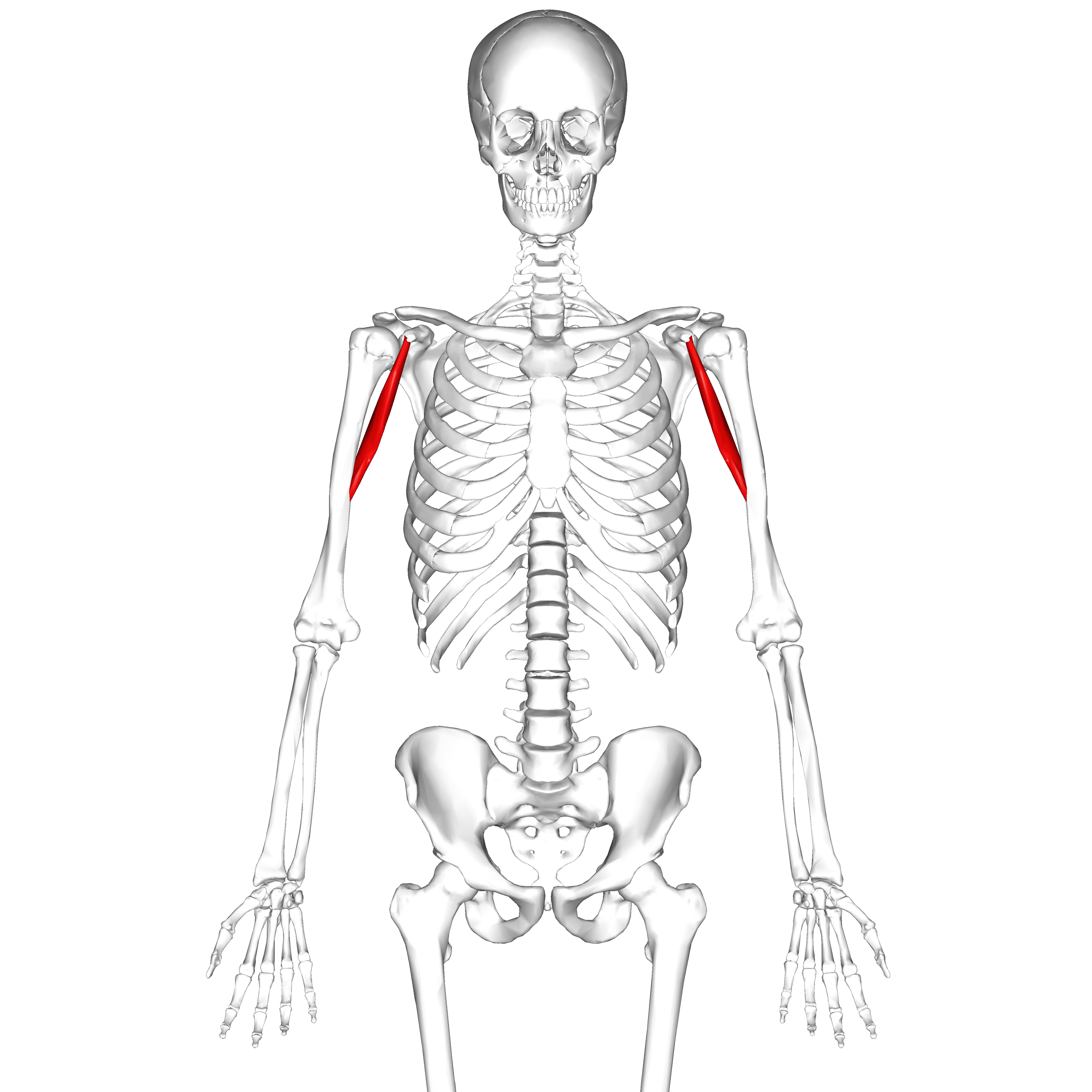
- Origin: the TIP of the coracoid process, shared with the short head of biceps as the CONJOINED TENDON; insertion: the middle third of the MEDIAL surface of the humeral shaft, opposite the deltoid tuberosity.
- Innervated by the musculocutaneous nerve (C5, C6, C7), which PIERCES the muscle in the great majority of people — 83.3 per cent in the largest cadaveric series (312 limbs). It is the classic example of a major peripheral nerve passing through the substance of the muscle it supplies, but the non-piercing variant is common enough that the nerve must be identified rather than assumed.
- The main musculocutaneous trunk enters the muscle a mean of 5.1 to 5.6 cm distal to the coracoid, but 29 to 33 per cent of nerves enter proximal to 5 cm, and small twigs enter as close as 1.7 to 2.2 cm.
- Actions: weak flexion and adduction of the shoulder; it does NOT cross the elbow and has no elbow action.
- The conjoined tendon is the medial retraction handle of the deltopectoral approach and the transferred structure in the Latarjet, where its sling effect on the inferior subscapularis is one of the three components of the triple-blocking mechanism.
- “Flatow and colleagues showed the classically taught 5 to 8 cm safe zone below the coracoid CANNOT be relied upon — 29 per cent of main trunks and 74 per cent of specimens counting twigs entered proximal to 5 cm.
- “A more recent cadaveric study proposed 3 cm rather than 5 cm as the working safe zone, because 91.7 per cent of specimens had a twig or trunk within 5 cm of the coracoid.
- “Musculocutaneous injury was the second commonest nerve injured in a series of 416 open Latarjet procedures (4 of 13 neurological injuries), with the axillary nerve first.
- “Coracobrachialis is described as the only muscle in the body routinely pierced by the major nerve that supplies it — a favourite one-liner.
Overview
Coracobrachialis is the smallest and least powerful of the three anterior arm muscles, and by function alone it would barely merit a page. Its importance is entirely topographical. It sits at the junction of the axilla and the arm, and three things happen there that matter to a surgeon:
- Its tendon of origin is fused with the short head of biceps to form the conjoined tendon — the structure that is retracted medially in every deltopectoral approach and transferred with the coracoid in a Latarjet.
- The musculocutaneous nerve pierces the muscle, a relationship unique among major peripheral nerves. The distance from the coracoid at which it does so defines the safe zone for anterior shoulder surgery.
- It forms part of the boundary between the axilla and the arm, running alongside the axillary and brachial vessels and the cords of the brachial plexus.
The Latarjet coracoid transfer is not simply a bone block, and examiners expect the three separate mechanisms. The coracobrachialis is central to the second.
- Bony effect. The transferred coracoid extends the anteroinferior glenoid arc, restoring the articular surface and increasing the critical distance the humeral head must travel to dislocate. This addresses glenoid bone loss.
- Sling effect. The conjoined tendon — coracobrachialis plus the short head of biceps — passes through the subscapularis split and acts as a dynamic anteroinferior sling. In abduction and external rotation, the position of apprehension, the tendon tensions the inferior third of the subscapularis and pushes the humeral head posteriorly. This is widely believed to be the most important of the three mechanisms, and it is entirely dependent on placing the split at the junction of the upper two-thirds and lower third of the subscapularis.
- Capsular effect (Bankart effect). The coracoacromial ligament stump, left attached to the lateral coracoid, is repaired to the anterior capsule and glenoid rim, restoring the capsulolabral restraint.
Why this matters clinically: the sling effect explains why a Latarjet works even in patients with an intact glenoid, and why it remains effective in contact athletes and in patients with capsular deficiency where a soft-tissue Bankart repair fails. It also explains why a split placed too superiorly, or a graft placed too high on the glenoid, loses the sling and produces a mechanically inferior construct.
Three Muscles, Two LigamentsCoracoid Attachments
Hook:Pectoralis minor is medial, the conjoined tendon is at the tip, the coracoacromial ligament is lateral, and the coracoclavicular ligaments are on top.
Attachments, Innervation and Relations
Origin
- Tip of the coracoid process of the scapula, arising with the short head of biceps brachii as the conjoined tendon.
- Within the conjoined tendon, coracobrachialis occupies the medial and posterior portion and the short head of biceps the lateral and anterior portion. Distally the two separate; the short head continues as a tendon, coracobrachialis becomes fleshy.
- A cadaveric study in a Sri Lankan population found the muscle to arise from the coracoid tip and additionally from the lateral, posterior and medial aspects of the short head of biceps tendon — the two are more intimately fused than a simple side-by-side arrangement.
Insertion
- Middle third of the medial surface of the humeral shaft, on and just anterior to the medial border, roughly opposite the deltoid tuberosity on the lateral side.
- The insertion is a linear ridge, typically 5 to 7 cm long, lying between the origin of the medial head of triceps posteriorly and the brachialis anteriorly.
- The nutrient foramen of the humerus lies near this insertion, directed distally.
Length and Dimensions
- The muscle is roughly 17 to 19 cm in overall length in an adult, with the muscle belly occupying most of that.
- It does not cross the elbow — an important negative. Coracobrachialis has no elbow action whatsoever, unlike the other two muscles of the anterior arm compartment.
Action and Biomechanics
Primary Actions
- Weak flexion of the shoulder (forward elevation in the sagittal plane).
- Weak adduction of the shoulder.
- No elbow action — it does not cross the elbow.
Biomechanical Role
- Coracobrachialis is best understood as a stabiliser and a synergist, not a prime mover. Its cross-sectional area is small and its moment arms for both flexion and adduction are modest.
- It acts to resist inferior and lateral displacement of the humeral head during loaded adduction, effectively a soft-tissue tether from the coracoid to the mid-humerus.
- It is described as helping to maintain the humeral head against the glenoid during activities in which the arm is loaded in adduction and flexion.
Synergists and Antagonists
- Prime movers
- Anterior deltoid, pectoralis major (clavicular head), biceps (long head)
- Coracobrachialis contribution
- Weak synergist
- Antagonists
- Posterior deltoid, latissimus dorsi, teres major
- Prime movers
- Pectoralis major, latissimus dorsi, teres major
- Coracobrachialis contribution
- Weak synergist
- Antagonists
- Deltoid, supraspinatus
- Prime movers
- Inferior glenohumeral ligament complex, subscapularis
- Coracobrachialis contribution
- Conjoined tendon sling effect AFTER Latarjet transfer
- Antagonists
- The dislocating force itself
- Prime movers
- Brachialis, biceps, brachioradialis
- Coracobrachialis contribution
- NONE — it does not cross the elbow
- Antagonists
- Triceps, anconeus
What Happens When It Fails
- Isolated coracobrachialis paralysis (for example from avulsion of the proximal twigs) is functionally silent. Deltoid and pectoralis major more than compensate.
- A full musculocutaneous palsy presents as weak elbow flexion (grade 3, brachioradialis-powered), weak supination and numbness of the lateral forearm — the coracobrachialis component contributes nothing detectable to the clinical picture.
- Loss of the conjoined tendon sling after a failed or malpositioned Latarjet is a different matter: it removes one of the three anti-dislocation mechanisms and is a recognised cause of recurrent instability after coracoid transfer.
The coracoid, the conjoined tendon and the subscapularis form a functional unit whose crowding produces subcoracoid impingement.
- Normal coracohumeral distance is roughly 8.7 to 11 mm on axial imaging; less than 6 mm is taken as stenotic.
- Provocative position: shoulder flexion plus adduction plus internal rotation — the throwing follow-through and the cross-body reach.
- Mechanism: the lesser tuberosity and the upper subscapularis are abraded against the coracoid tip and the undersurface of the conjoined tendon. Burkhart described this as the roller-wringer effect: the subscapularis tendon is compressed and wrung against the coracoid like cloth through a mangle, producing articular-sided partial tearing of the upper subscapularis.
- Consequence: subcoracoid impingement is strongly associated with upper subscapularis tears and biceps pulley lesions, and coracoplasty (resection of the posterolateral coracoid tip to restore a coracohumeral distance of around 7 mm) is a recognised adjunct at the time of subscapularis repair.
Surface Anatomy and Examination
Locating the Coracoid
- The coracoid process is the key landmark. It is palpable in the deltopectoral groove approximately 2 to 3 cm inferior to the clavicle, just medial to the anterior deltoid border, and is best felt with the arm at the side and the patient relaxed.
- It is often described as the lighthouse of the shoulder — every anterior shoulder structure is described in relation to it, and it is the fixed point from which the musculocutaneous safe zone is measured.
- It is tender to press in almost everyone, so tenderness alone is a poor sign; compare with the other side.
Palpating the Muscle
- With the arm abducted to about 90 degrees and externally rotated, the anterior axillary wall is put on stretch. Ask the patient to adduct and flex against resistance. The coracobrachialis is palpable as a firm cord in the medial part of the anterior axillary wall, running from the coracoid down the medial arm.
- It is deep to and medial to the short head of biceps; the two are hard to separate near the coracoid and easier to separate in the mid-arm.
Clinical Tests and What They Mean
- How to perform
- Arm forward-flexed to 60 degrees and adducted; resist
- Positive finding
- Weakness with a palpable defect or absent cord
- What it means
- Coracobrachialis or conjoined tendon disruption
- False positives
- Deltoid and pectoralis major substitution masks the deficit almost completely
- How to perform
- Elbow 90 degrees, forearm supinated, resist flexion
- Positive finding
- Grade 3 or less, with a visible brachioradialis cord in mid-prone
- What it means
- Musculocutaneous nerve palsy
- False positives
- Pain inhibition; distal biceps rupture with intact lacertus
- How to perform
- Light touch over the lateral forearm distal to the elbow crease
- Positive finding
- Reduced or absent
- What it means
- Musculocutaneous nerve involvement (lateral cutaneous nerve of forearm)
- False positives
- Overlap with the superficial radial territory at the margins
- How to perform
- Passive shoulder flexion to 90 degrees with adduction and internal rotation
- Positive finding
- Reproduction of deep anterior shoulder pain
- What it means
- Subcoracoid impingement; consider coracohumeral distance on imaging
- False positives
- Acromioclavicular joint pain (more superficial and localised); anterior labral pathology
- How to perform
- Standard subscapularis tests
- Positive finding
- Weakness
- What it means
- Associated upper subscapularis tear, which coexists with subcoracoid impingement
- False positives
- Pain inhibition; stiffness prevents the lift-off position
Regional Anaesthesia Relevance
- The infraclavicular block targets the cords of the brachial plexus lateral, posterior and medial to the axillary artery, deep to pectoralis major and minor and immediately medial and deep to coracobrachialis. The coracoid is the standard surface landmark for the classical coracoid approach to this block.
- The musculocutaneous nerve frequently leaves the lateral cord early and is already within coracobrachialis by the time the block is placed at the axilla. This is the anatomical reason an axillary block classically misses the musculocutaneous distribution unless the nerve is targeted separately within the substance of the muscle.
Complications
Denervation and Donor Morbidity
- Loss of the proximal twigs to coracobrachialis causes no clinically detectable deficit; the muscle is functionally redundant.
- Complete musculocutaneous transection costs biceps, brachialis and lateral forearm sensation, and is the deficit that matters. Elbow flexion falls to approximately grade 3 (brachioradialis).
- Coracobrachialis is not a useful donor — too small, too short a pedicle, pedicle shared with the nerve.
Late Sequelae
- Anterior shoulder stiffness — over-tensioning the anterior structures at closure, or scarring of the conjoined tendon to the subscapularis, limits external rotation.
- Subcoracoid crowding after a medially placed graft or a hypertrophic conjoined tendon can produce a secondary subcoracoid impingement.
Clinical Relevance
Mechanisms
- Setting
- Deltopectoral approach, Latarjet, shoulder arthroplasty
- Typical severity
- Neurapraxia; usually recovers
- Management
- Intermittent release of retraction; observe with serial examination
- Setting
- Latarjet, Bristow
- Typical severity
- Neurapraxia to axonotmesis
- Management
- Identify and protect the nerve and twigs before mobilising the graft
- Setting
- Trauma
- Typical severity
- Usually neurapraxia; often with axillary nerve involvement
- Management
- Reduce, document, observe; electrodiagnostics at 3 months if no recovery
- Setting
- Trauma
- Typical severity
- Neurotmesis
- Management
- Explore and repair or graft
- Setting
- Weight training, throwing athletes
- Typical severity
- Chronic exertional symptoms
- Management
- Activity modification; decompression is rarely required
Clinical Picture
- Motor: weak elbow flexion (biceps and brachialis lost; brachioradialis intact so flexion is around grade 3), weak supination, and absent or weak coracobrachialis (undetectable clinically).
- Sensory: reduced sensation over the lateral forearm in the distribution of the lateral cutaneous nerve of the forearm — the terminal continuation of the musculocutaneous nerve emerging lateral to the biceps tendon at the elbow.
- Reflex: diminished or absent biceps jerk (C5, C6).
Prognosis and Salvage
- Traction neurapraxia after retraction recovers in the great majority of cases within weeks to a few months. In the Gartsman series of 416 Latarjet procedures, 11 of 13 nerve injuries recovered completely or near-completely.
- Persistent deficit at 3 to 6 months warrants electrodiagnostic testing.
- Salvage: the Oberlin transfer — a redundant fascicle of the ulnar nerve (typically a flexor carpi ulnaris fascicle) transferred to the biceps motor branch — is the standard for restoring elbow flexion, often augmented by a median nerve fascicle to the brachialis branch (the double fascicular transfer).
Surgical Relevance
The Workhorse Anterior Shoulder Approach
- Skin incision: from the tip of the coracoid, running distally and slightly laterally toward the deltoid insertion, roughly 10 to 15 cm.
- Interval: deltoid (axillary nerve) and pectoralis major (medial and lateral pectoral nerves) — a true internervous plane.
- Cephalic vein: lies in the groove. Convention is to take it laterally with the deltoid, because most of its tributaries come from the deltoid side; taking it medially requires ligating more branches. Either is acceptable if done deliberately.
- Clavipectoral fascia: incised lateral to the conjoined tendon, which opens the plane onto subscapularis.
- Conjoined tendon: retracted medially. This is the critical step for the musculocutaneous nerve.
- The three sisters: the anterior circumflex humeral vessels run transversely across the inferior border of subscapularis; identify, coagulate or ligate them rather than tearing them.
- Subscapularis: managed by tenotomy, peel or lesser tuberosity osteotomy depending on the procedure.
Structures at Risk with Distances
- Location
- Enters coracobrachialis on its deep surface
- Distance from a landmark
- Main trunk mean 5.1 to 5.6 cm below the coracoid; twigs as close as 1.7 to 2.2 cm; 33 per cent of trunks and 92 per cent of specimens with twigs are within 5 cm
- How to protect
- Retract the conjoined tendon itself medially; release retraction intermittently; assume the nerve is within 3 cm
- Location
- Inferior border of subscapularis into the quadrangular space
- Distance from a landmark
- Approximately 1 to 2 cm below the inferior border of subscapularis; as little as 0.7 cm from the surgical neck posteriorly
- How to protect
- Palpate it with a finger sweeping along the inferior subscapularis before inferior capsular release
- Location
- Crossing the inferior subscapularis
- Distance from a landmark
- At the inferior border of subscapularis, at the level of the surgical neck
- How to protect
- Identify and ligate deliberately; they mark the inferior extent of the subscapularis
- Location
- Deltopectoral groove
- Distance from a landmark
- Superficial, in the groove
- How to protect
- Take it laterally with the deltoid; ligate tributaries as needed
- Location
- Medial and deep to coracobrachialis
- Distance from a landmark
- Immediately medial to the conjoined tendon proximally
- How to protect
- Never lever a retractor medially past the conjoined tendon
Guidelines, Registries & Global Practice
Anatomical Variation Across Populations
- Piercing rate: the musculocutaneous nerve pierces coracobrachialis in approximately 80 to 90 per cent of limbs across published series; the largest single series, of 312 Sri Lankan limbs, reported 83.3 per cent. Non-piercing variants leave the nerve running superficially and, counterintuitively, more exposed.
- Twig anatomy is not symmetrical. In matched cadaveric pairs, the presence of twigs differed between sides in 41.7 per cent and the number in 75.0 per cent of pairs — the contralateral limb is not a guide.
- Entry distance scales with limb length, approximately arm length divided by five, so absolute safe-zone figures translate poorly across populations of differing stature. This is a genuine limitation of Western cadaveric data applied globally.
- Communicating branches between the musculocutaneous and median nerves are reported in up to 20 to 30 per cent of limbs and account for atypical clinical pictures after injury.
Side-by-Side Guidance
- Position relevant to coracobrachialis and the anterior approach
- Emphasise identification rather than assumption of the musculocutaneous nerve during coracoid transfer, and formal palpation of the axillary nerve before inferior capsular release.
- Position relevant to coracobrachialis and the anterior approach
- Recommend that coracoid transfer for instability be performed by surgeons with adequate case volume, given the complication profile and the technical demands of graft positioning.
- Position relevant to coracobrachialis and the anterior approach
- Describes taking the cephalic vein laterally with the deltoid, incising the clavipectoral fascia lateral to the conjoined tendon, and ligating the anterior circumflex humeral vessels at the inferior subscapularis border.
- Position relevant to coracobrachialis and the anterior approach
- Argue that arthroscopic visualisation permits direct identification of the musculocutaneous and axillary nerves during graft passage; opponents note a demanding learning curve and comparable outcomes with the open technique in experienced hands.
Registry and Outcome Signals
- Coracoid transfer procedures are not tracked in arthroplasty registries, so outcome data come from institutional series and systematic reviews rather than national registries. The best complication data available are consecutive single-centre series, of which the 416-procedure Gartsman series is among the largest.
- Reported recurrence rates after Latarjet are consistently low (generally in the low single figures to around 5 per cent at medium-term follow-up) and lower than soft-tissue repair in the setting of significant glenoid bone loss, which is the principal reason the operation persists despite its complication profile.
- Long-term glenohumeral arthritis after coracoid transfer is the principal late concern and is strongly associated with lateral graft overhang, reinforcing the technical imperative of flush placement.
High- versus Limited-Resource Practice
- Well-resourced settings: CT with three-dimensional reconstruction and en-face glenoid views to quantify bone loss, arthroscopic assessment, and a choice between open and arthroscopic coracoid transfer.
- Limited-resource settings: the open Latarjet remains an excellent, equipment-light, single-stage operation requiring only two screws and no implants beyond that. It is arguably the most resource-efficient definitive solution for recurrent anterior instability with bone loss, and its global use reflects that.
- Where CT is unavailable, plain Bernageau or Garth apical oblique views and examination under anaesthesia are reasonable substitutes for assessing glenoid bone loss.
Related pages: Musculocutaneous Nerve Anatomy is the nerve that pierces this muscle and the reason the page exists, with Brachial Plexus Anatomy for its lateral cord origin and Nerve Injury and Regeneration for why most of these palsies are neurapraxic and recover. Latarjet Procedure is where the conjoint tendon is transferred and where the 3 cm rule matters most, driven by Anterior Shoulder Instability and Glenoid Bone Loss; Axillary Nerve Anatomy covers the nerve injured even more often than this one in Gartsman's series. The conjoint tendon's partners and neighbours are Proximal Biceps Ruptures for the short head that shares the coracoid tip, Brachialis Anatomy for the muscle this one hands the musculocutaneous nerve on to, and Subscapularis Anatomy with Subscapularis Tears for the tendon lying immediately deep to the conjoint tendon. Subcoracoid Impingement is the pathology of that same narrow space.
MCQ Practice Points
Q: Which two muscles form the conjoined tendon? A: Coracobrachialis (medial) and the short head of biceps brachii (lateral), arising together from the tip of the coracoid.
Q: What is anatomically unique about coracobrachialis? A: The musculocutaneous nerve PIERCES it — the only major peripheral nerve routinely passing through the substance of a muscle it supplies. It perforates the muscle in around 83 per cent of limbs.
Q: How far below the coracoid does the musculocutaneous nerve enter coracobrachialis? A: Main trunk mean 5.1 to 5.6 cm (range 1.7 to 8.2 cm); twigs as close as 1.7 cm. Use 3 cm, not 5 cm, as the working safe zone.
Q: Where does coracobrachialis insert? A: The middle third of the medial surface of the humeral shaft, opposite the deltoid tuberosity. It does not cross the elbow.
Q: What are the root values for coracobrachialis? A: C5, C6 and C7 via the musculocutaneous nerve — note the C7 contribution, which distinguishes it from biceps and brachialis (predominantly C5, C6).
Q: Where is the subscapularis split made in a Latarjet and why? A: At the junction of the upper two-thirds and lower third. This places the conjoined tendon low enough for the sling effect on the inferior subscapularis, avoids the nerves entering superiorly, and stays above the axillary nerve.
Q: What four structures pierce the clavipectoral fascia? A: Cephalic vein, thoracoacromial artery, lateral pectoral nerve and lymphatics.
Q: What attaches to the coracoid process? A: Pectoralis minor (medial), the conjoined tendon (tip), the coracoacromial ligament (lateral) and the coracoclavicular ligaments — conoid and trapezoid (superior).
Q: What coracohumeral distance defines subcoracoid stenosis? A: Less than 6 mm on axial imaging (normal approximately 8.7 to 11 mm). Coracoplasty aims to restore about 7 mm.
Q: Why does an axillary brachial plexus block classically miss the musculocutaneous distribution? A: The nerve has already left the sheath and entered coracobrachialis proximal to the axilla, so it must be blocked separately within the muscle.
Exam Viva Scenarios
Practise clinical reasoning and management decisions out loud
“You are performing an open Latarjet. Your registrar asks how far below the coracoid it is safe to work before the musculocutaneous nerve is at risk. What do you tell them?”
“A 23-year-old rugby player has recurrent anterior instability with 25 per cent glenoid bone loss. You recommend a Latarjet. He asks how it works. Explain the mechanisms, and tell me which one depends on coracobrachialis.”
“Three weeks after an open Latarjet, your patient reports numbness on the outside of the forearm and difficulty bending the elbow. What has happened, how do you assess it, and what do you do?”
Anatomy
- Origin: TIP of the coracoid with the short head of biceps (conjoined tendon)
- Insertion: middle third of the MEDIAL humeral shaft
- Does NOT cross the elbow - no elbow action
- Forms part of the lateral wall of the axilla
Innervation
- Musculocutaneous nerve C5, C6, C7 (note the C7)
- The nerve PIERCES the muscle in about 83 per cent
- Main trunk mean 5.1-5.6 cm below the coracoid
- Twigs as close as 1.7 cm - use a 3 cm safe zone
Latarjet
- Triple block: bony arc + conjoined tendon SLING + CA ligament capsular repair
- Subscapularis split at upper two-thirds / lower third junction
- Graft flush with articular surface at 3-5 o'clock
- Nerve injury 3.1 per cent; musculocutaneous 4 of 13 in a 416-case series
Deltopectoral Approach
- Interval: deltoid (axillary) / pectoralis major (pectoral nerves)
- Cephalic vein taken LATERALLY with the deltoid
- Clavipectoral fascia incised LATERAL to the conjoined tendon
- Anterior circumflex humeral 'three sisters' at the inferior subscapularis
Pathology
- Subcoracoid impingement: coracohumeral distance under 6 mm
- Roller-wringer effect on the upper subscapularis
- Ogawa I (base, unstable) vs II (tip, stable) coracoid fracture
- Axillary block misses the musculocutaneous territory
Evidence Base
An Anatomic Study of the Musculocutaneous Nerve and Its Relationship to the Coracoid Process
- Cadaveric study measuring the distance from the coracoid process to the entry of the musculocutaneous nerve into coracobrachialis
- The main nerve trunk entered a mean of 56 mm below the coracoid, range 31 to 82 mm
- Small nerve twigs proximal to the main trunk entered as close as 17 mm below the coracoid, mean 31 mm
- Twenty-nine per cent of main trunks entered proximal to 5 cm below the coracoid; this rose to 74 per cent when proximal twigs were counted
- The frequently cited 5 to 8 cm safe zone cannot be relied upon to describe a safe zone
Relationship of the Musculocutaneous Nerve and Its Twigs to the Coracoid Process: An Operative Exposure
- Twenty-four fresh-frozen shoulders (12 matched pairs) dissected after coracoid osteotomy, replicating the Latarjet exposure
- Twigs were present in 70.8 per cent of specimens, with marked side-to-side discrepancy in both presence (41.7 per cent) and number (75.0 per cent) within matched pairs
- The most proximal twigs lay a mean of 33.5 plus or minus 8.1 mm from the coracoid (range 21.9 to 47.6 mm)
- The main trunk lay a mean of 51.1 plus or minus 14.4 mm from the coracoid (range 16.7 to 71.9 mm)
- In 33.3 per cent the trunk entered within 5 cm of the coracoid, rising to 91.7 per cent when twigs were counted; the authors propose 3 cm as the working safe zone
Immediate and Early Complications of the Open Latarjet Procedure: A Large Consecutive Case Series
- Retrospective review of 416 open Latarjet procedures in 400 patients performed by three surgeons at a single high-volume centre
- Overall complication rate 5.0 per cent (21 complications in 19 procedures)
- Thirteen neurological injuries (3.1 per cent): axillary nerve in 7, MUSCULOCUTANEOUS nerve in 4, suprascapular nerve in 2
- Eleven of the 13 nerve injuries recovered completely or near-completely at final follow-up
- Six infections (1.4 per cent); increased age was associated with a higher complication rate, while previous surgery was not
Coracobrachialis Muscle: Morphology, Morphometry and Gender Differences
- Three hundred and twelve cadaveric upper limbs examined in an adult Sri Lankan population
- The muscle arose from the coracoid tip and additionally from the lateral, posterior and medial aspects of the short head of biceps tendon
- The musculocutaneous nerve perforated coracobrachialis in 83.33 per cent of limbs; in 50 per cent it pierced the middle third, and in none did it pierce the lower third
- Mean distance from the coracoid to the nerve entry point was 50.62 mm
- Entry distance correlated positively with arm length, and could be predicted as arm length divided by five
Structural Integrity and Clinical Outcomes After Arthroscopic Repair of Isolated Subscapularis Tears
- Prospective series of 17 consecutive patients undergoing all-arthroscopic repair of the subscapularis tendon; mean follow-up 29 months
- Average relative Constant score improved from 58 per cent to 96 per cent and the UCLA score from 16 to 32 points
- CT arthrography showed the repair completely intact in 15 of 17 patients, with partial re-rupture in two
- Progression of fatty infiltration of the subscapularis was not observed in any patient
- Mean interval from symptom onset to surgery was 24 months; four patients had complete separation of the subscapularis from the lesser tuberosity