NR4A3-Rearranged Soft-Tissue Sarcoma
- EXTRASKELETAL MYXOID CHONDROSARCOMA (EMC) is a RARE soft-tissue SARCOMA that, DESPITE its name, does NOT show true cartilaginous (hyaline-cartilage) differentiation - it is a distinct entity of uncertain differentiation, defined molecularly rather than by cartilage formation.
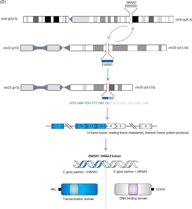
- It is characterised by NR4A3 GENE REARRANGEMENT - most commonly the EWSR1-NR4A3 fusion, with FUS-NR4A3 and other rarer variant partners (e.g. TAF15-NR4A3) - and this NR4A3 rearrangement is the molecular hallmark used to confirm the diagnosis.
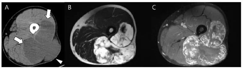
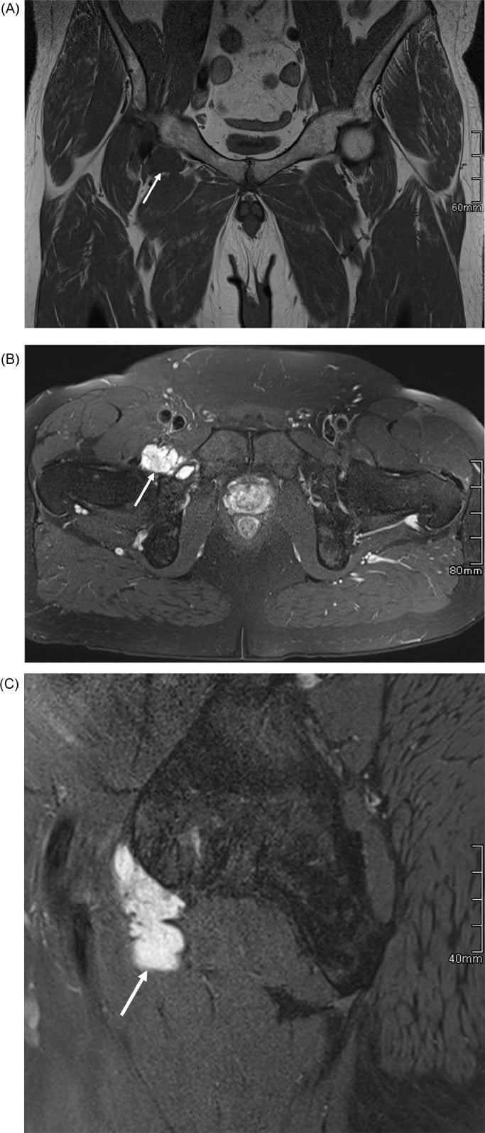
- It typically presents as a DEEP soft-tissue MASS of the PROXIMAL LIMBS and limb GIRDLES (especially the THIGH) in adults, often slowly enlarging; the imaging is of a deep, often lobulated, myxoid (T2-bright) soft-tissue mass.
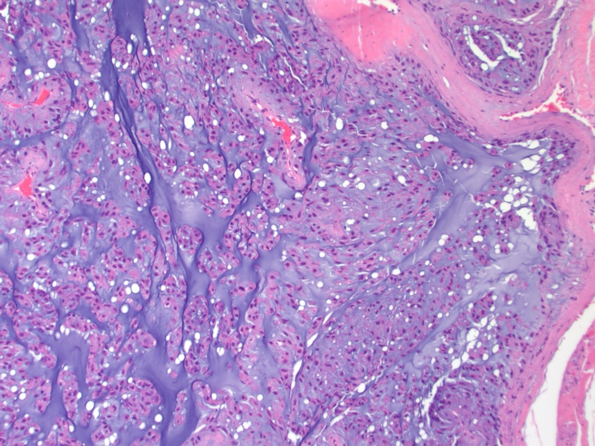
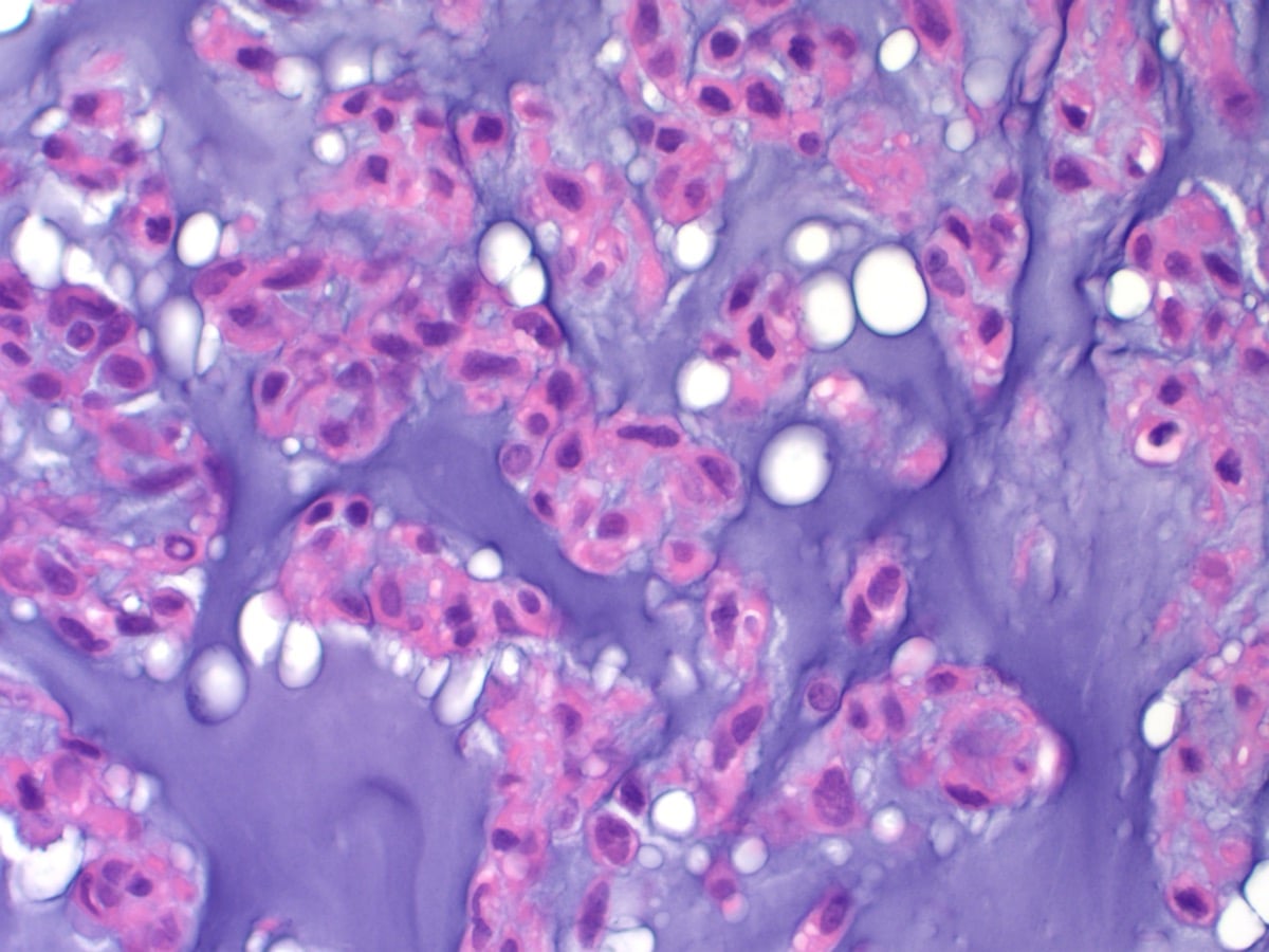
- HISTOLOGY shows a MYXOID matrix with a MULTILOBULAR architecture and CORDS, clusters or reticular arrays of relatively uniform eosinophilic OVOID-to-short-SPINDLE cells; immunohistochemistry is variable (e.g. variable CD117/S100), and morphological variants (rhabdoid, cellular/'high-grade', spindle) and rare non-EWSR1 fusions exist, so the molecular NR4A3 finding is valuable.
- PUT NUMBERS ON 'INDOLENT BUT RELAPSING' - AND NOTE THAT 'INDOLENT' UNDERSELLS IT. In a 117-case series with a median follow-up of 9 years (up to 22), estimated survival was 90 PERCENT at 5 years but 70 PERCENT at 10 and 60 PERCENT at 15; LOCAL RECURRENCE occurred in 48 percent (40 of 83), and 58 percent of those recurred MORE THAN ONCE; METASTASES occurred in 46 percent (35 of 76). The authors concluded that the high eventual death rate DISTINGUISHES EMC FROM LOW-GRADE SARCOMAS - so 'indolent' describes the tempo, not the eventual outcome, and prolonged surveillance follows from that.
- THE PROGNOSTIC FACTORS ARE CLINICAL, NOT HISTOLOGICAL. On multivariate analysis the adverse factors were OLDER AGE, LARGER TUMOUR SIZE and LOCATION IN THE PROXIMAL EXTREMITY OR LIMB GIRDLE - which is where 80 percent of these tumours arise, so the classic site is also the adverse one. METASTASIS reduced survival but LOCAL RECURRENCE did NOT. Critically, HISTOLOGIC GRADING WAS OF NO PROGNOSTIC VALUE in this series, so a report describing a 'cellular' or 'high-grade' variant should not be read as a worse prognosis; the cellular non-myxoid foci (seen in 29 percent) matter for RECOGNITION, since they mimic Ewing sarcoma, synovial sarcoma, fibrosarcoma and rhabdoid tumour.
- MANAGEMENT follows soft-tissue-sarcoma principles: MRI of the primary, a properly PLANNED BIOPSY at the treating sarcoma unit, and WIDE surgical RESECTION as the mainstay; EMC is relatively CHEMO/RADIO-RESISTANT, so radiotherapy/chemotherapy have a limited (selective) role, and LONG-TERM follow-up is essential because of late recurrence/metastasis - all within a specialist sarcoma multidisciplinary team.
- “Extraskeletal myxoid chondrosarcoma (EMC) = rare soft-tissue sarcoma; DESPITE the name it is NOT true cartilage. Defined by NR4A3 rearrangement (most often EWSR1-NR4A3; FUS/TAF15 variants).
- “Deep mass of the proximal limbs/girdles (thigh). Histology: myxoid + multilobular + cords/clusters of eosinophilic ovoid-spindle cells.
- “Indolent but RELAPSING long-term - local recurrence + LATE metastases (lung). Wide resection is the mainstay; chemo/radio-resistant; PROLONGED surveillance; sarcoma-centre care.
- “Quote the long tail: survival 90% at 5 years, 70% at 10, 60% at 15; local recurrence 48% (often repeated), metastases 46% (n=117, median follow-up 9 years). Adverse factors are older age, larger size and proximal-limb site - HISTOLOGIC GRADE has NO prognostic value, and local recurrence did not predict death.
Despite 'chondrosarcoma', EMC is not true cartilage - it is a distinct soft-tissue sarcoma defined by NR4A3 rearrangement (most often EWSR1-NR4A3).
Indolent but relapsing over the long term - local recurrence and late metastases (lung). Needs prolonged surveillance; it is chemo/radio-resistant (surgery is the mainstay).
Overview & Epidemiology
Extraskeletal myxoid chondrosarcoma (EMC) is a rare soft-tissue sarcoma of uncertain differentiation - despite its name it shows no true cartilage differentiation and is defined molecularly by NR4A3 rearrangement. It accounts for only a small minority of soft-tissue sarcomas and has no established population incidence figure. The demographics come from the largest series (Meis-Kindblom 1999, n=117):
- Sex: male-to-female ratio 2 to 1
- Age: median 52 years (range 6-89) - a tumour of adults
- Size: median 7 cm (range 1.1-25)
- Site: all deep (deep subcutis or deeper); 80 percent in the proximal extremities or limb girdles, 20 percent in the trunk; the thigh is the classic location
The defining clinical fact is the tempo: an indolent but relentlessly relapsing course, quantified under "The Long Tail" below.
What It Is, Genetics & Histology
EMC is a rare soft-tissue sarcoma that, despite its name, does not show true cartilage differentiation; it is a distinct entity defined by NR4A3 gene rearrangement (most often EWSR1-NR4A3, with FUS-NR4A3 and rarer variants). It presents as a deep mass of the proximal limbs/girdles (especially the thigh) in adults. Histology shows a myxoid, multilobular architecture with cords/clusters of eosinophilic ovoid-to-short-spindle cells (variable CD117/S100), and morphological/fusion variants exist - so the molecular NR4A3 finding is valuable. Its course is indolent but relapsing over the long term, with late recurrence/metastasis.
- Detail
- Uncertain - NOT true (hyaline) cartilage despite the name
- Detail
- NR4A3 rearrangement (EWSR1-NR4A3 most common; FUS/TAF15 variants)
- Detail
- Deep proximal limbs/limb girdles (especially thigh), adults
- Detail
- Myxoid, multilobular; cords/clusters of eosinophilic ovoid-spindle cells
- Detail
- Indolent but relapsing; local recurrence + late metastasis (lung)
- Detail
- Wide resection (mainstay); chemo/radio-resistant; long surveillance
Pathophysiology & Molecular Basis
NR4A3 (also known as CHN, NOR1 or TEC) is a nuclear orphan-receptor transcription factor on chromosome 9q22. In EMC a chromosomal translocation fuses NR4A3 to a strong transactivation partner - classically EWSR1 via t(9;22)(q22;q12) (about two-thirds of cases, first characterised by Clark 1996), with variant partners TAF15, TCF12 and FUS, and recently reported novel fusions such as ACTB-NR4A3 and FUS-NR4A2 (Chen 2026). The fusion protein replaces the RNA-binding domain of the partner with the entire NR4A3 protein, placing an intact orphan nuclear receptor under an aberrantly strong promoter and driving constitutive, dysregulated transcription - the oncogenic mechanism.
Two consequences matter clinically. First, the tumour's lineage is genuinely uncertain - the "chondrosarcoma" label is historical morphology, not biology, which is why EMC sits in the WHO chapter on tumours of uncertain differentiation. Second, because EWSR1 is promiscuous (Ewing sarcoma, clear cell sarcoma, DSRCT, angiomatoid fibrous histiocytoma), an EWSR1 break-apart result alone is not diagnostic - the NR4A3 partner must be demonstrated.
Clinical Presentation
- A deep, slowly enlarging, usually painless soft-tissue mass in an adult (median age 52), most often in the thigh or another proximal limb/limb-girdle site.
- Growth is indolent - patients may report a mass present for years - which is exactly why the eventual behaviour surprises the unwary.
- Symptomatic presentations relate to size and site: mechanical discomfort, a palpable lump, occasionally venous or neural compression; systemic symptoms are uncommon at presentation.
- Any deep mass larger than 5 cm should be treated as a sarcoma until proven otherwise and referred before any excision - in the largest series most initial excisions were intralesional or marginal, a pattern that feeds the 48 percent local-recurrence rate.
Investigations
- MRI of the primary is the imaging mainstay: a deep, lobulated, T2-bright (myxoid) intramuscular mass, typically with peripheral and septal enhancement after contrast, and low T1 signal (see the image gallery above).
- Planned biopsy at the treating sarcoma unit, along a track the surgeon can excise.
- Molecular confirmation is the clincher: demonstrate NR4A3 rearrangement (most often EWSR1-NR4A3) by FISH, RT-PCR or NGS. Do not rely on an EWSR1 break-apart result alone.
- Immunohistochemistry supports but does not define: CD117 positive in a proportion (~30%), variable S100, INI1 retained, keratin/EMA and brachyury negative - the profile that separates EMC from chordoma, myoepithelioma, myxoid liposarcoma, myxofibrosarcoma and low-grade fibromyxoid sarcoma.
- Staging: chest imaging (CT) for the lungs, the dominant metastatic site, with cross-sectional imaging of the primary; metastases can appear late, so staging is repeated through surveillance.
The Myxoid Differential and the Immunoprofile
- The immunoprofile. EMC is characteristically CD117 (c-KIT)-positive in a proportion (about 30%), variably S100-positive, INI1 (SMARCB1)-retained, and negative for keratins/EMA and brachyury - which is the key to the differential.
- The myxoid-tumour differential. A tumour with cords/strands of cells in a myxoid matrix mimics several entities, separated by immunostains and fusion:
- Chordoma / myoepithelioma - keratin/EMA-positive (chordoma also brachyury-positive); EMC is keratin-negative.
- Myxoid liposarcoma - DDIT3 (FUS-DDIT3) fusion, "chicken-wire" vasculature and lipoblasts.
- Myxofibrosarcoma - curvilinear vessels and pleomorphism; no NR4A3.
- Low-grade fibromyxoid sarcoma - FUS-CREB3L2, MUC4-positive.
- Intramuscular myxoma / ossifying fibromyxoid tumour - benign; no NR4A3.
- The clincher. The NR4A3 rearrangement, with the keratin-/brachyury-negative, often-CD117-positive profile, confirms EMC and separates it from these mimics.
Q: How do you distinguish EMC from the other myxoid soft-tissue tumours?
A: EMC is CD117 (c-KIT)-positive in ~30%, variably S100-positive, INI1-retained, and keratin/EMA- and brachyury-NEGATIVE. That profile plus the NR4A3 rearrangement separates it from chordoma/myoepithelioma (keratin/EMA+, chordoma brachyury+), myxoid liposarcoma (DDIT3 fusion, lipoblasts, chicken-wire vessels), myxofibrosarcoma (curvilinear vessels, pleomorphism), and low-grade fibromyxoid sarcoma (FUS-CREB3L2, MUC4+).
The Long Tail, Quantified - and Why 'Indolent' Misleads
The word "indolent" describes how slowly this tumour moves, not how it ends. In a series of 117 cases with a median follow-up of 9 years (range 2 months to 22 years), estimated survival was 90 percent at 5 years, 70 percent at 10 and 60 percent at 15. Local recurrence occurred in 48 percent (40 of 83) and more than half of those patients recurred repeatedly; metastases occurred in 46 percent (35 of 76). The authors' own conclusion is the one to quote: the high rate of local recurrence, the prolonged survival even after metastasis, and the eventual high rate of death distinguish EMC from low-grade sarcomas. A five-year disease-free interval is therefore an interim result, not a discharge.
- Effect on survival
- Adverse, on multivariate analysis
- Note
- One of only three independent factors
- Effect on survival
- Adverse, on multivariate analysis
- Note
- Median size in the series was 7 cm (range 1.1 to 25)
- Effect on survival
- Adverse, on multivariate analysis
- Note
- Also the commonest site - 80 percent arose there, 20 percent in the trunk
- Effect on survival
- Adverse
- Note
- Yet survival after metastasis was prolonged in some patients
- Effect on survival
- NOT associated with worse survival
- Note
- Common (48 percent) and often repeated, but did not predict death
- Effect on survival
- NO prognostic value
- Note
- Prognosis is dictated by clinical features, not by grading
Q: The report describes a cellular, non-myxoid, 'high-grade-looking' area. Does that worsen the prognosis?
A: No - histologic grading was of no prognostic value in the largest series with long follow-up, where prognosis was dictated by age, size and site. Cellular, solid, non-myxoid foci were found in 29 percent of cases and resemble chondroblastoma, Ewing sarcoma, synovial sarcoma, fibrosarcoma and rhabdoid tumour - so their importance is diagnostic, not prognostic: recognising them prevents the tumour being misclassified as one of those mimics, which would lead to quite different treatment. Note too the demographics the name does not convey - a male-to-female ratio of 2 to 1, median age 52, median size 7 cm. The series is retrospective and predates routine NR4A3 testing, so some cases were classified morphologically.
Management
- Diagnosis: MRI of the primary (deep, lobulated, myxoid/T2-bright mass); planned biopsy at the treating sarcoma unit; confirm NR4A3 rearrangement (e.g. EWSR1-NR4A3) on molecular testing.
- Surgery: wide resection with clear margins is the mainstay.
- Setting: specialist sarcoma multidisciplinary team; avoid unplanned excision of an undiagnosed deep mass. In the largest series most initial excisions were intralesional or marginal, which is precisely the pattern that produces the 48 percent local-recurrence rate.
The pathway that gets this right starts before the diagnosis is known: a deep mass larger than 5 cm should travel the route set out in soft-tissue masses and sarcoma referral, with imaging interpreted as in MRI of soft-tissue lesions and tissue obtained according to biopsy principles. The two myxoid tumours most often confused with it have their own pages - myxofibrosarcoma and, for the myxoid subtype, liposarcoma - and the bone tumour whose name it borrows is chondrosarcoma, with which it shares nothing but a word.
Two points define safe practice in extraskeletal myxoid chondrosarcoma. First, the name is misleading: despite 'chondrosarcoma', it is not a true cartilage tumour but a distinct soft-tissue sarcoma defined by NR4A3 rearrangement, so the diagnosis rests on the molecular finding (most often the EWSR1-NR4A3 fusion) together with the characteristic myxoid, multilobular histology, and morphological/fusion variants mean the molecular result is particularly useful. Second, and clinically most important, its behaviour is indolent but relapsing over a long horizon: patients may do well for years, yet local recurrence and late metastases (often pulmonary) occur, so short-term disease-free status is not reassurance - prolonged surveillance, including chest imaging, is essential. Because EMC is relatively resistant to chemotherapy and radiotherapy, wide surgical resection with clear margins is the mainstay of treatment, with adjuvant therapy playing only a limited, selective role, and management should be at a specialist sarcoma centre with a planned biopsy before any definitive surgery.
Complications & Prognosis
The complications of EMC are the disease's own long tail plus the consequences of treating a deep limb mass:
- Local recurrence - 48 percent in the largest series (40 of 83), and 58 percent of those recurred more than once; most initial excisions in that series were intralesional or marginal, so unplanned excision is the driver to avoid. Notably, local recurrence did not independently predict death.
- Metastasis - 46 percent (35 of 76), often late and predominantly pulmonary; survival after metastasis can still be prolonged, which is unusual among sarcomas.
- Treatment morbidity - wide resection of a deep proximal mass can sacrifice muscle compartments or neurovascular structures; adjuvant therapy, when used, adds toxicity with limited proven benefit (pazopanib's grade 3 events were hypertension 35%, raised ALT 23%, raised AST 19% - no grade 4 events).
- Prognosis - estimated survival 90 percent at 5 years, 70 at 10, 60 at 15 (Meis-Kindblom, median follow-up 9 years). Adverse factors on multivariate analysis are clinical: older age, larger size, proximal extremity/limb-girdle site. Histologic grading has no prognostic value.
The NR4A3 Fusion: Translocation, and Why EWSR1 Alone Is Not Enough
- The gene and the translocation. NR4A3 (also called NOR1 / CHN / TEC) is a nuclear orphan-receptor transcription factor on chromosome 9q22. The commonest fusion is EWSR1-NR4A3 from t(9;22)(q22;q12) (about two-thirds of cases), which places NR4A3 under the strong EWSR1 promoter and drives aberrant transcription. Variant partners include TAF15-NR4A3 (t(9;17)), TCF12-NR4A3 (t(9;15)) and FUS-NR4A3, plus rarer novel fusions (e.g. ACTB-NR4A3, FUS-NR4A2).
- Why the NR4A3 partner matters - not just EWSR1. EWSR1 is a promiscuous fusion gene rearranged in many sarcomas - Ewing sarcoma (EWSR1-FLI1), clear cell sarcoma (EWSR1-ATF1), desmoplastic small round cell tumour (EWSR1-WT1), angiomatoid fibrous histiocytoma (EWSR1-CREB1) and others - so an EWSR1 rearrangement alone is not specific; it is the NR4A3 partner (on RT-PCR/FISH/NGS) that defines EMC.
Q: What is the defining molecular abnormality of EMC, and why must the NR4A3 partner be identified?
A: EMC is defined by NR4A3 rearrangement - most often EWSR1-NR4A3 from t(9;22)(q22;q12) (~two-thirds), with TAF15-NR4A3, TCF12-NR4A3 and FUS-NR4A3 variants. NR4A3 (NOR1) is a nuclear orphan-receptor transcription factor on 9q22; the fusion drives aberrant transcription. Because EWSR1 is a promiscuous fusion gene (Ewing, clear cell sarcoma, DSRCT, AFH), EWSR1 rearrangement alone is not specific - it is the NR4A3 partner that clinches EMC.
Mnemonics & Memory Aids
MYXOID
Hook:MYXOID: Myxoid/multilobular, Y not truly cartilage, NR4A3 (X) fusion, Often deep thigh, Indolent but relapsing, Do wide resection + surveillance.
ASP
Hook:ASP - the three multivariate adverse factors in EMC are all CLINICAL: Age, Size, Place (proximal). Histologic grade is NOT among them.
CHORD
Hook:CHORD - the myxoid differential: separate by keratin/brachyury status and by which fusion (DDIT3, CREB3L2, or NR4A3) is present.
Clinical Decision Scenarios
Practise clinical reasoning and management decisions out loud
“A deep thigh sarcoma is reported as extraskeletal myxoid chondrosarcoma. What does the name actually mean, and how would you manage it?”
What it is
- Rare soft-tissue sarcoma; NOT true cartilage despite the name
- Defined by NR4A3 rearrangement (EWSR1-NR4A3 most common; FUS/TAF15 variants)
- Uncertain differentiation
Presentation & histology
- Deep mass of proximal limbs/limb girdles (thigh), adults
- Myxoid matrix, multilobular architecture
- Cords/clusters of eosinophilic ovoid-to-short-spindle cells (variants exist)
Behaviour
- Survival 90 percent at 5 years, 70 at 10, 60 at 15 (n=117, median follow-up 9 years)
- Local recurrence 48 percent (often repeated); metastases 46 percent - often late, to lung
- Adverse: older age, larger size, proximal-limb site. Grade has NO prognostic value; local recurrence did not predict death
- Short-term disease-free status is not reassurance
Management
- Planned biopsy + NR4A3 confirmation; MRI primary
- Wide resection is the mainstay (chemo/radio-resistant)
- Prolonged surveillance (incl. chest); sarcoma-centre care
Evidence & Key Studies
A series of extraskeletal myxoid chondrosarcomas with rare morphological and molecular variations
- Extraskeletal myxoid chondrosarcoma is a rare soft-tissue sarcoma characterised by a myxoid matrix, multilobular architecture, and eosinophilic ovoid-to-short-spindle cells arranged in cords, clusters or reticular patterns, with NR4A3 gene rearrangement.
- Five variant cases in adults (24-59 years, median 49) in the dorsal region, buttock, thigh, paravertebral region and elbow (4.0-16.0 cm, median 6.5); morphological variants included solid, rhabdoid, biphasic and spindle-cell patterns; all showed variable CD117 expression.
- NGS identified EWSR1-NR4A3, two FUS-NR4A3 fusions, and the novel fusions FUS-NR4A2 and ACTB-NR4A3, expanding the genetic spectrum; none of the five patients had local recurrence or metastasis at follow-up.
The defining features of extraskeletal myxoid chondrosarcoma - the myxoid matrix, multilobular architecture, cords/clusters of eosinophilic ovoid-to-short-spindle cells, the NR4A3 rearrangement (EWSR1-NR4A3 most common, with variant fusion partners), the adult age and deep proximal/limb-girdle sites, and the morphological variants - come from the cited Chen series. The fact that EMC does not show true cartilage differentiation despite its name, its indolent-but-relapsing long-term behaviour with late metastasis, its chemo/radio-resistance, and wide resection with prolonged surveillance as management are standard, well- established teaching. The survival rates, recurrence and metastasis proportions, demographics, prognostic factors and the finding that histologic grading carries no prognostic value come from the Meis-Kindblom series, which is retrospective, single-centre and predates routine NR4A3 testing. No randomised trial compares any adjuvant regimen in EMC, no systemic agent has an established survival benefit, and no surveillance interval or duration has been validated - so although the late-relapse pattern justifies long follow-up, no schedule is quoted here as though it were evidence-based.