Two Heads, Two Nerves, and the Alibi for Every Confusing Thumb
- Two heads with two different nerves: the SUPERFICIAL head from the recurrent motor branch of the median nerve and the DEEP head from the deep branch of the ulnar nerve, both C8-T1.
- Insertion onto the radial side of the base of the thumb proximal phalanx through the RADIAL sesamoid, with an extension into the extensor apparatus.
- The flexor pollicis longus tendon passes BETWEEN the two heads on its way to the thumb.
- Because of the dual supply, FPB is a UNRELIABLE test of median motor function - abductor pollicis brevis is the muscle to test.
- The recurrent motor branch is variable: in Poisel's 100 cadaver hands - the source of the figures usually credited to Lanz, who quotes them - it was extraligamentous in 46 per cent, subligamentous in 31 per cent and transligamentous in 23 per cent.
- “The Riche-Cannieu anastomosis, a communication between the deep ulnar branch and the recurrent median branch within the thenar eminence, produces the all-ulnar hand and can preserve thumb function despite a complete median lesion.
- “A cadaveric study of 60 limbs found the superficial head median-innervated in 70 per cent with double innervation in 30 per cent, and the deep head doubly innervated in 65 per cent.
- “Divide the transverse carpal ligament at its ULNAR margin and keep the incision in line with the radial border of the ring finger - the recurrent branch lies radial.
- “Recurrent motor branch injury produces loss of opposition with entirely NORMAL sensation - a devastating and easily misattributed complication.
Overview
Flexor pollicis brevis (FPB) is the middle muscle of the thenar eminence, lying between abductor pollicis brevis superficially and adductor pollicis in the deep plane. It flexes the thumb metacarpophalangeal joint and contributes the flexion component of opposition. Anatomically it is unremarkable; clinically and in examinations it is one of the most consequential muscles in the hand, for a single reason: it has two heads with two different nerves.
The superficial head is supplied by the recurrent motor branch of the median nerve; the deep head is supplied by the deep branch of the ulnar nerve. This is the classical exception that explains why a patient with an apparently complete median nerve lesion can still flex the thumb, why an ulnar lesion can be accompanied by unexpected thumb weakness, and why electrodiagnostic findings in the thenar eminence so often confuse. Layered on top of that is the Riche-Cannieu anastomosis, a communication between the two nerves within the thenar eminence, which in its extreme form produces an all-ulnar hand.
The muscle also defines the terrain of the most commonly performed operation in hand surgery. The recurrent motor branch of the median nerve enters the superficial head, and its course relative to the transverse carpal ligament varies enormously between individuals. Every rule about incision placement in carpal tunnel release exists to respect that variability.
When a thumb behaves in a way that does not fit a single nerve lesion, flexor pollicis brevis is nearly always the explanation. Three overlapping mechanisms:
- The two heads genuinely have two nerves. A cadaveric study of 60 limbs from 30 cadavers found the superficial head innervated by the median nerve in 70 per cent, with DOUBLE innervation in 30 per cent. The deep head was absent in 14 per cent, had double innervation in 65 per cent, was supplied by the deep ulnar branch alone in 17.5 per cent and by the recurrent median branch alone in 3.6 per cent. The authors concluded that the pattern that should be regarded as normal is a median superficial head and a deep head receiving both ulnar and median supply.
- The Riche-Cannieu anastomosis. A communication between the deep branch of the ulnar nerve and the recurrent (thenar) branch of the median nerve, running within the substance of the thenar muscles. Where it is substantial, all or part of thenar function is retained from the ulnar nerve despite a complete median lesion - the all-ulnar hand. It has been found in a meaningful minority of hands at dissection: one anatomical study of 68 palmar regions from 34 cadavers found an ulnar-median anastomosis in 13.
- Its cousins elsewhere. A Martin-Gruber anastomosis (median to ulnar, in the forearm) and a Marinacci anastomosis (ulnar to median, in the forearm) produce the same category of surprise at a more proximal level. Berrettini communications between common digital nerves do it for sensation.
Two practical consequences that examiners want said out loud:
- Never test median motor function with FPB. Use abductor pollicis brevis - palmar abduction of the thumb perpendicular to the plane of the palm, against resistance - because APB is the most consistently and purely median-innervated thenar muscle.
- Preserved thumb function does not exclude a complete median nerve division. Test sensation, test APB specifically, and if the picture remains inconsistent, name the anastomosis rather than doubting the injury.
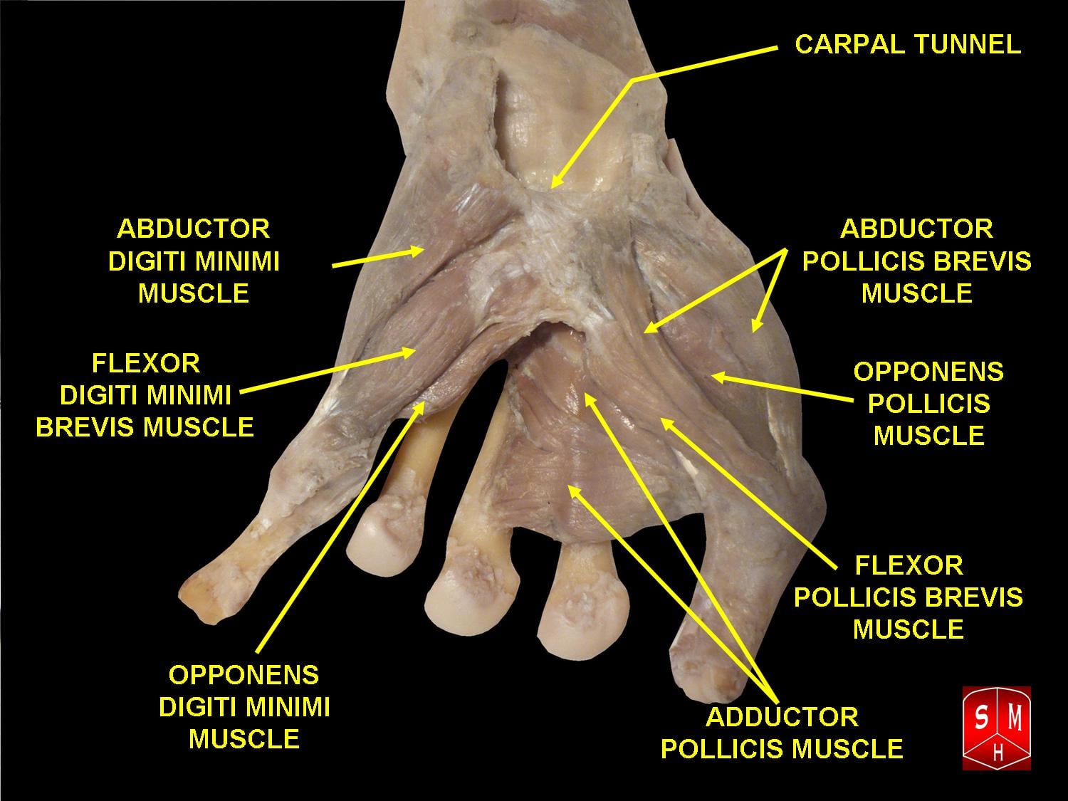
Attachments, Innervation and Relations
Origin - two heads
Superficial head
- The distal border of the flexor retinaculum (transverse carpal ligament).
- The tubercle of the trapezium.
- It is the larger head and lies superficially, ulnar to abductor pollicis brevis.
Deep head
- The trapezoid and capitate, and the adjacent palmar ligaments of the distal carpal row.
- It is smaller, sometimes considered part of the adductor pollicis complex, and is absent in a proportion of hands - reported absent in 14 per cent of 60 dissected limbs.
The relationship between the heads
- The two heads converge on a common insertion, and the flexor pollicis longus tendon passes BETWEEN them on its way from the carpal tunnel to the thumb. This is the anatomical basis of thumb zone III, the thenar segment in which a divided FPL retracts and must be retrieved.
Insertion
- The radial side of the base of the proximal phalanx of the thumb, through the RADIAL SESAMOID of the metacarpophalangeal joint.
- A slip continues into the extensor apparatus of the thumb, giving FPB a secondary role in interphalangeal extension in the same way the lumbricals act in the fingers.
- The sesamoid rule: the radial sesamoid receives flexor pollicis brevis and the ulnar sesamoid receives adductor pollicis. This matters in thumb metacarpophalangeal collateral injury, where the sesamoid position on radiographs indicates the integrity of the volar plate and sesamoid complex.
The thenar layers - superficial to deep
- Abductor pollicis brevis - most superficial and most radial; purely median in almost all hands.
- Flexor pollicis brevis, superficial head - deep and ulnar to APB; median (recurrent branch).
- Opponens pollicis - deep to APB, inserting along the shaft of the thumb metacarpal; median.
- Flexor pollicis brevis, deep head - deeper still; ulnar (deep branch).
- Adductor pollicis - deepest and most ulnar, with transverse and oblique heads; ulnar (deep branch).
Transligamentous in 23 per cent.
- Nearly one in four branches pierces the transverse carpal ligament rather than passing distal to it.
- A blade passed through the radial half of the ligament, or a blind percutaneous release, divides it.
- Result: loss of opposition with entirely NORMAL sensation - and a patient whose numbness resolved, so the complication is easily missed.
Two rules, always.
- Incision in line with the radial border of the ring finger, ulnar to the palmaris longus axis.
- Divide the transverse carpal ligament at its ULNAR margin, immediately radial to the hook of the hamate.
- Identify the distal edge of the ligament and the fat of the superficial palmar arch before finishing; never cut what you cannot see.
Action and Biomechanics
Actions
- Flexion of the thumb metacarpophalangeal joint - its principal action, and the one action for which it has no true rival (adductor pollicis flexes the MCP joint but with an adduction vector).
- Flexion and medial rotation of the first metacarpal, contributing the flexion component of opposition.
- Assists thumb interphalangeal extension through its slip into the extensor apparatus.
- Stabilises the metacarpophalangeal joint during pinch by tensioning the radial sesamoid and volar plate complex.
The thumb muscle balance
- Opposition is a composite movement: palmar abduction (abductor pollicis brevis), pronation/medial rotation (opponens pollicis) and flexion (flexor pollicis brevis), with the extrinsic muscles positioning and stabilising.
- Pinch is a couple: adductor pollicis and the first dorsal interosseous generate key pinch force; flexor pollicis longus flexes the terminal joint; FPB stabilises the metacarpophalangeal joint so that force is transmitted to the pulp rather than collapsing the joint.
- When the intrinsics fail: the thumb collapses into the plane of the palm and pinch is performed by flexor pollicis longus alone, producing the Froment sign - thumb interphalangeal flexion as FPL substitutes for adductor pollicis - and the Jeanne sign, metacarpophalangeal hyperextension from loss of the intrinsic flexion moment.
Numbers examiners ask for
- Flexor Pollicis Brevis
- Superficial head median (recurrent), deep head ulnar (deep branch)
- Comparator
- APB and opponens are median; adductor pollicis is ulnar
- Significance
- FPB is the classic dually innervated muscle
- Flexor Pollicis Brevis
- 70 per cent
- Comparator
- Double innervation 30 per cent
- Significance
- Even the superficial head is not reliably pure median
- Flexor Pollicis Brevis
- 14 per cent of dissected limbs
- Comparator
- -
- Significance
- Its absence is itself a variant
- Flexor Pollicis Brevis
- 46 per cent
- Comparator
- -
- Significance
- The classical and safest pattern
- Flexor Pollicis Brevis
- 31 per cent
- Comparator
- -
- Significance
- Runs beneath the ligament before turning radially
- Flexor Pollicis Brevis
- 23 per cent
- Comparator
- -
- Significance
- PIERCES the ligament - the variant that gets cut
- Flexor Pollicis Brevis
- Radial sesamoid and radial base of the proximal phalanx
- Comparator
- Adductor pollicis takes the ULNAR sesamoid
- Significance
- Sesamoid position on radiographs reflects the complex
What its loss produces
- Loss of the superficial head (median lesion): weakness of thumb metacarpophalangeal flexion and of the flexion component of opposition. In practice this is masked, because the ulnar-innervated deep head and adductor pollicis substitute - which is precisely why FPB is useless as a test of median function.
- Loss of the deep head (ulnar lesion): contributes to the weak key pinch of ulnar palsy alongside adductor pollicis and the first dorsal interosseous, and to the Froment and Jeanne signs.
- Loss of both (a combined lesion or a C8-T1 lesion): a flail, unstable thumb metacarpophalangeal joint with pinch performed entirely by the long flexor.
- Spastic overactivity (cerebral palsy, stroke): FPB is one of the deforming forces in the thumb-in-palm deformity, together with adductor pollicis, the first dorsal interosseous and flexor pollicis longus.
Surface Anatomy and Examination
Palpation and inspection
- The thenar eminence is the muscle mass at the base of the thumb. Abductor pollicis brevis forms its superficial radial bulk; flexor pollicis brevis lies ulnar and deep to it, and the two cannot be reliably separated by palpation.
- Wasting is best seen by looking at the thenar eminence in profile against the light, comparing sides, and by looking for loss of the normal convexity and for prominence of the first metacarpal.
- The radial sesamoid is palpable on the volar-radial aspect of the thumb metacarpophalangeal joint and marks the FPB insertion.
How to test the thenar muscles properly
- How to perform
- Hand flat and supinated; the patient lifts the thumb vertically out of the plane of the palm against resistance applied to the proximal phalanx
- Positive finding
- Weakness, or absent palpable APB contraction
- What it means
- Median motor deficit - recurrent motor branch or more proximal
- False positives
- Substitution by abductor pollicis longus, which abducts in the PLANE of the palm - watch the direction of movement
- How to perform
- Ask for thumb MCP flexion with the IP joint relaxed
- Positive finding
- Weakness
- What it means
- Little - the deep head is ulnar and adductor pollicis substitutes
- False positives
- Dual innervation makes a normal result meaningless for median function
- How to perform
- Pinch paper between thumb and index; the examiner pulls
- Positive finding
- Thumb IP joint flexes as FPL substitutes for adductor pollicis
- What it means
- Ulnar motor weakness
- False positives
- Any painful thumb condition weakening key pinch
- How to perform
- Observe the thumb MCP joint during key pinch
- Positive finding
- MCP hyperextension
- What it means
- Ulnar intrinsic weakness with loss of the intrinsic flexion moment
- False positives
- Constitutional MCP hyperlaxity - compare sides
- How to perform
- Ask the patient to touch the thumb tip progressively down the fingers to the base of the little finger
- Positive finding
- Reduced score
- What it means
- Composite loss of abduction, pronation and flexion
- False positives
- Basal joint arthritis or first web contracture limits opposition mechanically
- How to perform
- Static or moving two-point discrimination over the thumb, index and middle pulp
- Positive finding
- Increased threshold
- What it means
- Median SENSORY involvement - distinguishes a nerve trunk lesion from an isolated recurrent motor branch injury
- False positives
- Callus and cold fingers raise thresholds
The single most important examination discipline
Test abductor pollicis brevis, not flexor pollicis brevis, for median motor function. APB is the most consistently and purely median-innervated thenar muscle. Place the hand flat and supinated and ask the patient to point the thumb at the ceiling against resistance; palpate the muscle belly as they do it. A normal FPB tells you nothing, because the deep head is ulnar and, in 30 per cent of hands, so is part of the superficial head.
Recognising an isolated recurrent motor branch injury
- Loss of palmar abduction and opposition with a wasted thenar eminence.
- Completely normal sensation in the median territory - because only the motor branch is involved.
- A history of carpal tunnel release, a penetrating injury to the proximal palm, or a mass in the thenar region.
- The combination of motor loss with intact sensation after a carpal tunnel release is the presentation of an iatrogenic recurrent branch injury, and it is frequently attributed to slow recovery rather than recognised.
Complications
Iatrogenic recurrent motor branch injury
- The complication that defines this muscle. It produces loss of opposition and thenar wasting with entirely NORMAL sensation, most often after carpal tunnel release, and particularly after a limited-incision, percutaneous or endoscopic technique in a patient with a transligamentous branch.
- Why it is missed: the patient's numbness has resolved, so both they and the surgeon attribute the weakness to slow recovery.
- Avoidance: ring finger axis incision; division of the ligament at its ulnar margin; direct visualisation; open technique where anatomy is atypical or revision is planned.
- Management: if recognised intraoperatively, primary repair under magnification. If recognised late, opponensplasty - the transfer choice depending on which motors remain innervated.
Other complications of carpal tunnel release
- Incomplete release, most often distally, leaving persistent symptoms.
- Palmar cutaneous branch neuroma from an incision placed too radially or crossing the crease transversely - producing a hypersensitive scar and inability to weight-bear through the heel of the hand.
- Median nerve laceration, the catastrophic version of the same error.
- Bowstringing of the flexor tendons and loss of grip strength after wide release, usually transient; pillar pain is common and self-limiting.
- Ulnar neurovascular injury if the division strays too far ulnar past the hook of the hamate.
Transfer-specific complications
- Wrong pulley position in opponensplasty - a pulley placed too distally or radially produces palmar abduction without pronation, so the patient can lift the thumb but cannot rotate the pulp to meet the fingertips.
- Choosing a denervated donor - an FDS or palmaris longus transfer attempted in a high median palsy simply does not work.
- Donor swan neck after whole-tendon FDS harvest from a lax finger - leave one slip attached distally.
- Over- or under-tensioning, producing a fixed abducted thumb or a transfer with no useful excursion.
Thumb-in-palm surgery complications
- Over-release producing an unstable, hyperabducted thumb that cannot be brought to the fingers for grasp - functionally worse than the original deformity.
- Under-correction from releasing only one of the deforming forces.
- First web contracture recurrence where the skin deficiency was not addressed with a Z-plasty or flap.
- Recurrent motor branch injury during a dissection that crosses its territory directly.
Diagnostic complications
- Misinterpreting a Riche-Cannieu anastomosis as either an intact nerve or a severe lesion, and acting on the electrodiagnostic result rather than the clinical picture.
- Testing FPB rather than APB and wrongly concluding that median motor function is intact.
Clinical Relevance
Thenar wasting in carpal tunnel syndrome
- Advanced median compression produces wasting of the median-innervated thenar muscles - abductor pollicis brevis, opponens pollicis and the superficial head of flexor pollicis brevis - while the ulnar-innervated deep head and adductor pollicis are spared.
- The clinical picture is a flattened, hollowed thenar eminence with loss of palmar abduction and opposition, with sensory symptoms that may by then have become constant or, in long-standing cases, have paradoxically diminished.
- Recovery of motor function after release is unreliable in the presence of established wasting and an absent motor response - which is the indication to consider a simultaneous opponensplasty.
The safe zone for carpal tunnel release
Every rule derives from the variability of the recurrent motor branch and of the median nerve itself:
- Incision in line with the radial border of the ring finger, that is, ULNAR to the palmaris longus axis proximally. This keeps away from the median nerve (deep and radial to palmaris longus), from its palmar cutaneous branch (which runs between palmaris longus and flexor carpi radialis) and from the recurrent motor branch (which lies radially).
- Do not cross the wrist crease transversely. Cross obliquely if extension is required.
- Divide the transverse carpal ligament at its ULNAR margin, immediately radial to the hook of the hamate. This is the single most protective step: it moves the plane of division away from both the median nerve and every configuration of the recurrent branch.
- Visualise the distal edge of the ligament and the fat pad of the superficial palmar arch to confirm complete distal release, and pass an instrument freely into the distal forearm to confirm proximal release.
- Be alert to the Lanz variants: accessory branches at the distal tunnel, a high division of the nerve (bifid median nerve, often with a persistent median artery) and accessory proximal branches. A bifid nerve is at particular risk during a limited-incision or endoscopic technique.
Endoscopic and limited-incision techniques
- These reduce scar tenderness and may speed return to work, but they rely on the surgeon not seeing the recurrent branch. In a transligamentous branch - 23 per cent of hands - the branch crosses the very structure being divided.
- Preoperative ultrasound identifies a bifid median nerve and a persistent median artery and is a reasonable adjunct where a limited technique is planned.
- Where anatomy is atypical, revision surgery is planned, or a mass is suspected, open release with direct visualisation is the safe choice.
Revision carpal tunnel release
- Indications include persistent, recurrent or new symptoms. New motor loss with intact sensation after a release should raise immediate suspicion of a recurrent branch injury.
- At revision the nerve is identified in virgin tissue proximal to the previous incision, then followed distally through the scar.
- A hypothenar fat pad flap is a recognised adjunct for coverage of a scarred median nerve; the ulnar bundle must be protected during its elevation.
If you take one thing from this page into a clinic: abductor pollicis brevis is the median motor test, not flexor pollicis brevis. Hand flat and supinated, thumb pointing at the ceiling, resist at the proximal phalanx, and palpate the muscle belly. Watch for substitution by abductor pollicis longus, which abducts the thumb in the plane of the palm rather than out of it.
Surgical Relevance
Open carpal tunnel release - the anatomy of each step
Incision. Longitudinal, in line with the radial border of the ring finger, from just distal to the distal wrist crease toward Kaplan cardinal line, 3 to 4 cm. Never transverse across the crease.
- Why this line: it is ulnar to the palmaris longus axis, and therefore ulnar to the median nerve (deep and radial to palmaris longus), ulnar to the palmar cutaneous branch (which runs between palmaris longus and flexor carpi radialis) and ulnar to every configuration of the recurrent motor branch.
Superficial dissection. Through skin and subcutaneous fat to the palmar fascia, then through the palmar fascia in line with the incision. Small palmar cutaneous twigs crossing the field should be preserved where possible.
Division of the transverse carpal ligament.
- Divide at its ULNAR margin, immediately radial to the hook of the hamate, under direct vision.
- Protect the contents with a blunt instrument passed beneath the ligament.
- Confirm complete DISTAL release by seeing the fat pad of the superficial palmar arch beyond the distal edge of the ligament; confirm complete PROXIMAL release by passing an instrument freely into the distal forearm.
Inspection. Look for the Lanz variants - a bifid nerve, a persistent median artery, accessory branches at the distal tunnel - and for a space-occupying lesion such as tenosynovium, a ganglion or an anomalous muscle belly.
Closure. Skin only. Do not repair the ligament.
If the recurrent branch is seen
- An extraligamentous branch curving back over the distal edge is the common and reassuring finding.
- A transligamentous branch encountered piercing the ligament must be protected and the ligament divided around it, ulnar to its course.
- If a branch is inadvertently divided, repair it primarily under magnification at the same sitting - this is far more likely to give a useful result than delayed reconstruction, and delayed diagnosis is the usual reason it is not done.
Nearly one in four recurrent motor branches pierces the transverse carpal ligament. You cannot know in advance which patient that is. Two rules make the variation irrelevant: keep the incision in line with the radial border of the ring finger, and divide the ligament at its ULNAR margin, immediately radial to the hook of the hamate. A recurrent branch injury produces loss of opposition with completely normal sensation - and because the numbness has resolved, both surgeon and patient may take months to realise what has happened.
OAF plus a Deep HalfThenar Muscles and Their Nerves
Hook:Three and a half thenar muscles are median; the deep head of FPB and adductor pollicis are ulnar - which is why FPB can never test median function.
Guidelines, Registries & Global Practice
Anatomical variation across populations
- Dual innervation of flexor pollicis brevis is the rule rather than the exception: a 60-limb cadaveric series found double innervation of the superficial head in 30 per cent and of the deep head in 65 per cent, with the deep head absent altogether in 14 per cent.
- Median-to-ulnar communications within the hand were found in 13 of 68 dissected palms in one anatomical series, providing the substrate for the Riche-Cannieu pattern.
- The Lanz course variants of the thenar branch - the eponym is Lanz's, the percentages are Poisel's: extraligamentous 46 per cent, subligamentous 31 per cent, transligamentous 23 per cent - are quoted worldwide and have not been meaningfully overturned by later series, though the exact proportions differ between studies.
- Bifid median nerve with a persistent median artery is a recognised variant that clusters together and raises the risk of injury during limited-incision techniques; where preoperative ultrasound is available, it identifies both.
Differences in described technique between sources
- Position on carpal tunnel and thenar practice
- Supports surgical release as effective for carpal tunnel syndrome; recommends against routine use of a single technique, with the choice of open versus endoscopic left to surgeon and patient after discussion of the risk profile.
- Position on carpal tunnel and thenar practice
- Recommend a structured non-operative trial in mild disease, and open release as the default with endoscopic technique reserved for surgeons with specific training.
- Position on carpal tunnel and thenar practice
- Emphasises the ring finger axis incision and division of the transverse carpal ligament at its ulnar margin, with explicit warning about the transligamentous recurrent branch.
- Position on carpal tunnel and thenar practice
- Emphasises assessment of voluntary control and sensation before thumb-in-palm surgery, and correction of all three components - release, augmentation and joint stabilisation.
Evidence positions worth stating
- Endoscopic versus open release: meta-analyses show comparable symptom relief with faster early return to work after endoscopic release, and no clear difference in serious complications, though the technique inherently forgoes direct visualisation of a transligamentous recurrent branch. The defensible position is either technique in trained hands, with open release where anatomy is atypical, a mass is suspected or the surgery is revision.
- Simultaneous opponensplasty: there is no randomised evidence establishing when a transfer should be added to release. Published series support adding a Camitz or modified Camitz at the time of release in severe thenar wasting with absent motor response, where recovery after decompression alone is unlikely.
- Nerve conduction studies do not override clinical examination in a hand with a suspected anastomosis. Where the electrodiagnostic picture is internally contradictory, the anastomosis should be named and the clinical findings - specifically APB power and median sensation - relied upon.
High- versus limited-resource practice
- Well-resourced settings: preoperative ultrasound for bifid nerves and persistent median arteries, electrodiagnostic confirmation, endoscopic release, formal hand therapy after transfers, and microsurgical repair of a divided recurrent branch.
- Limited-resource settings: open carpal tunnel release requires no special equipment and remains the safest technique anywhere. The two anatomical rules - ring finger axis incision and division at the ulnar margin - cost nothing and prevent the one complication that ruins the operation. Opponensplasty likewise needs no implant, and the Huber (abductor digiti minimi) transfer is particularly attractive where thenar bulk as well as function is desired. Clinical assessment of APB power is a complete substitute for electrodiagnostics in the great majority of cases.
MCQ Practice Points
Q: What supplies each head of flexor pollicis brevis? A: Superficial head - the recurrent motor branch of the MEDIAN nerve. Deep head - the deep branch of the ULNAR nerve. Both C8-T1.
Q: Which thenar muscle should be used to test median motor function? A: Abductor pollicis brevis - palmar abduction out of the plane of the palm. FPB is unreliable because of its dual supply.
Q: How often is the superficial head purely median? A: About 70 per cent, with double innervation in 30 per cent. The deep head is doubly innervated in about 65 per cent and is absent in about 14 per cent of limbs.
Q: What is the Riche-Cannieu anastomosis? A: A communication between the deep branch of the ulnar nerve and the recurrent branch of the median nerve within the thenar eminence, which in its extreme form produces the all-ulnar hand.
Q: What are the proportions of thenar branch course variants? A: Extraligamentous 46 per cent, subligamentous 31 per cent, transligamentous 23 per cent in 100 cadaver hands.
Q: Where is the transverse carpal ligament divided and why? A: At its ULNAR margin, immediately radial to the hook of the hamate - away from the median nerve and from every configuration of the recurrent motor branch.
Q: Which sesamoid receives flexor pollicis brevis? A: The RADIAL sesamoid. Adductor pollicis inserts through the ulnar sesamoid.
Q: What passes between the two heads of flexor pollicis brevis? A: The flexor pollicis longus tendon.
Q: Loss of opposition with entirely normal sensation after carpal tunnel release - diagnosis? A: Iatrogenic injury to the recurrent motor branch of the median nerve, classically in a transligamentous variant.
Q: Which opponensplasty donors are available in a HIGH median nerve palsy? A: Extensor indicis proprius or abductor digiti minimi (Huber). FDS and palmaris longus are denervated.
Q: Which muscles produce the thumb-in-palm deformity? A: Adductor pollicis, the first dorsal interosseous and flexor pollicis brevis, with flexor pollicis longus if the interphalangeal joint is also flexed.
Exam Viva Scenarios
Practise clinical reasoning and management decisions out loud
“A 27-year-old sustained a deep laceration at the volar wrist. At exploration the median nerve was found completely divided and was repaired. At review two weeks later he has dense sensory loss in the median territory, but he can flex his thumb metacarpophalangeal joint, oppose reasonably well and has a well-preserved thenar eminence. Your registrar wonders whether the nerve was really divided. What do you say?”
“Six months after an uncomplicated endoscopic carpal tunnel release, a 55-year-old woman reports that her numbness resolved completely but she now struggles to pick up a cup and her thumb 'lies flat'. Examination shows a wasted thenar eminence, absent palmar abduction and completely normal sensation. What has happened?”
“A 12-year-old with spastic hemiplegic cerebral palsy holds his thumb flexed and adducted across the palm. His mother cannot clean the web space and he cannot grasp a cup. How do you assess him and what does surgery involve?”
Anatomy
- Superficial head: flexor retinaculum and trapezium
- Deep head: trapezoid and capitate; absent in about 14 per cent
- Insert: radial sesamoid and radial base of the thumb proximal phalanx
- FPL tendon passes BETWEEN the two heads
- Adductor pollicis takes the ULNAR sesamoid
Dual Innervation
- Superficial head: recurrent motor branch of the MEDIAN nerve
- Deep head: deep branch of the ULNAR nerve
- Superficial head doubly innervated in 30 per cent
- Deep head doubly innervated in 65 per cent
- Test APB, never FPB, for median motor function
Anastomoses
- Riche-Cannieu: deep ulnar to recurrent median, in the HAND
- All-ulnar hand - thenar function despite a median lesion
- Martin-Gruber: median to ulnar in the FOREARM
- Marinacci: ulnar to median in the FOREARM
Lanz Variants
- Extraligamentous 46%, subligamentous 31%, transligamentous 23%
- Group II accessory distal branches; III high division; IV proximal branches
- Approach the median nerve from the ULNAR side
- Bifid nerve travels with a persistent median artery
Safe Zones and Reconstruction
- Incision in line with the radial border of the ring finger
- Divide the TCL at its ULNAR margin, radial to the hook of hamate
- Motor loss with NORMAL sensation = recurrent branch injury
- Low median palsy: FDS (Bunnell) or Camitz opponensplasty
- High median palsy: EIP or Huber transfer
Evidence Base
Flexor Pollicis Brevis Muscle: Anatomical Study and Clinical Implications
- Anatomical dissection of 60 limbs from 30 cadavers examining the nature, incidence and innervation of flexor pollicis brevis
- The superficial head was innervated by the median nerve in 70 per cent and had double innervation in 30 per cent
- The deep head was ABSENT in 14 per cent of limbs
- The deep head had double innervation in 65 per cent, supply from the deep branch of the ulnar nerve alone in 17.5 per cent and from the recurrent branch of the median nerve alone in 3.6 per cent
- The authors conclude that the pattern to be regarded as normal is a median-innervated superficial head with a deep head receiving both ulnar and median supply
Anatomical Variations of the Median Nerve in the Carpal Tunnel
- 246 operatively explored carpal tunnels with 29 variations in the course of the median nerve identified
- Accessory branches at the distal portion of the tunnel in 18 hands, high division of the median nerve in 7 and accessory branches proximal to the tunnel in 4
- Four groups defined: I variation in the course of the thenar branch, II accessory distal branches, III high divisions, IV accessory proximal branches
- IN THE PAPER'S OWN WORDS, the widely quoted thenar branch percentages are POISEL'S, not Lanz's: 'the thenar branch variations in 100 cadaver hands (Poisel) were extraligamentous in 46 percent, subligamentous in 31 percent, and transligamentous in 23 percent'
- Lanz's own material is the 246 operative explorations; the 46/31/23 split is quoted from a separate cadaveric series and is routinely mis-attributed to this paper
- The findings emphasise the importance of approaching the median nerve from the ULNAR side when opening the carpal tunnel
Variations in the Motor Nerve Supply of the Thenar and Hypothenar Muscles of the Hand
- Sixty-eight palmar regions from 34 adult cadavers of both sexes aged 40 to 70 years were dissected
- The distribution of the muscular branches of the median and ulnar nerves and the motor innervation of the thenar and hypothenar muscles were mapped
- The structure of the flexor pollicis brevis was examined in all 68 hands
- An anastomosis between the ulnar and median nerves was found in 13 of the 68 hands
Cannieu-Riche Anastomosis of the Ulnar to Median Nerve in the Hand: Case Report
- A very long Cannieu-Riche anastomosis was observed in a male cadaver, running between the proximal portion of the deep branch of the ulnar nerve destined for adductor pollicis and a ramus of the recurrent branch of the median nerve to the SUPERFICIAL HEAD of flexor pollicis brevis
- The clinical relevance is possible preservation of all or part of thenar muscle function from the ulnar nerve in the presence of a median nerve lesion
- Ignorance of the anomaly can produce obscure clinical, surgical and electroneuromyographic findings
- The report reviews the incidence, the double innervation and the clinical significance and proposes a classification of the types of this communication
Opponensplasty Using Palmaris Longus Tendon and Flexor Retinaculum Pulley in Patients with Severe Carpal Tunnel Syndrome
- Twelve cases of long-standing severe carpal tunnel syndrome with severe thenar muscle atrophy treated by modified Camitz opponensplasty with a pulley at the ulnar remnant of the flexor retinaculum
- Mean grade of thenar atrophy improved from 3 preoperatively to 0.83 at final follow-up
- Maximal palmar abduction improved from 3.6 to 6.8 cm and spatial rotation from 54 to 83 degrees
- Kapandji tip opposition improved from 65 to 85 per cent symmetry
- All patients were satisfied or very satisfied
Minimally Invasive Modified Camitz Opponensplasty for Severe Carpal Tunnel Syndrome
- Twenty-eight hands treated by a minimally invasive modified Camitz passing the palmaris longus beneath the abductor pollicis brevis fascia through palm and thumb incisions only, compared with 10 conventional procedures
- Thumb palmar abduction, pinch power and grip strength improved significantly in both groups
- The minimally invasive group had no transferred tendon bowstringing, no painful wrist scars and no palmar cutaneous branch injuries
- The conventional group had four painful wrist scars and nine cases of bowstringing