The Only Muscle That Truly Opposes
- The only thenar muscle inserting into the whole radial border of the first metacarpal shaft rather than into the proximal phalanx.
- It is the only muscle capable of true active axial pronation of the thumb ray — the component of opposition that cannot be substituted.
- Supplied by the recurrent (thenar) motor branch of the median nerve, C8-T1, arising from the radial side of the nerve at or just distal to the transverse carpal ligament.
- Lanz classified the variants of this branch: extraligamentous (commonest), subligamentous, and transligamentous — the last pierces the flexor retinaculum and is the one divided in carpal tunnel release.
- Loss of opposition, not loss of sensation, is the functional catastrophe of median nerve palsy — the thumb becomes an immobile post.
- “The recurrent motor branch arises at the distal edge of the transverse carpal ligament and turns back radially like a hook toward the thenar muscles.
- “Kapandji score grades opposition 1 to 10 by where the thumb pulp can reach; 10 is the distal palmar crease of the hand, and a crawling thumb does not count.
- “Camitz uses palmaris longus with a strip of palmar fascia — it restores palmar abduction, not true pronation, but it is quick and can be done at the same sitting as carpal tunnel release.
- “Huber transfer (abductor digiti minimi) is the choice in the congenital hypoplastic thumb because it adds bulk to a flat thenar eminence as well as motion.
Overview
Opponens pollicis is the deepest of the three superficial thenar muscles, lying beneath abductor pollicis brevis and radial to the superficial head of flexor pollicis brevis. It is small — roughly 3-4 cm long — but it is the only muscle in the body whose sole purpose is to rotate a metacarpal about its own long axis.
That single property is what makes the human hand human. The examiner's question is rarely "where does it attach"; it is "why can no other muscle replace it, and what will you transfer when it is gone".
Opposition is a composite motion of the thumb ray occurring principally at the trapeziometacarpal (TMC) saddle joint, comprising:
- Palmar abduction — the thumb metacarpal moves anteriorly away from the plane of the palm, driven mainly by abductor pollicis brevis (with abductor pollicis longus at the extreme).
- Flexion and adduction across the palm — driven by flexor pollicis brevis and, terminally, adductor pollicis.
- Axial pronation (internal rotation) of the metacarpal — around 90 degrees of combined rotation through the arc, so that the thumb pulp comes to face the finger pulps. Opponens pollicis is the only muscle that generates this actively.
The saddle geometry of the TMC joint supplies obligatory conjunct rotation, so some pronation occurs passively as the metacarpal flexes and abducts. That passive component is why a Camitz transfer, which supplies only palmar abduction, still restores useful function despite not reproducing opponens pollicis.
Examiner's follow-up: "So if conjunct rotation happens anyway, why does anyone need an opponensplasty?" Because passive conjunct rotation requires the metacarpal to be actively driven into flexion-abduction against resistance. A paralysed thenar eminence cannot drive it, so the thumb sits supinated and adducted in the plane of the palm and the patient pinches side-to-side rather than pulp-to-pulp. The transfer restores the drive; the joint supplies the rotation.
LOAF plus the deep halfThenar Muscles and Their Nerves
Hook:Only the deep head of flexor pollicis brevis breaks the rule — everything else in the thenar eminence proper is recurrent median.
DVPMBefore Any Opponensplasty — DVPM
Hook:Every row of this page's complications table is one of these four missed: absent donor, wrong vector, unaddressed joint, or an unconsidered cost to the donor side.
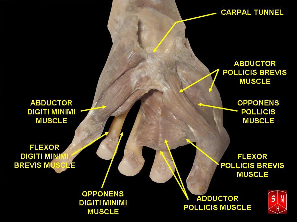
Attachments, Innervation and Relations
Origin
- The tubercle (crest) of the trapezium.
- The adjacent flexor retinaculum (transverse carpal ligament) on its palmar surface.
- Some fibres arise from the trapezial ridge in continuity with the origin of abductor pollicis brevis.
Insertion
- The entire length of the radial border and adjacent palmar surface of the shaft of the first metacarpal, from just distal to the base to the neck.
- This is the defining feature. Abductor pollicis brevis and flexor pollicis brevis both insert into the base of the proximal phalanx (via the radial sesamoid and the extensor hood); opponens pollicis alone inserts into the metacarpal shaft, which is precisely why it rotates the ray rather than moving the joint distal to it.
Architecture
- The fibres run distally, laterally and dorsally, curving around the metacarpal in a spiral — the anatomical signature of a rotator.
- Fibre length is short (of the order of 2-3 cm) with a modest cross-sectional area; it is a positioning muscle, not a strength muscle. This matters when selecting a donor: an opponensplasty needs excursion and vector, not raw power.
- The recurrent motor branch most commonly recurs around the distal edge of the transverse carpal ligament and then runs radially within a zone roughly 1 cm distal and radial to that edge. Kaplan's cardinal line — from the apex of the first web space, parallel to the proximal palmar crease, toward the hook of hamate — approximates the level of the distal ligament edge and the superficial palmar arch.
- Rule of the open release: keep the incision in line with the radial border of the ring finger, ulnar to the axis of the thenar crease, and divide the ligament at its ulnar-most extent against the hook of hamate. Every millimetre radial increases the risk to a transligamentous branch and to the palmar cutaneous branch.
- Palmar cutaneous branch of the median nerve: arises 5-7 cm proximal to the wrist crease between palmaris longus and flexor carpi radialis. Injury produces a painful neuroma at the base of the palm — a frequently overlooked cause of persistent "pillar pain" after release.
- Superficial palmar arch: lies approximately 1 cm distal to the distal edge of the transverse carpal ligament, at Kaplan's cardinal line. Blind distal division of the ligament risks it.
- Endoscopic release: the transligamentous variant (Lanz Ic) is the one genuinely at risk, because the blade divides a ligament with a nerve inside it. Adequate visualisation of the distal ligament edge before cutting is the mitigation.
Action and Biomechanics
Actions in each plane
- Contribution of opponens pollicis
- Primary and effectively sole active generator
- Co-actors
- Passive conjunct rotation of the TMC saddle joint
- What is lost if it fails
- Pulp-to-pulp pinch; the patient converts to key pinch
- Contribution of opponens pollicis
- Secondary contributor
- Co-actors
- Abductor pollicis brevis (primary), abductor pollicis longus
- What is lost if it fails
- Thumb lies in the plane of the palm — the ape hand
- Contribution of opponens pollicis
- Contributes at the metacarpal
- Co-actors
- Flexor pollicis brevis, adductor pollicis
- What is lost if it fails
- Cannot reach the ulnar digits
- Contribution of opponens pollicis
- Deepens the distal transverse arch on the radial side
- Co-actors
- Opponens digiti minimi on the ulnar side
- What is lost if it fails
- Flat palm, poor conformity to spherical objects
Moment arm and excursion
- Opponens pollicis has a short fibre length (of the order of 2-3 cm) and therefore small excursion. Its moment arm for pronation about the metacarpal long axis is small in absolute terms, but it is the only one available.
- A donor for opponensplasty must supply an excursion of roughly 3-5 cm with a vector directed from the pisiform toward the thumb metacarpophalangeal joint. This vector line is the practical rule for every opponensplasty, whatever the donor.
The transverse arches
- The hand has a proximal transverse arch (the fixed distal carpal row) and a distal transverse arch (mobile, at the metacarpal heads, keystone the third metacarpal head), plus longitudinal arches along each ray.
- Opponens pollicis and opponens digiti minimi are the two muscles that actively deepen the distal transverse arch by rotating the border rays toward each other — the "cupping" that lets a flat palm grasp a sphere.
- Loss of both (combined low median and ulnar palsy) produces the flat, simian hand in which nothing can be cupped.
Synergists and antagonists
- Synergists: abductor pollicis brevis, flexor pollicis brevis, abductor pollicis longus at the extreme of palmar abduction, and opponens digiti minimi from the other side of the palm.
- Antagonists: extensor pollicis longus (which supinates and extends the thumb ray), extensor pollicis brevis, and adductor pollicis (which supinates the metacarpal as it adducts it).
What happens when it fails
- Ape (simian) hand: the thumb falls into the plane of the palm, supinated and adducted, pulled there by the unopposed extensor pollicis longus and adductor pollicis.
- The patient substitutes with key pinch (thumb pulp to the radial side of the index), which is ulnar-dependent and preserved. This is why a median palsy patient still records measurable pinch and why pinch dynamometry alone underestimates the deficit — Kapandji scoring, not pinch strength, documents opposition.
- Secondary changes: the first web narrows over months, and the trapeziometacarpal joint stiffens in adduction and supination, converting a soft deficit into a fixed one. Splinting the thumb in palmar abduction from the outset is not optional.
Surface Anatomy and Examination
Palpation and inspection
- The thenar eminence is the fleshy mound at the base of the thumb. Opponens pollicis lies deep to abductor pollicis brevis and is directly palpable only along the radial border of the first metacarpal shaft with the abductor relaxed.
- Wasting is best appreciated by looking from the ulnar side with the hands held palm to palm, comparing the contour of the two eminences, and by looking for flattening of the thenar crease with prominence of the first metacarpal.
- In median palsy, wasting affects abductor pollicis brevis and opponens pollicis; the ulnar half of the eminence (deep head of flexor pollicis brevis, adductor pollicis) is spared, so the eminence flattens radially rather than disappearing.
Testing opposition
The standard median motor test.
- Hand flat, dorsum on the table, palm up.
- Ask the patient to point the thumb vertically at the ceiling — pure palmar abduction.
- Resist at the thumb proximal phalanx while palpating the thenar eminence.
- Why this and not opposition: abductor pollicis brevis is the most consistently recurrent-branch innervated muscle, and the movement cannot be faked by extensor pollicis longus or adductor pollicis.
- False positive: allowing the wrist to extend or the hand to lift off the table lets abductor pollicis longus substitute.
Pulp-to-pulp, nail-to-nail.
- Ask the patient to touch the thumb pulp to the pulp of the little finger, then to the base of the little finger.
- Watch the thumbnail: in true opposition it rotates to face the palm. If the nail stays in the plane of the palm, the patient is reaching, not opposing.
- False positive: flexor pollicis longus plus adductor pollicis can drag the thumb across the palm in a flat, supinated posture that superficially resembles opposition.
Kapandji opposition score
The Kapandji score grades opposition by the most distal point the thumb pulp can reach, using the hand itself as the reference system so that no angles need measuring. As published, the scale runs 1 to 10, with 0 recorded when the thumb cannot reach even point 1:
- Point reached by the thumb pulp
- Cannot reach point 1 — no useful opposition
- Point reached by the thumb pulp
- Lateral (radial) side of the middle phalanx of the index finger
- Point reached by the thumb pulp
- Lateral (radial) side of the distal phalanx of the index finger
- Point reached by the thumb pulp
- Tip of the index finger
- Point reached by the thumb pulp
- Tip of the middle finger
- Point reached by the thumb pulp
- Tip of the ring finger
- Point reached by the thumb pulp
- Tip of the little finger
- Point reached by the thumb pulp
- Distal interphalangeal crease of the little finger
- Point reached by the thumb pulp
- Proximal interphalangeal crease of the little finger
- Point reached by the thumb pulp
- Proximal (metacarpophalangeal) crease of the little finger
- Point reached by the thumb pulp
- Distal palmar crease of the hand — full opposition
- A score of 7 or more is generally considered functional; the usual target after opponensplasty is 7-9.
- Kapandji's own caveat: the test is only valid if the earlier stages are genuinely achievable — a thumb crawling across the palm is not opposition. Watch the thumbnail rotate.
- Kapandji also described a paired counter-opposition (reposition) test, using the opposite hand as the reference while the thumb is actively drawn up from the table.
- It is reproducible, needs no equipment, and is the outcome measure quoted in almost every opponensplasty series — which is exactly why examiners expect it.
Named clinical tests around the thenar eminence
- How to perform
- Hand flat, palm up; thumb points to the ceiling against resistance
- Positive finding
- Weakness with palpable soft thenar mass
- What it means
- Recurrent median motor branch dysfunction
- False positives
- Wrist extension or hand lifting lets abductor pollicis longus substitute
- How to perform
- Thumb pulp walked from index to the base of the little finger
- Positive finding
- Score less than 7
- What it means
- Functionally inadequate opposition
- False positives
- Hypermobile joints allow a high score with weak muscles
- How to perform
- Wrist held in maximal passive flexion for 60 seconds
- Positive finding
- Paraesthesia in the median distribution
- What it means
- Median neuropathy at the wrist
- False positives
- Cervical radiculopathy; anxiety-driven reporting
- How to perform
- Direct even thumb pressure over the carpal tunnel for 30 seconds
- Positive finding
- Median paraesthesia reproduced
- What it means
- Median neuropathy at the wrist; generally more sensitive than Phalen or Tinel
- False positives
- Pressure over the palmar cutaneous branch alone
- How to perform
- Oppose thumb to little finger with the wrist gently flexed
- Positive finding
- Visible or palpable tendon at the mid-wrist
- What it means
- Confirms a donor for Camitz transfer or graft
- False positives
- Obesity and oedema hide the tendon; check both limbs
- How to perform
- Key pinch on a card against resistance; watch the thumb IP joint
- Positive finding
- IP joint flexes
- What it means
- Adductor pollicis (ulnar) weakness — not a median test
- False positives
- Painful thumb MCP joint causes guarding
Electrodiagnosis
- Abductor pollicis brevis is the standard recording muscle for median motor conduction. A distal motor latency greater than approximately 4.2-4.5 ms across the wrist is the conventional threshold for median neuropathy at the wrist, although laboratories differ.
- Fibrillations and reduced recruitment in abductor pollicis brevis with a normal first dorsal interosseous localises to the median nerve; the reverse localises to the ulnar nerve. Both abnormal points to a lower trunk or C8-T1 lesion.
Complications
- Mechanism
- Transligamentous (Lanz Ic) branch cut during release, or incision too radial
- Prevention
- Incision in line with the radial border of the ring finger; divide the ligament at its ulnar extent; visualise the distal edge
- Salvage
- Early re-exploration and direct repair; late opponensplasty
- Mechanism
- Incision crossing the wrist crease or over the thenar eminence
- Prevention
- Longitudinal incision ulnar to the thenar crease, not crossing the crease
- Salvage
- Neuroma excision with transposition into pronator quadratus or bone
- Mechanism
- Blind distal division of the ligament at Kaplan's cardinal line
- Prevention
- Direct visualisation of the distal ligament edge before completing division
- Salvage
- Direct repair, or ligation if the deep arch is patent
- Mechanism
- Transfer routed radial to the pisiform line
- Prevention
- Construct a formal pulley at the pisiform; test the vector before insertion
- Salvage
- Revision with re-routing through a correct pulley
- Mechanism
- FDS harvest leaves the PIP volar plate unsupported
- Prevention
- Leave a distal FDS slip attached or choose another donor in hypermobile patients
- Salvage
- PIP volar plate capsulodesis or FDS tenodesis
- Mechanism
- FDP tethered by scar or an over-tight distal FDS harvest
- Prevention
- Harvest FDS at the mid-palm or PIP level under direct vision
- Salvage
- Release the tether; tenolysis
- Mechanism
- Failure to repair the EDC hood at the index MCP
- Prevention
- Close the extensor hood defect at harvest
- Salvage
- Side-to-side tenodesis of the EIP stump to EDC
- Mechanism
- Transfer inserted into an unstable MCP joint
- Prevention
- Assess and stabilise the MCP before transferring
- Salvage
- Volar plate capsulodesis or MCP arthrodesis
- Mechanism
- Set with the thumb short of full palmar abduction
- Prevention
- Tension with the wrist neutral and thumb maximally abducted and pronated
- Salvage
- Lengthening or re-insertion
- Mechanism
- Sacrifice of abductor digiti minimi
- Prevention
- Counsel the patient; the functional cost is usually minor
- Salvage
- Rarely needs treatment
- Mechanism
- The transferred abductor digiti minimi pivots about the PISIFORM, and a muscle left attached proximally can compress the ulnar nerve exactly where Guyon's canal is tightest (Cawrse 2003)
- Prevention
- Island the muscle completely on its neurovascular pedicle without re-inserting the proximal end - or inspect and decompress the nerve at the pisiform before closing
- Salvage
- Decompression at the pisiform; release the tethering proximal attachment
Clinical Relevance
Low median nerve palsy (wrist level)
- Deficit: abductor pollicis brevis, opponens pollicis, superficial head of flexor pollicis brevis and the radial two lumbricals; sensory loss over the radial three and a half digits (palmar cutaneous territory spared if the lesion is within the tunnel).
- Posture: the ape hand — thumb adducted and supinated in the plane of the palm.
- Causes: carpal tunnel syndrome, laceration at the wrist, distal radius fracture with acute carpal tunnel syndrome, iatrogenic recurrent branch division.
High median nerve palsy (elbow or proximal forearm)
- Adds loss of flexor pollicis longus, flexor digitorum profundus to index and middle, flexor digitorum superficialis, pronator teres and pronator quadratus.
- Benediction (pointing) hand on attempted fist: index and middle fail to flex. Note carefully that this is the high median posture on active fist, and it must not be confused with the ulnar claw, which is a resting posture of the ring and little fingers in ulnar palsy.
- Opponensplasty in high median palsy must use a donor that is not itself median-innervated: extensor indicis proprius or abductor digiti minimi (Huber). Palmaris longus and ring flexor digitorum superficialis are median-supplied and unavailable.
Carpal tunnel syndrome
- Thenar wasting is a late finding, indicating axonal loss, and predicts incomplete recovery of opposition after release.
- A patient with established wasting should be counselled that release relieves pain and prevents progression but may not restore opposition, and that opponensplasty (commonly a Camitz at the time of release) is a reasonable adjunct.
- Acute carpal tunnel syndrome after a distal radius fracture is a surgical emergency; unremitting pain with progressive median paraesthesia mandates urgent release, not observation.
Surgical Relevance
Opponensplasty — selecting the transfer
- 1Step 1 — Is the basal joint supple and stable?
Passive opposition must be full and the trapeziometacarpal joint stable. A stiff or unstable basal joint must be dealt with first — no transfer will move a stiff joint.
If unstable and congenital, consider pollicisation instead
- 2Step 2 — Is the first web supple?
Release any adduction contracture before or at the time of the transfer.
A tight web defeats every opponensplasty
- 3Step 3 — Is the lesion low or high median?
Low median palsy leaves palmaris longus and ring FDS available; high median palsy does not.
The donor must be innervated by an intact nerve
- 4Step 4 — Is palmaris longus present?
Absent in a substantial minority of limbs with wide population variation. Confirm by opposing thumb to little finger with the wrist gently flexed, on both sides.
Camitz requires palmaris longus
- 5Step 5 — What is the clinical setting?
Advanced carpal tunnel syndrome with wasting: Camitz at the time of release. Congenital thumb hypoplasia: Huber. Post-traumatic low median palsy in a young high-demand patient: ring FDS. High median palsy or absent palmaris longus: EIP.
Setting drives donor selection
- 6Step 6 — Route the transfer correctly
The vector must run from the region of the pisiform to the thumb metacarpophalangeal joint. FDS transfers use a pulley at the pisiform, most often a distally based slip of flexor carpi ulnaris; EIP passes around the ulnar border of the wrist.
Vector, not donor power, determines the result
- 7Step 7 — Insertion and tension
Insert into the abductor pollicis brevis tendon and continue into the dorsoulnar extensor hood at the thumb MCP joint; tension with the wrist neutral and the thumb in maximal palmar abduction and pronation.
Thumb spica in opposition for 4 weeks, then graded re-education
- Donor
- Palmaris longus extended with a strip of palmar aponeurosis
- Route and pulley
- Straight subcutaneous line from the palm to the thumb MCP; no formal pulley
- Best used when
- Advanced carpal tunnel syndrome with thenar wasting, at the same sitting as release
- Weakness
- Restores palmar ABDUCTION rather than true pronation - Park's paper states plainly that a Camitz 'provides little benefit to thumb flexion and pronation'. Adding a pulley at the ulnar remnant of the retinaculum buys the rotation: in 12 hands, abduction 3.6 to 6.8 cm, spatial rotation 54 to 83 degrees, Kapandji 65 to 85 per cent of the other hand
- Donor
- Abductor digiti minimi rotated on its neurovascular pedicle
- Route and pulley
- Turned over like a page from the ulnar to the radial side of the palm, subcutaneously
- Best used when
- Congenital thumb hypoplasia, Blauth II and IIIA; restores thenar bulk as well as motion
- Weakness
- Demanding pedicle dissection; costs little-finger abduction, and can compress the ulnar nerve at the pisiform if left attached proximally. Expect motion, not power - key pinch averaged 40 per cent of normal in Upton's series. Where the palm is flat and tight, take hypothenar skin as a myocutaneous flap
- Donor
- Flexor digitorum superficialis of the ring finger
- Route and pulley
- Around a pulley at the pisiform, usually a distally based slip of flexor carpi ulnaris
- Best used when
- Low median palsy with a normal ulnar nerve, in a hand needing strength
- Weakness
- Risk of donor swan-neck at the PIP joint and of quadriga; unavailable in high median palsy
- Donor
- EIP tendon
- Route and pulley
- Around the ulnar border of the wrist, then subcutaneously across the palm
- Best used when
- High median palsy, combined palsy, or absent palmaris longus
- Weakness
- Modest excursion; must repair the index EDC hood to avoid an extension lag
- Donor
- EDM
- Route and pulley
- Around the ulnar border of the wrist
- Best used when
- Alternative when EIP is unavailable or already used
- Weakness
- Small tendon; risks little-finger extension lag
- Donor
- Middle FDS
- Route and pulley
- Pisiform pulley
- Best used when
- When the ring FDS is required elsewhere
- Weakness
- Same donor risks as Bunnell with greater functional consequence
Technical points that separate a good transfer from a poor one
- The pulley is everything. The line of pull must approximate the native thenar resultant — from the pisiform to the thumb MCP joint. Routed too radially it produces flexion without pronation; too dorsally, extension.
- Insertion: the most reproducible insertion is into the abductor pollicis brevis tendon combined with the dorsoulnar aspect of the extensor hood at the thumb MCP. The dorsoulnar limb supplies the pronation; abductor pollicis brevis alone supplies only abduction.
- Never transfer into a hyperextending MCP joint — the transfer will simply hyperextend it further. Stabilise (volar plate capsulodesis) or fuse first.
- Tension: set with the wrist neutral and the thumb in full palmar abduction and pronation. Over-tensioning produces a fixed abducted thumb that cannot reach the index for key pinch — a functionally worse hand than before.
- Rehabilitation: thumb spica in opposition for 4 weeks, then protected active motion with re-education using the donor's original command (for EIP, "point the index" becomes "oppose the thumb").
Distances and safe zones in the thenar region
- Recurrent motor branch: arises within approximately 1-2 cm distal to the distal wrist crease, at or just beyond the distal edge of the transverse carpal ligament; the branch itself is only about 1 cm long before entering muscle, which is why direct repair works so well when it is recognised early.
- Kaplan's cardinal line: from the apex of the first web space parallel to the proximal palmar crease to the hook of hamate. The superficial palmar arch lies at or just proximal to it; the recurrent branch crosses just proximal and radial to its midpoint.
- Palmar cutaneous branch: arises 5-7 cm proximal to the wrist crease.
- Superficial palmar branch of the radial artery: enters the proximal thenar eminence approximately 1 cm distal to the radial styloid level.
- Safe incision: longitudinal, in line with the radial border of the ring finger, from the distal wrist crease distally; do not cross the wrist crease transversely.
Recurrent motor branch injury — recognition and salvage
- Recognition: immediate post-operative inability to abduct the thumb from the plane of the palm, with preserved sensation, after a carpal tunnel release. Do not attribute this to pain or swelling beyond the first fortnight.
- Immediate re-exploration is indicated for a suspected sharp division recognised early; direct repair gives good results because the branch is purely motor, short, and the target muscle is close.
- Late salvage: opponensplasty. Because the median trunk itself is intact in this setting, palmaris longus and ring FDS remain available donors.
Other operations in which the muscle matters
- Trapeziectomy for basal joint arthritis: the opponens origin from the trapezium is elevated; failure to appreciate a fixed adducted metacarpal converts a good trapeziectomy into a poor functional result.
- Thenar flap for fingertip reconstruction: raised over abductor pollicis brevis and opponens pollicis; keep the flap in the subdermal plane and site it at the proximal thenar crease to reduce donor scar tenderness and PIP flexion contracture.
- Pollicisation: the index finger is transposed on its neurovascular pedicle and the intrinsics are reassigned — the first dorsal interosseous becomes the new abductor pollicis brevis and the first palmar interosseous becomes the new adductor. There is no true opponens in a pollicised thumb; opposition depends on positioning the new metacarpal in roughly 120-140 degrees of pronation and about 40 degrees of palmar abduction at the time of surgery.
Guidelines, Registries & Global Practice
Variation prevalence across populations
- The transligamentous recurrent motor branch was found in 23% of the 100 cadaver hands Lanz cited (Poisel), and is reported across a range in other series depending on whether the observation is cadaveric or operative. No population has been shown to be free of it, so the surgical rule is universal.
- Palmaris longus absence shows marked geographic variation, commonly quoted around 10-15% in European populations and considerably lower in several East Asian series. This has direct practical consequence: a Camitz-first policy is more dependable in some populations than others, and the tendon must always be confirmed clinically rather than assumed.
- The Martin-Gruber anastomosis is present in approximately 15-20% of limbs across most populations and confounds nerve conduction interpretation everywhere.
Differences in described practice
- AAOS clinical practice guidance on carpal tunnel syndrome supports surgical release for patients with persistent symptoms, and notes that established thenar atrophy predicts incomplete motor recovery.
- BOA and BSSH guidance similarly recommend release for confirmed carpal tunnel syndrome and treat thenar wasting as a reason to expedite surgery rather than to withhold it.
- Divergence on adjunctive opponensplasty: performing a Camitz at the time of release in patients with established wasting is routine in units where patients present late, and uncommon where waiting times are short and advanced wasting is rare. Neither position rests on randomised evidence.
- Endoscopic versus open release: both are internationally accepted. The point relevant to this page is that the endoscopic technique places a transligamentous branch at particular risk; several meta-analyses report marginally higher rates of transient nerve injury with endoscopic release, with low permanent injury rates for both.
Global burden of opposition loss
- Leprosy remains the single largest global cause of intrinsic hand palsy requiring opponensplasty, concentrated in South Asia, Brazil and parts of Africa. Reconstruction programmes there rely on ring flexor digitorum superficialis and extensor indicis proprius transfers because they need no implants and no microsurgery.
- Congenital thumb hypoplasia is managed within specialist paediatric hand services worldwide; access rather than technique is the limiting factor, and pollicisation in particular is concentrated in a small number of high-volume centres.
- Tendon transfer is a technology-independent operation — no implant, no imaging, no power tools. This is why opponensplasty remains one of the highest-value reconstructive operations available in limited-resource settings.
MCQ Practice Points
Q: Which thenar muscle inserts into the metacarpal shaft rather than the proximal phalanx? A: Opponens pollicis — the whole radial border of the first metacarpal. That is why it rotates the ray.
Q: Which Lanz variant of the recurrent motor branch is most dangerous in carpal tunnel release? A: Transligamentous (Ic) — the branch pierces the transverse carpal ligament and is divided when the ligament is cut.
Q: What does a Kapandji score of 10 mean? A: The thumb pulp reaches the distal palmar crease of the hand — full opposition.
Q: Where does the palmar cutaneous branch of the median nerve arise? A: 5-7 cm proximal to the wrist crease, between palmaris longus and flexor carpi radialis. It does not pass through the carpal tunnel.
Q: Does a Camitz transfer restore true opposition? A: No. It restores palmar abduction; pronation comes only from the conjunct rotation of the trapeziometacarpal joint.
Q: What distinguishes Blauth IIIA from IIIB thumb hypoplasia? A: Trapeziometacarpal joint stability. IIIA is stable and reconstructable; IIIB is unstable and requires pollicisation.
Q: A patient with a completely divided median nerve at the wrist can still oppose. Why? A: Riche-Cannieu anastomosis — an ulnar-to-median motor communication in the palm producing the all-ulnar hand.
Q: Which muscle do you test to assess median motor function at the wrist, and why? A: Abductor pollicis brevis — consistently recurrent-branch innervated, and palmar abduction cannot be faked by extrinsic tendons.
Q: How far distal to the transverse carpal ligament is the superficial palmar arch? A: Approximately 1 cm, at the level of Kaplan's cardinal line.
Q: What is the target vector for an opponensplasty? A: Pisiform to the thumb metacarpophalangeal joint. Route the transfer around a pulley to reproduce it.
Exam Viva Scenarios
Practise clinical reasoning and management decisions out loud
“A 58-year-old woman had an endoscopic carpal tunnel release six weeks ago. Her night pain has resolved but she cannot lift her thumb off the table. Sensation is normal. What has happened and what do you do?”
“A 2-year-old presents with a small thumb. There is a flat thenar eminence, a narrow first web and a lax ulnar collateral ligament. When you stress the base of the thumb metacarpal it feels stable. What is the diagnosis and what is your plan?”
“A 26-year-old sustained a laceration at the elbow with median nerve division, repaired 18 months ago. He has recovered protective sensation and some long flexor function but has no thumb opposition. How would you reconstruct?”
Anatomy
- Origin: trapezium tubercle and flexor retinaculum
- Insertion: radial border of the whole first metacarpal shaft
- The only active pronator of the thumb ray
- Lies deep to abductor pollicis brevis
Nerve
- Recurrent motor branch of the median nerve, C8-T1
- Lanz Ia extraligamentous — commonest and safest
- Lanz Ib subligamentous
- Lanz Ic transligamentous — the one you cut
Examination
- Test abductor pollicis brevis, not opposition
- Watch the thumbnail rotate — true opposition pronates
- Kapandji 1-10; 7 or more is functional
- Bilateral painless wasting is not carpal tunnel syndrome
Reconstruction
- Camitz (palmaris longus): late carpal tunnel with wasting
- Huber (abductor digiti minimi): congenital hypoplasia, adds bulk
- Bunnell (ring FDS): low median palsy, pisiform pulley
- EIP: high median palsy or absent palmaris longus
- Vector: pisiform to thumb MCP joint
Evidence Base
Anatomical Variations of the Median Nerve in the Carpal Tunnel
- 246 carpal tunnels explored AT OPERATION, in which 29 variations in the course of the median nerve were found
- Of those 29: accessory branches at the distal tunnel in 18, high division of the median nerve in 7, and accessory branches proximal to the tunnel in 4
- Four groups: I variation in the course of the thenar branch; II accessory branches distally; III high division; IV accessory branches proximally
- THE FAMOUS PERCENTAGES ARE NOT LANZ'S OWN OPERATIVE DATA: extraligamentous 46 per cent, subligamentous 31 per cent and TRANSLIGAMENTOUS 23 PER CENT come from POISEL's 100 cadaver hands, which Lanz quotes
- The operative conclusion he draws: approach the median nerve from the ULNAR side when opening the carpal tunnel
Clinical Test of Apposition and Counter-Apposition of the Thumb
- Deliberately avoids measuring angles - the HAND ITSELF is the reference system, which is why the score needs no goniometer
- Scores 1 to 6 as the thumb tip reaches the index second phalanx, index third phalanx, index tip, middle tip, ring tip and little finger tip
- Then 7 to 10 as it travels proximally down the little finger: DIP crease, PIP crease, proximal digital crease, and finally the distal palmar crease
- THE VALIDITY CAVEAT, IN THE AUTHOR'S OWN WORDS: the test 'is valid only if the first stages are possible: a CRAWLING THUMB IN THE PALM IS NOT AN OPPOSITION MOTION'
- The paper also describes a counter-opposition (reposition) test, using the opposite hand as the reference