Ipsilateral Fractures Above & Below the Elbow
- A 'floating elbow' is the combination of IPSILATERAL fractures ABOVE and BELOW the elbow on the same limb, so the elbow joint is left flail/'floating' between them - classically a displaced SUPRACONDYLAR humerus fracture with an ipsilateral FOREARM fracture in CHILDREN, or a humeral (shaft/distal) plus forearm fracture in ADULTS.
- It is an indicator of HIGH-ENERGY trauma and carries a HIGHER incidence of OPEN fractures, NEUROVASCULAR injury (e.g. with supracondylar fractures - brachial artery, median/anterior interosseous and radial nerves) and the NEED for open reduction than isolated fractures.
- COMPARTMENT SYNDROME is a key risk - the high-energy mechanism, swelling, and combined fractures (and potential vascular injury) put the forearm at risk - so maintain a high index of suspicion and monitor closely.
- Management requires OPERATIVE STABILISATION of BOTH fractures: a flail elbow cannot be managed in a cast, and rigid fixation of both fractures allows early motion and protects against the complications. In children this is emergency reduction and percutaneous K-wire stabilisation of the supracondylar fracture plus reduction/fixation of the forearm; in adults, plate or nail fixation of the humerus and (usually plate) fixation of the forearm.
- Thorough NEUROVASCULAR assessment (before and after reduction) and treatment of any OPEN wounds (debridement, antibiotics) are essential; outcomes are good with prompt aggressive operative management, though they are worse than for isolated fractures because of the associated injuries.
- “Floating elbow = ipsilateral fractures above AND below the elbow - a marker of HIGH-ENERGY trauma.
- “Expect (and look for) open fractures, neurovascular injury and COMPARTMENT SYNDROME - higher than isolated fractures.
- “Treat by stabilising BOTH fractures operatively (you cannot cast a flail elbow); early motion follows rigid fixation.
Definition & Patterns
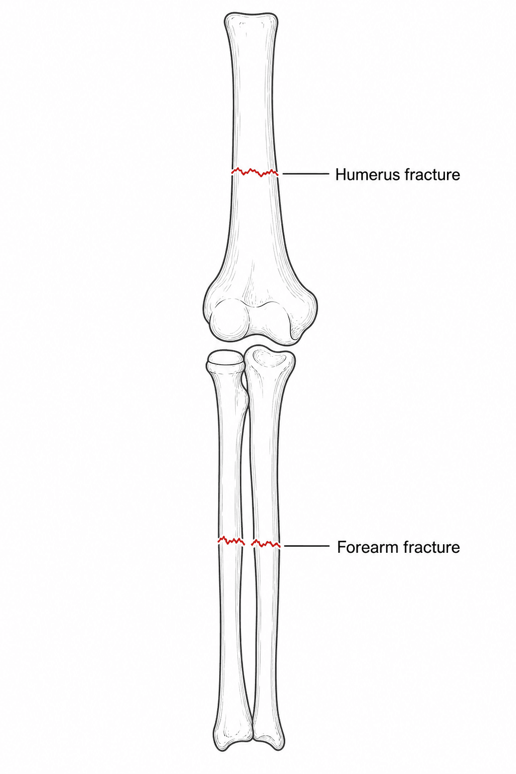
The injury. A floating elbow is the combination of ipsilateral fractures on both sides of the elbow, one above it in the humerus and one below it in the forearm. The elbow joint is left flail, "floating" between the two. Whatever the exact bones, the injury reflects substantial energy transmitted through the limb.
Children and adults. The classic paediatric pattern is a completely displaced supracondylar humerus fracture with an ipsilateral forearm fracture, either of the distal radius or of both bones. In adults the analogous injury is an ipsilateral humeral fracture, of the shaft or the distal humerus, with a forearm fracture.
Associated Injuries & Assessment
A marker of energy. Compared with isolated fractures, the floating elbow carries higher rates of open fractures, neurovascular injury and the need for open reduction. The brachial artery and the median, anterior interosseous, radial and ulnar nerves are at risk, especially with a displaced supracondylar fracture. The high-energy mechanism, the swelling, the two fractures and possible vascular injury together put the forearm at risk of compartment syndrome, and the limb is often one of several injuries in a polytrauma patient.
Assessment. The aim is to find everything the energy has done:
- A full neurovascular examination before and after any reduction: pulses, perfusion and every nerve
- Inspection for open wounds, and assessment of the soft-tissue swelling and the compartments
- Radiographs of the whole limb (humerus, elbow, forearm and wrist), so that the second fracture is not missed
- Treatment as a high-energy, potential polytrauma patient, on ATLS principles where relevant
Compartment syndrome: what "a high index of suspicion" means
Maintain a high index of suspicion and monitor closely, but telling a candidate to be vigilant is not an answer. The examinable content is how the diagnosis is made and what is done about it.
In the child, the five Ps are useless. Pain, pallor, paraesthesia, paralysis and pulselessness are late signs, and pulselessness is often absent altogether. Compartment pressure rises well above capillary filling pressure long before it occludes a major artery, so a palpable pulse never excludes the diagnosis.
The early signs. In a child these are the three A's, with the local signs alongside them:
- Increasing analgesia requirement, the most useful single sign: a child needing escalating opioid after fixation is declaring itself
- Anxiety
- Agitation
- Pain on passive extension of the fingers
- A tense, swollen forearm
- Pain disproportionate to the injury
Measure when the clinical picture is unreliable. In the obtunded, intubated, very young or regionally blocked patient the clinical signs cannot be trusted, so keep a low threshold for measuring pressures and measure if in doubt. The threshold is a delta P of less than 30 mmHg: the diastolic blood pressure minus the measured compartment pressure. An absolute pressure alone is misleading in a hypotensive patient, which is why the differential is used.
Where to measure. Measure in the compartment nearest the fracture and in the deep volar compartment, which is where the pressure is highest and where flexor digitorum profundus dies.
Fasciotomy is the treatment, and it is time-critical. For the forearm:
- A volar release through an extended Henry-type or curved ulnar-based incision, decompressing the superficial and, crucially, the deep flexor compartment
- A carpal tunnel release with it
- A separate dorsal incision for the extensor and mobile wad compartments if they remain tight after the volar release
- Wounds left open, a re-look at 48 hours, and closure or grafting when the swelling permits
Beware the regional block. A regional block can mask the developing syndrome, which is a real argument against dense regional anaesthesia in exactly this injury.
The pulseless hand: pink is not the same as white
With a displaced supracondylar component, assess the vascular status carefully. A pulseless hand after a high-energy elbow injury, whether pale or pink, demands urgent reduction and reassessment.
A pulseless hand comes in two forms, and they are managed differently.
The white (or dusky) pulseless hand is an unperfused hand: cool, pale, with sluggish or absent capillary refill. This is limb-threatening ischaemia. Reduce and stabilise the fracture immediately and, if perfusion does not return at once, explore the brachial artery with vascular surgical support. Do not observe it.
The pink pulseless hand is perfused but has no palpable radial pulse. It is warm and pink, with capillary refill, presumably supported by collateral flow, and it is the genuinely contested one. After reduction and fixation the reasonable position is admission and close serial observation by an experienced team, with documented perfusion checks. Persistent pulselessness, any deterioration or an inability to observe reliably should prompt vascular assessment and exploration rather than indefinite watching.
Why pink is not reassuring. The concern is not the missing pulse itself but the occult ischaemia and the developing compartment syndrome behind it, and there is a well-recognised subgroup whose collateral flow proves inadequate. A pink pulseless hand with a rising analgesia requirement or a tense forearm is a compartment syndrome until proven otherwise.
Management
The conventional rule. The teaching is operative stabilisation of both fractures, because a flail elbow between two fractures cannot be controlled in a cast. Rigid fixation of both converts the flail elbow into a stable segment, reduces complications and allows early motion, which is the single best protection against the elbow stiffness this injury otherwise causes.
How firm the rule is. "Fix both" is a safe default, but say it honestly in a viva. The CORTICES systematic review (Baghdadi, J Pediatr Orthop 2020; 11 studies, 433 children) found compartment syndrome in only 2%, a pulseless limb in 4% (every one regained a pulse after reduction), and good-to-excellent outcomes in 83-100%. It concluded that mandatory fixation of both fractures is not supported by evidence.
The real hazard is the cast. The documented historical hazard is the tight circumferential cast over a swelling limb (Ring 2001: the compartment events clustered in the casted forearms), not the un-pinned forearm fracture. The defensible position is therefore to pin the displaced supracondylar fracture urgently, stabilise the forearm when it is unstable, and never trap a swelling limb in a tight cast.
Which fracture first. There is no absolute rule. A common strategy is to stabilise the more proximal (humeral or supracondylar) fracture first, which restores a stable proximal segment, a lever against which the forearm can then be reduced and fixed. Some surgeons instead fix whichever fracture is simpler or more unstable first.
For every patient. Whatever the age:
- Treat open wounds with debridement, antibiotics and tetanus cover
- Monitor the compartments, and repeat the neurovascular assessment
- Have a plan for fasciotomy and for vascular repair if needed
Damage control. In a grossly contaminated open injury or a physiologically unstable polytrauma patient, temporary spanning external fixation or K-wires buy time, with definitive fixation once the soft tissues and the patient allow. The principles are developed in Damage Control Orthopaedics.
Implants, and one position that must be avoided
In the child. The supracondylar fracture is treated by emergency closed (or open) reduction and percutaneous K-wire stabilisation: two or three lateral-entry wires, or crossed wires accepting the ulnar nerve risk of the medial pin. The forearm fracture is conventionally stabilised too, rather than cast as it might be in isolation, because a cast fails to abolish the flail segment and needs a circumferential dressing over a maximally swelling limb. It is fixed with flexible intramedullary nails, K-wires or a plate, depending on level and age.
Flexion beyond about 90 degrees markedly raises compartment pressure and reduces brachial artery flow, and in a limb already at high risk this is how an avoidable Volkmann's contracture is produced. This is precisely why the supracondylar fracture is pinned rather than held reduced by hyperflexion in a cast, and why the post-operative splint sits in less than 90 degrees with the dressing loose and split.
In the adult: the humerus. Both fractures are usually plated. For the humerus, plate fixation is generally preferred to intramedullary nailing in this setting: it gives rotational control, permits direct reduction and, where the fracture is distal or intra-articular, is the only option. An antegrade nail adds shoulder morbidity and controls rotation less well in a limb where the forearm is also unstable.
When a nail is reasonable. Nailing remains reasonable for a segmental or pathological midshaft fracture, or in the polytrauma patient where speed and soft-tissue preservation dominate. With a distal-third humeral fracture the radial nerve must be identified or deliberately protected, and again at plate removal.
In the adult: the forearm. Restore radial bow, length, alignment and rotation with plate fixation of both bones, to allow early rehabilitation. Malunion here costs pronation and supination, which is the function the patient will actually notice.
Then move it. Once both fractures are fixed, begin early supervised motion to protect against stiffness and loss of forearm rotation.
Outcomes
Generally good, less predictable. With prompt, aggressive operative management outcomes are generally good: in the paediatric series cited below, 10 of 12 children achieved good or excellent results after emergency reduction and K-wire stabilisation of both fractures. Read that as a small single-centre series rather than a reliable rate. The injury is rare enough that the literature is case series and reports, with no comparative trials.
What drives the result. Because of the associated injuries (open fractures, nerve injury, compartment syndrome), results are less predictable than for isolated fractures, and early recognition and treatment of those complications drives the outcome. Children generally do better than adults, whose stiffness, articular involvement and higher rate of open injury make a full functional recovery less likely.
Complications of the Floating Elbow
The feared complication: Volkmann's ischaemic contracture. This is the devastating end-stage of an unrecognised forearm compartment syndrome, with a fixed flexion contracture of the wrist and fingers and intrinsic loss. It is the reason for the relentless compartment vigilance, and the syndrome is covered in detail in Forearm Compartment Syndrome.
The commonest functional problem: stiffness. The flail joint, extensive surgery and any immobilisation predispose to elbow stiffness and lost forearm rotation. Rigid fixation of both fractures with early motion is the main preventive, developed in Elbow Stiffness & Contracture.
Nerve injury and recovery. Most nerve injuries are traction neurapraxias that recover over weeks to months. A nerve that is interposed in the fracture or lacerated, especially in an open injury, needs exploration; document the injury and follow it.
Malunion and nonunion. Paediatric supracondylar malunion can give cubitus varus, forearm malunion costs the rotation described above, and adults risk nonunion of the humeral or forearm fracture.
Other complications include heterotopic ossification around the high-energy elbow, re-displacement or loss of fixation, and vascular sequelae.
Guidelines, Registries & Global Practice
Guidance and the Evidence Reality
No society publishes a dedicated floating-elbow guideline; the injury is rare enough that the literature is case series plus two systematic reviews, and practice is extrapolated from supracondylar and forearm fracture principles. The honest viva summary:
- Paediatric: the displaced supracondylar component drives the urgency (standard CRPP teaching). The CORTICES review (433 children) found compartment syndrome in only 2% and does not support mandatory dual fixation - but a tight circumferential cast over a swelling limb is the documented hazard (Ring 2001).
- Adult: the Ditsios systematic review (32 studies) quantifies the associated-injury burden - 51.2% open fractures, 34.9% neural injury, 76.3% multiple-system injury - which is why adults fare worse and dual operative stabilisation is the norm.
- Polytrauma frameworks (ATLS, damage-control orthopaedics) govern sequencing when the limb is one of several injuries.
Registry and Resource Notes
- No registry captures floating elbow as an entity; outcomes come from single-centre series pooled in the reviews above.
- High-resource settings: percutaneous pinning, flexible nails, plating and compartment-pressure monitoring are routine. Limited-resource settings rely on K-wire fixation and clinical compartment assessment - the non-negotiables are the same everywhere: never a tight cast over a swelling forearm, and repeated neurovascular checks.
Mnemonics & Memory Aids
FLOAT
Hook:A FLOATing elbow: fractures both sides, look for nerve/vessel injury, fix both, watch compartments, high-energy.
BOTH
Hook:Fix BOTH, beware Open injury, the Tense forearm, and the Hand's perfusion.
PINKThe Pulseless Hand - PINK
Hook:PINK pulseless: Pink = watch, White = act, No pulse never clears the compartments, Know your delta-P.
Clinical Decision Scenarios
Practise clinical reasoning and management decisions out loud
“A child falls from a height and has a displaced supracondylar humerus fracture and an ipsilateral distal forearm fracture. What is this injury, why does it concern you, and how would you manage it?”
“How does the floating elbow differ in adults, and what general principles guide its treatment?”
Definition
- Ipsilateral fractures above AND below the elbow (elbow flail/'floating')
- Children: displaced supracondylar + forearm fracture (classic)
- Adults: humeral (shaft/distal) + forearm fracture
Why it matters
- Marker of high-energy trauma
- Higher rates of open fractures, neurovascular injury, need for open reduction
- Forearm compartment syndrome risk
Assessment
- Neurovascular exam before/after reduction; inspect for open wounds
- Radiograph the whole limb (don't miss the 2nd fracture)
- Assess compartments; treat as high-energy/polytrauma
Management
- Operative stabilisation of BOTH fractures (can't cast a flail elbow)
- Children: CRPP supracondylar + forearm reduction/fixation; Adults: plate/nail humerus + plate forearm
- Treat open wounds; monitor compartments (low threshold for fasciotomy); repair vascular injury; early motion
Evidence & Key Studies
Management of the floating elbow injury in children: simultaneous ipsilateral fractures of the elbow and forearm
- In 12 children with a completely displaced supracondylar humerus fracture plus an ipsilateral forearm fracture, all underwent emergency operative reduction and percutaneous K-wire stabilisation; 10 had good/excellent and 2 fair results.
- The incidence of open fractures, nerve injury and the need for open reduction was higher than for isolated supracondylar fractures.
- The floating elbow is an indicator of a high-energy injury and requires aggressive operative management.
Pediatric floating elbow (the cast-warning series)
- Retrospective review of 16 children with floating elbow over 9 years at a tertiary children's hospital, confirming substantial swelling and compartment-syndrome potential - particularly with circumferential cast immobilisation.
- Of 10 patients whose forearm fracture was treated with closed reduction and plaster, 2 developed compartment syndrome and 4 had incipient compartment syndrome that responded to splitting the cast; 3 of these required remanipulation of the distal radius.
- One compartment-syndrome patient developed Volkmann ischaemic contracture; 6 patients underwent stabilisation of both fractures.
Floating Elbow in Adults: A Systematic Review and Meta-Analysis
- Systematic review of 32 studies of floating elbow in patients aged 17 and older; 73.1% male, median age 33 years, median follow-up 19.5 months.
- THE ASSOCIATED-INJURY BURDEN THAT DEFINES THE ADULT INJURY: 51.2% of fractures were open, 34.9% of patients suffered neural injury, 24.4% had articular involvement, and 76.3% had multiple-system injuries.
- Confirms the adult floating elbow as a high-energy, complication-dense injury with outcomes driven by those associated injuries.
Pediatric Floating Elbow Injuries Are Not as Problematic as Once Thought (CORTICES systematic review)
- Systematic review (PRISMA) of 11 studies and 433 children with ipsilateral supracondylar plus forearm fractures; the mechanism was a fall in 96%, and CRPP of both fractures was the commonest treatment.
- Paediatric acute compartment syndrome occurred in only 8 of 433 patients (2%); a pulseless limb at presentation occurred in 4%, and ALL regained a pulse after reduction.
- Three iatrogenic ulnar nerve injuries were reported, all with medial pin placement; good-to-excellent results in 83-100% by modified Flynn criteria.
- The authors conclude that mandatory fixation of BOTH fractures is NOT supported by evidence, that compartment syndrome is much commoner with delayed treatment of displaced fractures, and that outcomes do NOT reflect the poor prognosis of adult floating elbows.
Paediatric bilateral 'floating elbow'
- Reports a rare bilateral floating elbow (ipsilateral supracondylar + distal forearm fractures bilaterally) managed by closed reduction and pinning, with uneventful union and no functional limitation.
- Emphasises vigilance for compartment syndrome in floating-elbow injuries.
- Concludes that early surgical fixation may give better results.
The high-energy nature, the higher rates of open fractures/nerve injury/open reduction, and the good results of emergency operative stabilisation come from the cited Harrington paediatric series, and the compartment-syndrome vigilance and early-fixation message from the cited Rangasamy report. The paediatric cast/compartment-syndrome data come from the cited Ring series, the pooled paediatric rates (2% compartment syndrome, 4% pulseless limb, 83-100% good/excellent) from the CORTICES systematic review, and the adult associated-injury burden (51.2% open, 34.9% neural injury, 76.3% multiple-system) from the Ditsios systematic review. The adult patterns and fixation principles are standard, well-established trauma teaching. The compartment-syndrome thresholds and fasciotomy technique, the pink-versus-white pulseless hand, the hyperflexion caution and the implant choices are standard, well-established trauma teaching. (See also Supracondylar Humerus Fracture, Both-Bone Forearm Fractures and Forearm Compartment Syndrome.)