A Common Injury with an Uncommon but Serious Tail of Infection
- Plantar puncture wounds are common and the GREAT MAJORITY heal uneventfully; only a small proportion - on the order of about 1.5-2% in published series - develop a deep infection that progresses to osteomyelitis or septic arthritis.
- The CLASSIC deep pathogen is PSEUDOMONAS AERUGINOSA, characteristically after a puncture THROUGH A RUBBER-SOLED SHOE (the warm, moist foam of a trainer/sneaker harbours Pseudomonas which is inoculated into the deep tissues); by contrast, early superficial CELLULITIS is usually due to Staphylococcus aureus or streptococci.
- A RETAINED FOREIGN BODY (nail fragment, wood, glass, sock/shoe debris) is a key driver of persistent infection and must be actively EXCLUDED - radio-opaque bodies (metal, glass, gravel) show on plain radiographs, while radiolucent material (wood, plastic) needs ULTRASOUND or MRI.
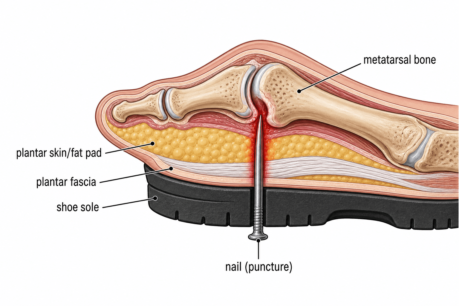
- TETANUS status must be checked and updated (puncture wounds are tetanus-prone); a full neurovascular and soft-tissue examination is needed, and the DEPTH and LOCATION matter - deep FOREFOOT punctures over the metatarsal heads/MTP joints carry the highest risk of bone and joint involvement.
- DIABETES (and peripheral neuropathy/vascular disease) markedly worsens the picture: infections are more often POLYMICROBIAL, may present late because of insensate feet, and carry a higher risk of chronic ulceration and AMPUTATION - the puncture can be the initiating injury of a diabetic foot infection.
- Management of a SIMPLE puncture is wound cleaning/irrigation, removal of any foreign body, tetanus prophylaxis and CLOSE follow-up; routine prophylactic antibiotics for clean simple punctures are NOT clearly beneficial (selective use only). Established DEEP infection or OSTEOMYELITIS requires SURGICAL DEBRIDEMENT with deep cultures and CULTURE-DIRECTED antibiotics (empirically covering Pseudomonas if deep/late), typically for several weeks.
- “Deep/late infection after a puncture through a trainer = Pseudomonas aeruginosa until proven otherwise; early cellulitis = S. aureus/Strep.
- “Always exclude a retained foreign body: plain film for radio-opaque, US/MRI for wood/plastic. Check tetanus.
- “Simple puncture: clean + foreign-body exclusion + tetanus + follow-up (no routine prophylactic antibiotics). Osteomyelitis: debride + culture-directed (anti-pseudomonal) antibiotics.
Superficial, within days - usually Staphylococcus aureus or streptococci. Treat with appropriate oral/IV antibiotics and review.
Abscess, septic arthritis or osteomyelitis - classically Pseudomonas aeruginosa, especially after a puncture through a rubber-soled shoe. Needs debridement + anti-pseudomonal, culture-directed antibiotics.
Mechanism & Why Some Get Infected
The typical injury is stepping on a nail. Most punctures are trivial, but several factors push a wound toward deep infection: a puncture through a rubber-soled shoe inoculates Pseudomonas aeruginosa from the warm, moist sole foam; a retained foreign body (nail fragment, wood splinter, glass, or a fragment of sock/shoe carried in) acts as a nidus; a deep puncture, and especially one over the forefoot (metatarsal heads / MTP joints), reaches bone and joint; delayed presentation, contamination, and host factors (diabetes, neuropathy, vascular disease, immunosuppression) all increase risk. Recognising these lets you stratify the wound that needs more than simple cleaning.
Assessment & Imaging
Take a focused history (object, footwear, time since injury, environment, immunisation and diabetic/vascular status) and examine the wound for depth, location, contamination and signs of infection, with a full neurovascular assessment. A retained foreign body must be actively excluded:
- Plain radiographs detect radio-opaque bodies (metal, glass, gravel) and any bony changes (though osteomyelitis is radiographically silent for ~10-14 days).
- Ultrasound or MRI is needed for radiolucent material (wood, plastic, thorn) and to define deep abscess, septic arthritis or early osteomyelitis. Tetanus: puncture wounds are tetanus-prone - check status and give a booster +/- immunoglobulin as indicated.
Management
- Simple, clean puncture (no infection, no foreign body): thorough cleaning/irrigation, removal of any superficial foreign material, tetanus prophylaxis, dressing, analgesia and clear safety-netting with follow-up. Routine prophylactic antibiotics are not clearly beneficial for clean simple punctures and are used selectively (e.g. heavily contaminated wounds, deep forefoot punctures, immunocompromised/diabetic hosts).
- Cellulitis: antibiotics covering S. aureus/streptococci, with review to ensure resolution.
- Deep infection / abscess / septic arthritis / osteomyelitis: surgical debridement with removal of any foreign body and deep tissue/bone cultures, followed by culture-directed antibiotics - started empirically to cover Pseudomonas when the infection is deep or late - typically for several weeks (published series report a mean of around five weeks for osteomyelitis). Recurrent or extensive bone destruction may need repeat debridement.
In a patient with diabetes or peripheral neuropathy, a puncture wound may present late (insensate foot), is more likely to be polymicrobial, and can be the initiating injury of a limb-threatening diabetic foot infection with a higher risk of amputation. Have a low threshold for imaging, deep cultures, surgical debridement and multidisciplinary (vascular/diabetic-foot) involvement. (See our Diabetic Foot / Charcot topics.)
Evidence & Key Studies
Calcaneal osteomyelitis caused by nail puncture wounds
- A small proportion (around 1.8%) of plantar puncture wounds become infected and progress to osteomyelitis.
- In six patients with calcaneal osteomyelitis after a heel nail-puncture, healthy patients grew a single pathogen, whereas diabetic patients grew MULTIPLE pathogens.
- The only amputation occurred in a diabetic patient; with diabetic neuropathy, the puncture may initiate chronic ulceration and raise amputation risk.
Pyogenic osteomyelitis after a plantar puncture wound: analysis of a series of 8 cases
- Osteoarticular infection followed plantar puncture in 1.65% of cases presenting to the emergency department; the mechanism was stepping on a nail in all cases.
- Cultures were monomicrobial in 6 of 8 cases, with Pseudomonas aeruginosa isolated in five - and a retained foreign body was found in one.
- Combined surgical and antibiotic treatment (mean antibiotic duration about five weeks) was used; initial proper wound treatment, foreign-body detection and follow-up are emphasised to prevent late infection.
According to PubMed, the infection/osteomyelitis rates (~1.65-1.8%), the predominance of Pseudomonas aeruginosa in deep infection, the polymicrobial pattern and amputation risk in diabetics, and the role of foreign-body detection and several-week antibiotic courses come from the cited Laughlin and Morales series. The Pseudomonas-through-a-rubber-sole association is long-established clinical teaching. (See also our Osteomyelitis, Septic Arthritis and Diabetic Foot topics.)
Clinical Decision Scenarios
Practise clinical reasoning and management decisions out loud
“A healthy adult stepped on a nail through their trainer 10 days ago and now has a painful, swollen, discharging forefoot. What is the likely diagnosis and organism, and how would you manage them?”
“How does a foot puncture wound differ in a patient with diabetes, and why does that change your management?”
Mnemonics & Memory Aids
PUNCTURE
Hook:Run through PUNCTURE for every foot puncture wound.
BUGS
Hook:BUGS reminds you which organism to expect and to culture deep.
Epidemiology
- Common injury (typically stepping on a nail); most heal uneventfully
- ~1.5-2% develop deep infection / osteomyelitis
- Highest risk: deep forefoot punctures over metatarsal heads/MTP joints
Microbiology
- Early cellulitis: S. aureus / streptococci
- Deep/late through rubber sole: Pseudomonas aeruginosa
- Diabetic: polymicrobial; higher amputation risk
Assessment
- Exclude foreign body: X-ray (radio-opaque), US/MRI (radiolucent - wood/plastic)
- Check tetanus status; full neurovascular exam
- Inflammatory markers/cultures if infected; MRI for abscess/osteomyelitis
Management
- Simple: clean/irrigate, remove FB, tetanus, follow-up (no routine prophylactic antibiotics)
- Cellulitis: anti-staph/strep antibiotics
- Deep/osteomyelitis: debride + deep cultures + culture-directed (anti-pseudomonal) antibiotics ~weeks