Fracture of Necessity | ORIF Required | DRUJ Instability | Adult vs Pediatric Difference
- Fracture of Necessity - virtually always requires ORIF in adults
- DRUJ instability is the key associated injury - always assess
- Anatomic radius reduction usually restores DRUJ stability
- Pediatric Galeazzi often treated conservatively (different than adults)
- Compare to Monteggia: Galeazzi = Distal (DRUJ), Monteggia = Proximal (radial head)
- “G for GRUJ (distal), M for MPRUJ (proximal) - mnemonic for location
- “Supination after fixation usually stabilizes DRUJ
- “Brachioradialis deforming force causes shortening
- “Check ulnar styloid - base fracture indicates DRUJ disruption
Overview and Introduction
A Galeazzi fracture-dislocation is a fracture of the radial shaft, typically at the junction of the middle and distal thirds, together with disruption of the distal radioulnar joint (DRUJ). Both components must be present for the diagnosis. The DRUJ disruption takes one of three forms:
- True dislocation
- Subluxation
- Ulnar styloid base fracture in place of a true dislocation, the Galeazzi equivalent
Its mirror image at the other end of the forearm, an ulnar shaft fracture with dislocation of the radial head, is the Monteggia fracture; the paediatric form of that injury is covered under Monteggia fractures in children.
Described by Riccardo Galeazzi in 1934. The phrase "fracture of necessity" is Campbell's (1941), not Hughston's - a common misattribution worth getting right. Hughston (1957) supplied the evidence behind the phrase in "Fracture of the distal radial shaft: mistakes in management", the series in which the great majority of non-operatively treated patients (widely cited as 35 of 38) had unsatisfactory results. So: Campbell named it, Hughston proved it.
Who. Galeazzi injuries make up 3-7% of all forearm fractures and are more common than Monteggia fracture-dislocations. Men outnumber women 3:1, and the peak age is 30-40 years.
Mechanism. A fall on the outstretched hand with the forearm pronated is the most common story. The others:
- Direct blow to the dorsoradial forearm
- Axial load on a hyperpronated wrist
- High-energy trauma, including motor vehicle accidents
- Sporting injury (cycling, contact sports)
Galeazzi versus Monteggia. Distal for Galeazzi, proximal for Monteggia, in both the bone and the joint:
- Galeazzi
- Radius (distal)
- Monteggia
- Ulna (proximal)
- Galeazzi
- DRUJ (distal)
- Monteggia
- Radiocapitellar (proximal)
- Galeazzi
- Ulna from radius (DRUJ)
- Monteggia
- Radial head
- Galeazzi
- Always ORIF
- Monteggia
- Usually ORIF
- Galeazzi
- Conservative possible under age 10
- Monteggia
- Often conservative
Anatomy and Pathophysiology
The joint. The ulnar head articulates with the sigmoid notch of the radius, and the radius rotates around the relatively fixed ulna through 180°. The articulation is shallow, with articular surface coverage of approximately 60°, so the joint relies heavily on soft tissue for its stability.
The stabilisers. With so little bony constraint, the soft tissues hold the ulnar head in the notch:
- TFCC (triangular fibrocartilage complex), the primary stabiliser
- Dorsal and volar radioulnar ligaments, the components of the TFCC
- Pronator quadratus
- Interosseous membrane (central band)
- ECU subsheath
- Ulnocarpal ligaments
Why the instability outlasts the fracture. The shallow sigmoid notch provides minimal bony constraint, so the TFCC and the interosseous membrane are what keep the joint reduced. In a Galeazzi fracture the interosseous membrane is disrupted and the TFCC is often torn, which is why DRUJ instability can persist even after the radius has been fixed.
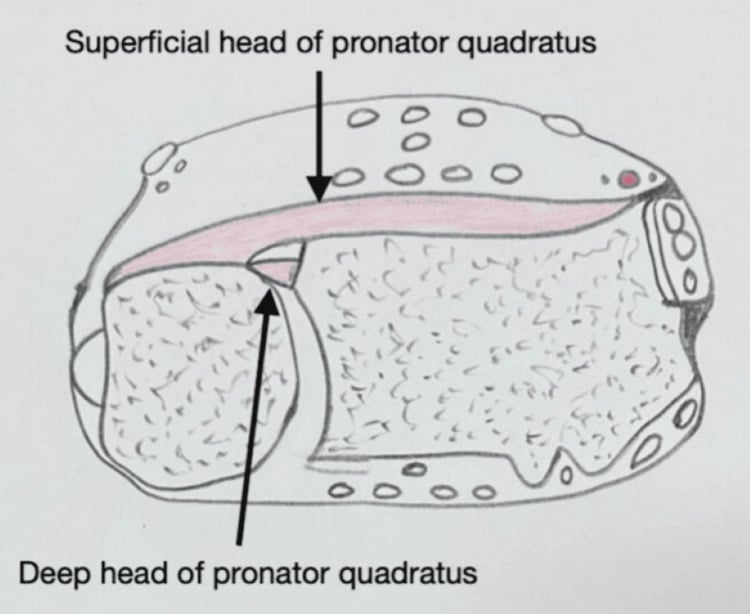
Deforming forces. The muscles decide where the fragments go. The proximal fragment is pulled proximally by biceps and supinator; its rotation varies with the level of the fracture, and it tends to supinate if the fracture is proximal to pronator teres. The distal fragment is shortened by brachioradialis, the primary deforming force, pronated by pronator quadratus and flexed by the wrist flexors, and the fracture typically angulates apex dorsal.
The result. A shortened radius with apex dorsal angulation and loss of the radial bow, and a DRUJ that is incongruent, with the ulnar head prominent.
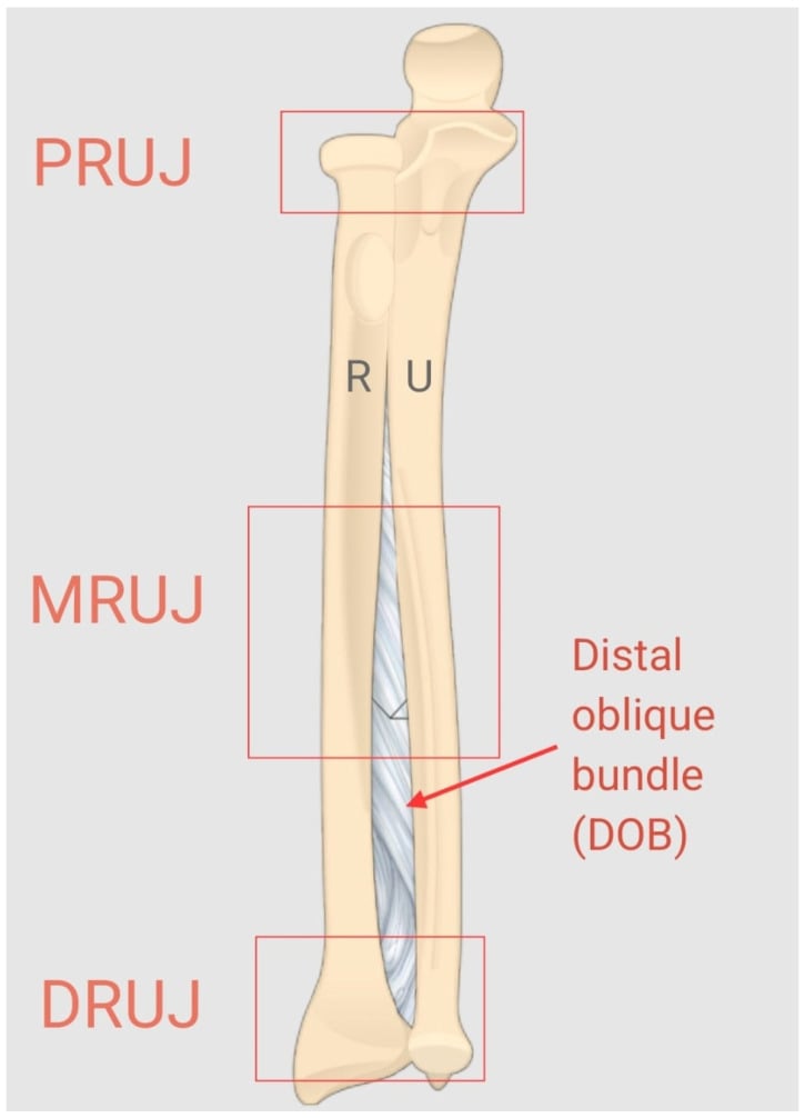
The interosseous membrane. The central band is the primary longitudinal stabiliser of the forearm; it runs obliquely from radius to ulna (proximal-distal) and transmits 80% of axial load from the radius to the ulna. It is disrupted in a Galeazzi fracture, which contributes to the radial shortening, may prevent closed reduction, and adds to the persistent DRUJ instability. Keep it in mind when the DRUJ is assessed after fixation.
Classification Systems
Two things are classified: where the radius broke, and how the DRUJ behaves once the radius is fixed. The first predicts the second.
By location. The classic fracture sits at the junction of the middle and distal thirds of the radius. The more distal the fracture, the higher the risk of DRUJ instability; a fracture in the proximal third should raise suspicion of an Essex-Lopresti injury, with complete disruption of the interosseous membrane and proximal migration of the radius.
- Location
- Junction middle/distal thirds
- Clinical Significance
- Classic Galeazzi pattern, high DRUJ instability (most common)
- Location
- True mid-shaft radius
- Clinical Significance
- Less DRUJ instability but still assess carefully
- Location
- Proximal third radius
- Clinical Significance
- Rare, consider Essex-Lopresti variant with IOM disruption
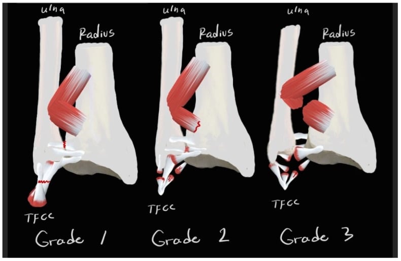
The numbers behind the gradient. Rettig and Raskin divided forty fractures at 7.5 cm from the mid-articular surface of the distal radius. Type I, within 7.5 cm, was unstable after radial fixation in 12 of 22; type II, more than 7.5 cm proximal, in only 1 of 18. Persistent instability was managed by K-wire transfixion in 10 and TFCC repair in 3. Anatomic radial reduction alone restores the joint in most mid-shaft injuries, and it is the distal-third fracture that will need something extra, so plan for it; either way the DRUJ is tested, not assumed.
Essex-Lopresti is the variant to know, because missing it is a classic exam disaster. The Essex-Lopresti injury is a triad of radial head fracture + complete longitudinal interosseous membrane (central band) disruption + DRUJ dissociation - that is, longitudinal instability of the whole forearm, allowing the radius to migrate proximally.
- Mechanism: high-energy axial load through the forearm.
- The trap: it is frequently missed because attention fixes on the radial head. Any radial head fracture with wrist/ulnar-sided pain, or with proximal radial migration, is Essex-Lopresti until proven otherwise.
- Assessment: check for DRUJ tenderness and a positive ballottement, and compare radial length to the contralateral wrist; the radius pull ("criss-cross") test under fluoroscopy (proximal radial translation of more than about 3 mm) confirms longitudinal instability.
- The critical principle: NEVER simply excise the radial head in a suspected Essex-Lopresti - the radial head is the last longitudinal stabiliser, and excision causes catastrophic proximal radial migration, ulnar impaction and chronic wrist pain. Instead preserve and fix the radial head, or replace it with a metal prosthesis (never a silastic spacer), then address the DRUJ (reduce and pin in supination). Acute interosseous-membrane repair is difficult and chronic cases are very hard to salvage - so recognition at presentation is everything.
Clinical Assessment
History. The mechanism is one of those in the Overview, most often the fall on the pronated hand. The symptoms:
- Forearm pain and swelling
- Wrist pain (DRUJ involvement)
- Visible deformity
- Inability to supinate or pronate the forearm
- Weakness of grip
Look. Deformity and swelling of the forearm, and swelling at the wrist, especially dorsally over the DRUJ. A dorsally prominent ulnar head means the DRUJ is dislocated. Check the skin for an open fracture and compare radial length with the opposite side.
Feel. Point tenderness at the radius fracture, tenderness dorsally over the DRUJ, and tenderness over the ulnar styloid, where a base fracture is being sought. Palpate along the interosseous membrane and assess the compartments.
Move. Supination and pronation are limited and painful, and the patient usually cannot rotate the forearm. Wrist motion is limited by pain.
Neurovascular examination. Each nerve, and the circulation:
- Median nerve: sensation in the first web space, thumb opposition
- Ulnar nerve: sensation over the little finger, finger abduction
- Radial nerve: wrist and finger extension, sensation over the dorsal first web space
- Vascular: radial and ulnar pulses, capillary refill
DRUJ stability test. The same test is used in clinic and again on the table after the radius is fixed:
- Stabilise the radius firmly with one hand
- Translate the ulna dorsally and volarly with the other hand
- Assess the amount of translation and compare with the contralateral side
- Test in supination, neutral and pronation
- Note the position of maximum stability (usually supination)
Reading the test. Increased translation compared with the opposite side is instability, and pain on testing indicates ligament injury. Dorsal prominence of the ulnar head is a DRUJ dislocation. The joint is usually most stable in supination.
Differential diagnosis. The mimics are separated by which bone is broken and which joint is disrupted.
- Distinguishing Features
- Distal-third radius shaft fracture PLUS DRUJ disruption
- Key Discriminator
- DRUJ widening/dorsal ulnar prominence with an isolated radius fracture
- Distinguishing Features
- Radius fracture with a congruent, stable DRUJ on imaging and exam
- Key Discriminator
- Normal DRUJ - the defining absence
- Distinguishing Features
- Proximal ULNA fracture PLUS radial head dislocation
- Key Discriminator
- Pathology is proximal (radiocapitellar), not distal (DRUJ)
- Distinguishing Features
- Fractures of both radius and ulna shafts
- Key Discriminator
- Ulna is also fractured; DRUJ usually intact
- Distinguishing Features
- Radial head fracture, complete IOM disruption and DRUJ dissociation
- Key Discriminator
- Proximal radial migration; tenderness along the whole IOM and elbow
- Distinguishing Features
- Metaphyseal distal radius fracture; DRUJ may be involved via ulnar styloid
- Key Discriminator
- Fracture is metaphyseal/intra-articular, not diaphyseal shaft
- Distinguishing Features
- DRUJ disruption without a radial shaft fracture
- Key Discriminator
- No diaphyseal radius fracture present
Investigations
Radiographs. The forearm series must include both the elbow and the wrist so that associated injuries are not missed, and the wrist gets its own views:
- Full-length forearm radiographs, AP and lateral, including both joints
- Dedicated wrist views, PA and lateral
- Contralateral forearm for comparison if needed
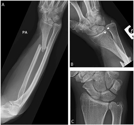
On the AP view. The radius fracture at the middle-distal junction, radial shortening relative to the ulna, and an ulnar styloid base fracture, which suggests DRUJ disruption. The joint itself shows as DRUJ widening: a difference of greater than 2 mm compared with the opposite wrist is abnormal.
On the lateral view. Dorsal subluxation of the ulnar head and loss of the normal DRUJ relationship, with apex dorsal angulation of the radius fracture. Volar displacement of the distal radius is possible.
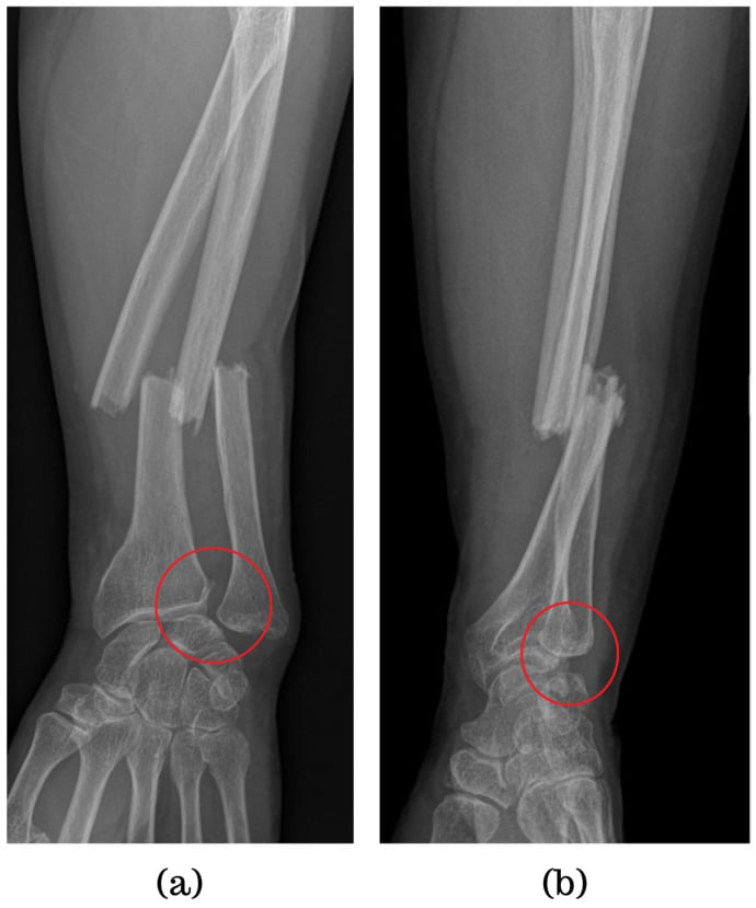
CT. It answers what plain films leave open: detailed sigmoid notch anatomy and the fracture fragments, quantification of DRUJ subluxation, associated fragments such as the ulnar styloid, and articular step-off. The indications:
- DRUJ congruity when plain films are unclear
- Complex intra-articular fracture patterns
- Pre-operative planning for comminuted fractures
- Post-operative assessment if the DRUJ reduction is questioned
- Chronic DRUJ instability
MRI. Rarely needed in the acute setting; it may be useful in delayed or chronic cases, where it shows TFCC tears (central perforations or peripheral detachments), IOM disruption, ligament injuries and cartilage damage. The indications:
- TFCC assessment (tears, quality)
- Chronic DRUJ instability
- Persistent unexplained symptoms after treatment
- IOM assessment in suspected Essex-Lopresti
Management Algorithm
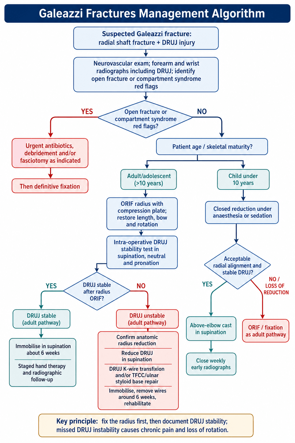
Why it is operated on. The Galeazzi is the "fracture of necessity" because operative treatment is mandatory in adults. Conservative treatment fails in most of them: 80% in Mikic's 1975 series of 125 patients, and the great majority in Hughston's. Both figures describe the same conclusion from different cohorts; do not blend them into one number.
The goals and the timing. An anatomic radius and a stable DRUJ. Surgery is typically performed within 24-48 hours, and the steps follow in order:
- ORIF of the radius with a compression plate (anatomic reduction)
- Assess DRUJ stability intra-operatively
- Stabilise the DRUJ if unstable (pin or repair)
- Immobilise in the position of stability (supination)
- Early mobilisation protocol
- Treatment
- ORIF radius with plate
- Key Points
- Fracture of necessity - always operative
- Treatment
- Consider closed reduction and cast
- Key Points
- Conservative possible if reduction acceptable
- Treatment
- ORIF radius with plate
- Key Points
- Treat as adult
- Treatment
- Supination cast 6 weeks
- Key Points
- Most common outcome (80%)
- Treatment
- Pin DRUJ or repair TFCC
- Key Points
- Occurs in 20% of cases
- Treatment
- Urgent debridement then ORIF
- Key Points
- Standard open fracture protocol
Children. A child under 10 years can often be treated with closed reduction and cast immobilisation in supination: the DRUJ is more likely to reduce and remain stable because of the periosteal sleeve and the remodelling potential. Older children and adolescents, over 10 years, should be treated like adults with ORIF, because remodelling potential decreases. Walsh's series supports this: results of conservative management were generally good in 41 children under 15, with a gradient that echoes the adult one, the more distal the radial fracture the more trouble.
When to cast a child. The indications for conservative treatment:
- Age under 10 years
- Acceptable closed reduction achieved (less than 10° angulation)
- DRUJ stable after reduction
- Compliant child and family
- Close follow-up available
How. Closed reduction under sedation or general anaesthesia, then an above-elbow cast in supination for 6 weeks. Weekly radiographs for the first 2-3 weeks watch for loss of reduction, and if reduction is lost the child proceeds to ORIF. Close monitoring is essential to the success of conservative treatment in a child.
Surgical Technique
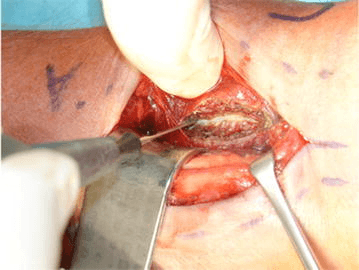
Planning. Review the full forearm radiographs including the DRUJ, plan the approach by the fracture location, template the plate size and length, and consent the patient for possible DRUJ stabilisation as well as the radius.
Positioning.
- Supine on the operating table
- Arm on a radiolucent hand table
- Tourniquet on the upper arm, which may be left uninflated if the DRUJ is to be checked without it
- C-arm positioned for AP and lateral views
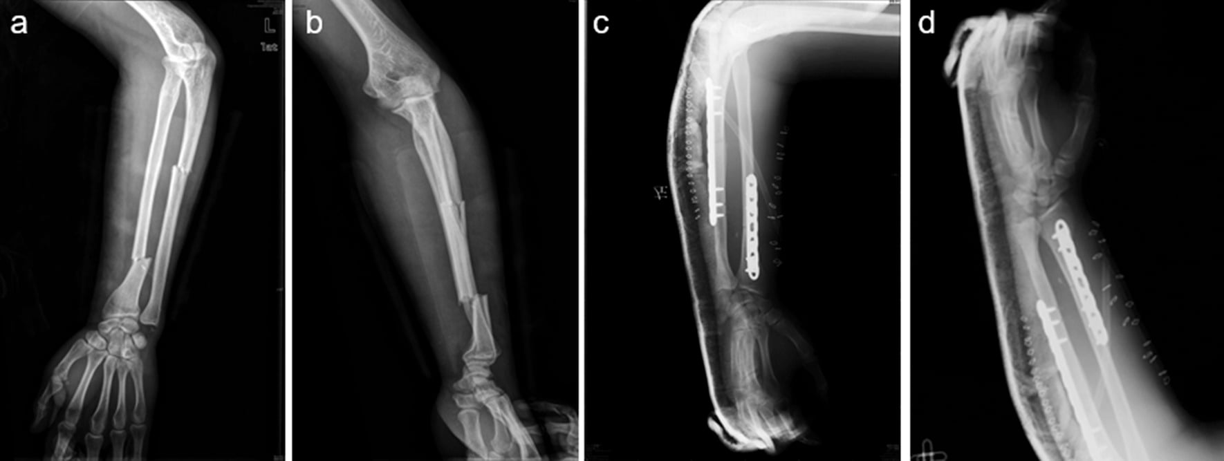
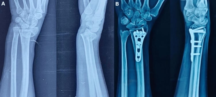
What the films should show afterwards. A radius restored to length, bow and rotation under a plate, and a concentrically reduced DRUJ.
Complications
Compartment syndrome. The volar and dorsal forearm compartments are at risk, and the first 48 hours after surgery are the time to watch. The diagnosis is clinical, pain out of proportion and pain on passive stretch, and the treatment is urgent fasciotomy.
Nerve and vessel injury. Each approach has its own nerve at risk, and nerve function is examined and documented after surgery:
- Superficial radial nerve: at risk with the volar approach, lying on brachioradialis
- PIN: at risk with the dorsal approach, emerging through supinator
- Median nerve: rare, but possible with the volar approach
- Posterior interosseous artery: can bleed with the dorsal approach
Wound problems. Infection carries a 1-2% risk; dehiscence and haematoma are the others.
Acute DRUJ instability. The most common early problem. It may only become apparent after the cast comes off, and it needs assessment and possibly secondary stabilisation.
Persistent DRUJ instability. The most common late complication, in 10-15%, caused by inadequate initial stabilisation, a missed TFCC tear or a malunion. The patient reports pain, weakness, clicking and a sensation of instability. Treatment follows the reconstructive ladder in the pearl below; a chronic ulnar styloid nonunion is fixed.
Malunion. Inadequate reduction or loss of fixation leaves a shortened radius, angular deformity and loss of the bow, which produce DRUJ incongruity, loss of rotation and pain. A symptomatic malunion is treated with a corrective osteotomy.
Nonunion. Rare with rigid plate fixation, less than 2%; inadequate fixation, infection and smoking are the risk factors, and the treatment is revision ORIF with bone graft.
Loss of motion. Loss of supination and pronation is the most common residual deficit, from DRUJ problems, malunion or soft-tissue contracture. Early motion and an anatomic reduction are what prevent it; avoid prolonged rigid immobilisation beyond 6 weeks.
Other late complications. Rarer, and mostly the generic ones:
- Heterotopic ossification (rare)
- Radioulnar synostosis (very rare)
- Post-traumatic arthritis (DRUJ or radiocarpal)
- Hardware prominence or irritation
- Chronic regional pain syndrome (CRPS)
A missed Galeazzi (or persistent post-fixation DRUJ instability) presents late with ulnar-sided wrist pain, weakness, painful clicking and loss of rotation - and the reconstructive ladder is examinable.
- Correct the bone first: a radial malunion (shortening or lost bow) must be addressed by corrective osteotomy before any DRUJ procedure, because residual incongruity perpetuates instability.
- Soft-tissue reconstruction (younger patient, congruent non-arthritic joint): anatomic dorsal-and-volar radioulnar ligament reconstruction with a tendon graft - the Adams-Berger procedure - restores the TFCC stabilisers.
- Salvage for the arthritic/incongruent DRUJ:
- Darrach (distal ulna resection): simple, reliable pain relief, best for the low-demand/elderly patient; risks painful radioulnar convergence and stump instability in the young and active.
- Sauve-Kapandji (DRUJ arthrodesis plus a proximal distal-ulna pseudarthrosis): preserves the ulnar buttress/support of the carpus while restoring forearm rotation - favoured in younger, higher-demand patients (can also develop proximal-stump instability).
- Ulnar head (or total DRUJ) arthroplasty: preserves load transfer and avoids the convergence problems of resection.
Exam point: fix the radius first, reconstruct the radioulnar ligaments (Adams-Berger) in the young congruent joint, and choose Darrach (low-demand) versus Sauve-Kapandji (young/high-demand) versus ulnar head replacement for the arthritic DRUJ.
Postoperative Care and Rehabilitation
The position. The forearm is immobilised in supination, where the radius and ulna come parallel. Supination tightens the volar radioulnar ligament and brings the radius over the ulna into the reduced position, taking the stress off the healing TFCC; in pronation the volar ligaments are lax and the DRUJ subluxates dorsally. Cadaveric studies show maximum DRUJ stability in supination.
Galeazzi Fracture Rehabilitation Protocol
Above-elbow cast or splint in supination with the elbow at 90° (if DRUJ stable) or sugar-tong splint. Elevation of limb. Finger ROM exercises (all joints) and grip strengthening encouraged. Wound checks at 2 weeks. Maintain shoulder and elbow mobility.
Convert to below-elbow cast in neutral or slight supination if DRUJ stable (elbow ROM allowed). Continue finger exercises. K-wires remain in place if DRUJ pinned. Radiographs at 6 weeks to assess healing.
Remove cast and K-wires. Begin active wrist flexion and extension and forearm rotation. Gentle strengthening if union confirmed, strengthening exercises from week 8. Hand therapy referral.
Progressive strengthening programme. Return to sport when ROM and strength recovered (usually 3-4 months). Hardware removal if symptomatic (typically 12+ months). Full recovery expected by 6 months.
If the DRUJ was pinned. An above-elbow cast in supination for 4-6 weeks, with the K-wires removed at 6 weeks in clinic, no anaesthetic needed, then protected motion for a further 2-4 weeks: 8-10 weeks of immobilisation in total.
If the TFCC was repaired. An above-elbow cast for 4 weeks, then a below-elbow cast for 2 more, then protected range-of-motion exercises and hand therapy: 6-8 weeks in total.
Therapy goals. Full recovery typically takes 3-6 months, and the targets the therapist works towards:
- Full pronation-supination (80° each way)
- Wrist flexion and extension of at least 60°
- Grip strength 90% of the opposite side
- Pain-free wrist and forearm function
- Return to pre-injury activities
Red flags during recovery. Watch for:
- Persistent DRUJ instability or clicking
- Progressive loss of motion after initial gains
- New neurological symptoms (weakness, numbness)
- Signs of compartment syndrome (severe pain, tense forearm)
- Wound complications (redness, drainage, dehiscence)
- Failure to progress with therapy
Follow-up. The standard schedule:
- 2 weeks: wound check and radiograph to ensure no loss of reduction
- 6 weeks: remove cast and K-wires, radiograph to assess union, begin wrist ROM
- 12 weeks: radiograph, assess ROM and strength
- 6 months: final assessment, return to full activities
Discharge. The fracture united, with bridging callus on 3 cortices; a DRUJ that is clinically stable; range of motion at least 80% of the opposite side; pain minimal or absent; and a patient satisfied with the function.
Outcomes and Prognosis
What the sources report. Conservative treatment in adults fails, and the verified figure for that is Mikic's, quoted under Management. Operative treatment is clearly superior: Mikic reports over half excellent with combined fixation, and that is the honest operative figure. Beware the widely repeated "92% success" for ORIF: it mirrors Hughston's 92% failure figure for conservative treatment and has no primary source. Operative treatment is the standard because conservative treatment fails, not because a precise success percentage has been established.
Favourable factors. In the patient and in the treatment:
- Younger age (under 50 years), or a child under 10 treated conservatively
- No smoking, good compliance with rehabilitation, no significant comorbidities
- Anatomic radius reduction achieved and the DRUJ stable after radius fixation
- Early surgery (under 1 week) with rigid plate fixation
- Early mobilisation protocol followed
Unfavourable factors. In the injury and in what went wrong:
- High-energy mechanism, open fracture or significant soft-tissue injury
- Comminuted fracture pattern or associated injuries
- Residual radius malreduction or loss of the radial bow
- Persistent DRUJ instability
- Delay to surgery (over 2 weeks) or inadequate fixation
Motion and strength. 95% achieve at least 80% of the opposite side's pronation-supination, wrist flexion and extension are usually full or near-full, and elbow motion should be full. Grip strength recovers to 85-95% of the opposite side, with full strength taking 4-6 months.
Return to activities. Graded by demand:
- Light activities: 6-8 weeks
- Heavy labour: 3-4 months
- Contact sports: 4-6 months
- Full unrestricted activity: 6 months
Residual symptoms. 10-15% have mild residual DRUJ discomfort, 5-10% a permanent minor loss of rotation, and fewer than 5% significant disability.
Guidelines, Registries & Global Practice
Global Epidemiology
Galeazzi fracture-dislocations are uncommon, classically quoted as roughly 3-7% of forearm fractures and far less frequent than isolated diaphyseal or distal radius fractures. They sit within the young-adult, higher-energy diaphyseal forearm fracture group described in the reference population-based epidemiology of adult fractures (Court-Brown & Caesar, Injury 2006; PMID 16814787). Typical mechanisms worldwide are a fall on the outstretched, pronated hand, a direct dorsoradial blow, road-traffic and sporting trauma, with a male predominance and a peak in the second-to-fourth decades.
- Position on Adult Galeazzi
- Diaphyseal radius fracture with DRUJ injury: anatomic ORIF with a 3.5 mm compression/locking plate; restore length, bow and rotation; assess and stabilise the DRUJ
- Evidence Level
- Expert consensus / Level IV-V
- Position on Adult Galeazzi
- Operative fixation is standard for displaced adult forearm-shaft fractures; non-operative care reserved for selected paediatric injuries
- Evidence Level
- Level IV-V consensus
- Position on Adult Galeazzi
- Open injuries: urgent debridement and combined ortho-plastic care; definitive ORIF once soft tissues allow
- Evidence Level
- Standard of care / guideline
- Position on Adult Galeazzi
- Concordant: 'fracture of necessity' - adult Galeazzi requires ORIF with intra-operative DRUJ assessment
- Evidence Level
- Expert consensus
- Position on Adult Galeazzi
- Closed reduction and casting reasonable in younger children with acceptable alignment and a reduced DRUJ; operative fixation for older children/adolescents and irreducible/unstable patterns
- Evidence Level
- Level IV
There is no high-level RCT or dedicated society guideline specific to Galeazzi fractures - the evidence base is case series and expert consensus (Level IV-V). Across AO, AAOS/OTA, BOA and European teaching the recommendation is uniform: anatomic radial ORIF plus intra-operative DRUJ assessment in adults. Differences are in emphasis (e.g. BOAST open-fracture pathways), not in principle.
Registry Evidence
National joint and fracture registries (NJR, AJRR, AOANJRR, the Swedish/SHAR and Norwegian registries) are arthroplasty-focused and do not capture Galeazzi-specific outcomes, so registry-level survival data are not available for this injury. The best available evidence remains operative case series reporting good-to-excellent results in the large majority of anatomically reduced adults, with residual DRUJ problems as the dominant late issue (see Evidence Base).
Global Practice Variation
- High-resource settings: routine plate ORIF on a radiolucent hand table with intra-operative fluoroscopy and formal DRUJ testing; hand-therapy-led rehabilitation.
- Limited-resource settings: the same biomechanical priorities apply, but elastic intramedullary nailing or external fixation may be used where plating implants or image intensifiers are scarce; anatomic radial length and DRUJ reduction remain the non-negotiable goals.
- Universal medicolegal point: the dominant pitfall worldwide is a missed DRUJ injury - every forearm radiograph must include the wrist, and DRUJ stability after radial fixation must be documented.
MCQ Practice Points
Q: What are the two essential components of a Galeazzi fracture-dislocation?
A: Fracture of the radius shaft (typically at junction of middle and distal thirds) PLUS DRUJ disruption (dislocation or subluxation of the distal radioulnar joint). Both components must be present for the diagnosis.
Q: Why is the Galeazzi fracture called the "fracture of necessity"?
A: Because operative treatment is necessary in adults for a good outcome - conservative management fails in about 80% of adults (Mikic, 125 patients), and Hughston's earlier series reached the same conclusion. Two traps in one question. The term was coined by Campbell in 1941, not Hughston - Hughston supplied the evidence. And there is no "92% success rate" for ORIF: that figure is simply Hughston's 92% failure rate mirrored, and it has no primary source. Say that operative treatment is clearly superior and quote the failure rate, which is real.
Q: In what position is the DRUJ most stable, and why is this clinically important for Galeazzi fractures?
A: The DRUJ is most stable in supination because this position tightens the volar radioulnar ligament and brings the radius over the ulna. After Galeazzi repair, the forearm is immobilized in supination to maximize DRUJ stability during healing. This is based on biomechanical studies.
Q: How does a Galeazzi fracture differ from a Monteggia fracture in terms of anatomic location?
A:
- Galeazzi = Radius fracture (distal) + DRUJ disruption (distal joint)
- Monteggia = Ulna fracture (proximal) + Radial head dislocation (proximal joint)
Mnemonic: G for GRUJ (distal), M for MPRUJ (proximal)
Q: How does treatment of Galeazzi fractures differ between children and adults?
A:
- Adults: Always require ORIF with plate fixation - conservative treatment fails in about 80% (Mikic)
- Children under 10: Can often be treated conservatively with closed reduction and cast in supination - Walsh reported generally good results in 41 children, though with a gradient: the more distal the radial fracture, the more trouble
- Children over 10: Should be treated like adults with ORIF
This age-based difference is due to remodeling potential in young children.
Q: What is the most common complication after Galeazzi fracture treatment and how can it be prevented?
A: Persistent DRUJ instability is the most common complication (10-15% of cases). Prevention strategies include:
- Achieving anatomic radius reduction (restoring length and bow)
- Testing DRUJ stability intraoperatively after radius fixation
- Immobilizing in supination for 6 weeks
- Pinning DRUJ if unstable (do not ignore instability)
Clinical Decision Scenarios
Practise clinical reasoning and management decisions out loud
“A 35-year-old man presents to the emergency department after falling from his bicycle onto his outstretched hand. X-rays show a fracture of the radius at the junction of the middle and distal thirds with DRUJ widening visible on the AP view. How do you manage this patient?”
“An 8-year-old girl fell on the playground. X-rays show a greenstick fracture of the distal radius with DRUJ widening. Her mother asks if surgery is needed. How do you counsel them and what is your treatment plan?”
“You have just completed ORIF of a Galeazzi fracture with anatomic reduction confirmed on fluoroscopy. However, when you test the DRUJ with the tourniquet down, it is grossly unstable in all positions of rotation. What is your approach and what are your options?”
Definition
- Radius fracture (middle-distal junction)
- PLUS DRUJ disruption (always present)
- Fracture of necessity = always ORIF in adults
- 3-7% of forearm fractures, peak age 30-40
Galeazzi vs Monteggia
- Galeazzi: Radius + DRUJ (distal)
- Monteggia: Ulna + radial head (proximal)
- G for GRUJ (distal), M for MPRUJ (proximal)
- Both usually need ORIF in adults
Surgical Approach
- ORIF radius FIRST (volar Henry approach)
- 3.5mm plate, 6+ cortices each side
- Restore length, bow (10-12°), rotation
- Test DRUJ stability after fixation
- 80% stable, 20% need additional procedure
DRUJ Management
- Test in supination, neutral, pronation
- Compare to opposite wrist (key!)
- Most stable in supination position
- If unstable: pin in supination 6 weeks
- Alternative: TFCC repair, styloid fixation
Immobilization
- Above-elbow cast in SUPINATION
- Supination tightens DRUJ ligaments
- 6 weeks total immobilization
- K-wires removed at 6 weeks if used
- Begin wrist ROM after cast removal
Pediatric Difference
- Under 10 years: Conservative OK (85% success)
- Closed reduction + cast in supination
- Weekly X-rays to monitor reduction
- Over 10 years: Treat like adults (ORIF)
- Remodeling potential key factor
Key Evidence & Outcomes
- Conservative in adults: FAILS in 80% (Mikic, 125 patients)
- ORIF: clearly superior - but there is NO '92% success' figure; it mirrors the failure rate
- Anatomic radius reduction → DRUJ stability
- Campbell 1941 NAMED it 'fracture of necessity'; Hughston 1957 proved it
- Complications: DRUJ instability (10-15%)
Exam Traps to Avoid
- Don't attempt conservative in adults
- Don't forget to assess DRUJ intraop
- Don't immobilize in pronation
- Don't ignore ulnar styloid base fracture
- Don't treat all children with surgery
Evidence Base
The 'Fracture of Necessity' Concept (Landmark)
- Classic case series of distal radial shaft fractures (Galeazzi pattern) documenting that closed/conservative management in adults frequently failed with loss of reduction and DRUJ malalignment
- Established that anatomic reduction and internal fixation of the radius is required for a good outcome in the adult Galeazzi injury
- The associated term 'fracture of necessity' reflects that operative fixation is essentially mandatory in the adult
Galeazzi Fracture-Dislocations - the largest classic series
- 125 patients: 14 children and 86 adults with the classic Galeazzi lesion, plus 25 with a special type - fracture of BOTH bones with distal radio-ulnar dislocation
- Conservative management was successful ONLY in children; in adults it failed in 80 per cent
- The radial fragments and the DRUJ dislocation are both highly unstable, most of all in the both-bone variant, so rigid internal fixation is needed for the DISLOCATION as well as the fracture
- With combined fixation, over half the results were excellent - the honest operative figure, and note it is 'over half excellent', not a 90-plus per cent success rate
Treatment-Oriented Classification & DRUJ Instability (Landmark)
- Forty Galeazzi fracture-dislocations treated with ORIF of the radial shaft; fracture location predicted DRUJ instability
- Type I (distal third, within 7.5 cm of the mid-articular distal radius): DRUJ unstable after radial ORIF in 12 of 22 cases (more than half)
- Type II (middle third, more than 7.5 cm from the mid-articular surface): only 1 of 18 was unstable after radial fixation
- Persistent intra-operative DRUJ instability was managed with K-wire transfixion (10 patients) or TFCC repair (3 patients)
DRUJ Bony Incongruity & Soft-Tissue Stability
- The sigmoid notch is shallow with a radius of curvature about 50% greater than the ulnar head, so bony architecture gives minimal inherent DRUJ stability (citing af Ekenstam & Hagert, Scand J Plast Reconstr Surg 1985)
- The TFCC, principally the dorsal and volar radioulnar ligaments, is the major soft-tissue stabiliser of the DRUJ
- Restoration of the radioulnar ligaments best re-establishes the normal DRUJ constraints and kinematics
Paediatric Galeazzi Fractures - Conservative Management
- Reviewed 41 children under 15 years with a radius fracture plus disruption of the inferior (distal) radio-ulnar joint
- Despite the DRUJ injury being initially unrecognised in 41% of cases and varied treatments, final results of conservative management were generally good
- The more distal the radial fracture, the greater the problems encountered
Ulnar Styloid Base Fractures and DRUJ Instability
- Retrospective review of 166 distal radius fractures correlating ulnar styloid fractures with DRUJ instability
- Every distal radius fracture complicated by DRUJ instability was accompanied by an ulnar styloid fracture
- An ulnar styloid base fracture and significant styloid displacement each increased the risk of DRUJ instability
Global Epidemiology of Forearm/Radius Fractures
- Population-based analysis of 5953 adult fractures from a single orthopaedic trauma unit, defining the distribution curves of all adult fractures
- Radial/forearm diaphyseal fractures (which include the Galeazzi pattern) follow a predominantly younger, higher-energy distribution
- Provides the reference epidemiological framework cited worldwide for adult fracture demographics by age and sex