Excessive Lateral Pressure Syndrome (ELPS)
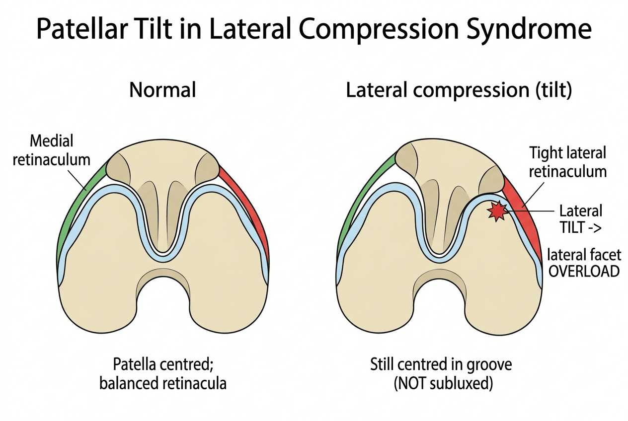
- ELPS is caused by a TIGHT LATERAL RETINACULUM that tilts the patella laterally and concentrates load on the LATERAL facet: anterior knee pain and, over time, lateral facet cartilage degeneration.
- The patella is TILTED, not subluxing or dislocating: a problem of PRESSURE/tilt, NOT of instability.
- Hallmark sign: a positive PATELLAR TILT TEST - the tight lateral retinaculum prevents the lateral edge being everted past the horizontal/neutral.
- AXIAL (skyline/Merchant) radiographs show an abnormal PATELLAR TILT ANGLE and a reduced/negative LATERAL PATELLOFEMORAL ANGLE; CT or MRI confirm tilt WITHOUT subluxation and exclude trochlear dysplasia or a high TT-TG.
- CONSERVATIVE first: stretch the lateral retinaculum and iliotibial band, strengthen the VMO and hip abductors/external rotators, taping/bracing to medialise/de-rotate the patella.
- ARTHROSCOPIC LATERAL RETINACULAR RELEASE (sometimes with lateral patelloplasty) for refractory, genuine ELPS; outcomes are worse with high-grade lateral facet cartilage damage.
- Release is NOT for INSTABILITY: a randomised trial found no benefit from adding it to MPFL reconstruction, and an inappropriate or over-aggressive release can WORSEN instability (medial patellar subluxation).
- “ELPS = tight LATERAL RETINACULUM → patellar TILT + excessive lateral pressure → anterior knee pain (TILT, not instability).
- “Positive PATELLAR TILT TEST (can't evert the lateral patella past neutral); skyline radiographs show abnormal patellar tilt angle / lateral patellofemoral angle.
- “Conservative first (stretch lateral structures, VMO/hip). Lateral retinacular release for refractory TILT/ELPS - NOT for instability (no benefit added to MPFL recon; can worsen instability).
Definition and Mechanism
What it is. Lateral patellar compression syndrome, also called excessive lateral pressure syndrome (ELPS), is caused by a tight lateral retinaculum that tilts the patella laterally. The tilt concentrates load on the lateral patellofemoral facet, pressing it hard against the lateral trochlea while the medial facet lifts and the medial retinaculum is stretched. That excessive lateral pressure produces anterior knee pain and, over time, lateral facet cartilage wear.
Tilted, not unstable. The defining point is that the patella is tilted, not subluxing or dislocating. This is a problem of pressure, not of instability.
Proving It Is Tight and Not Loose at the Bedside
The history. Anterior knee pain, worse with activity and with prolonged sitting, and tenderness over the lateral patellofemoral joint.
The patellar tilt test. With the knee extended and the quadriceps relaxed, try to lift (evert) the lateral edge of the patella. In ELPS the tight lateral retinaculum prevents the lateral edge being everted past the horizontal, or neutral. A positive tilt test is the hallmark sign.
Why the tilt test is not enough. Treatment turns entirely on separating a tight patella from an unstable one, and the tilt test alone does not do it, because a patella can be both tilted and lax. Two further examinations settle it, and they take seconds.
The patellar glide test quantifies the restraints in quadrants. With the knee flexed to about 20 to 30 degrees and the quadriceps relaxed, divide the patella into four longitudinal quadrants and translate it medially, then laterally, recording how many quadrants of its own width it moves:
- Medial glide of less than one quadrant means the lateral structures are tight. It supports excessive lateral pressure and is the finding a release or lengthening is designed to correct. Normal is around two quadrants.
- Lateral glide of three quadrants or more means the medial restraints are incompetent, and four quadrants is a frank dislocation. This is instability, and it makes a lateral release the wrong operation.
The two can coexist, which is exactly the trap: a tight lateral retinaculum in a patient who also has a deficient MPFL. Releasing that patella removes what little lateral checkrein remained.
Apprehension should be absent. Pushing the patella laterally with the knee near extension provokes a protective, alarmed response in a patient whose patella has dislocated. It produces nothing in excessive lateral pressure, where the complaint is pain from compression rather than fear of the patella leaving the groove.
A positive apprehension sign points away from this diagnosis and towards instability, and is the fastest single discriminator available.
Where apprehension is positive, or lateral glide is excessive, reach for the instability work-up (trochlear dysplasia, TT-TG distance, patellar height and MPFL competence), covered in Patellofemoral Instability and MPFL Injury.
Imaging: The Axial Angles That Define Tilt
The views. Axial radiographs (skyline/Merchant) are the key imaging. The angles are measured on an axial view, either a Merchant or Laurin skyline or an axial CT or MRI, with the knee in early flexion, and the examinable detail is knowing what each one measures:
- Lateral patellofemoral angle (Laurin). The angle between a line across the anterior aspects of the two femoral condyles and a line along the lateral patellar facet. Normally it opens laterally; in excessive lateral pressure the lateral tilt makes the lines parallel or the angle open medially, the classic Laurin sign of patellar tilt.
- Patellar tilt angle. The angle between the transverse (equatorial) axis of the patella and the posterior femoral condylar line. It increases as the patella tilts laterally, is commonly cited as abnormal beyond about twenty degrees, and is a direct measure of how tilted the patella sits in the trochlea.
Tilt or subluxation. Tilt is a rotational malposition, which is what the two angles above measure. Subluxation is translational, measured by the congruence angle (Merchant), which locates the patellar apex relative to the bisector of the sulcus angle.
ELPS is tilt with a normal congruence angle. If the congruence angle is abnormal, you are dealing with instability or maltracking instead.
When the congruence angle is abnormal. Patellar height (Insall-Salvati/Caton-Deschamps) and trochlear depth (sulcus angle) are checked alongside; both are developed in Patellar Height Abnormalities and Patellofemoral Instability.
CT and MRI. Cross-sectional imaging confirms lateral tilt without subluxation and assesses the lateral facet cartilage. It also excludes maltracking from trochlear dysplasia or a high TT-TG, either of which would change management.
Management
Conservative first. Activity modification and analgesia, with a rehabilitation programme that stretches the tight lateral structures (the lateral retinaculum and iliotibial band) and strengthens the vastus medialis obliquus (VMO) and the hip abductors and external rotators. Patellar taping or bracing is used to medialise the patella.
When to operate. Surgery is for refractory, genuine ELPS. Before offering a release, confirm all of:
- Failed conservative management
- A genuinely tight lateral retinaculum with a positive tilt test
- Abnormal tilt angles on skyline imaging
- No instability
What the release achieves. An arthroscopic lateral retinacular release, sometimes combined with a lateral patelloplasty, is an effective minimally invasive option: it corrects the tilt, relieves pain and improves function. Outcomes are poorer when the lateral facet already has high-grade cartilage damage.
The single most important judgement in lateral patellar compression syndrome is to confirm that the problem is TILT from a tight lateral retinaculum and NOT instability, because lateral retinacular release is the right operation for the former and the wrong one for the latter. A randomised trial showed no benefit from adding a routine lateral release to MPFL reconstruction in recurrent patellar dislocation, and an inappropriate or over-aggressive lateral release in an unstable or already-lax patella can worsen instability and even cause medial patellar subluxation. Treat instability with the appropriate medial-sided and bony procedures instead.
Lateral Release vs Lengthening: Technique and Complications
The release. The tight lateral retinaculum, both its superficial oblique and deep transverse fibres, is divided from around the superior pole down toward the inferior pole or tibial-tubercle level, arthroscopically (inside-out) or open, so that the tilted patella can be everted and re-centred. A lateral patelloplasty, as in the cited Wang series, adds contouring or chondral management of the worn lateral facet at the same sitting.
Why lengthening is gaining favour. A complete release sacrifices the lateral restraint, so many surgeons now prefer a controlled z- or step-cut lengthening of the retinaculum. It relieves the tight lateral tether and corrects tilt while preserving a lateral checkrein, reducing the risk of iatrogenic medial instability, particularly when there is any doubt about laxity. Release and lengthening alike are reserved for a genuinely tight, tilted, stable patella.
Complications.
- Haemarthrosis, the commonest, from bleeding from the lateral superior genicular vessels
- Iatrogenic medial patellar instability or subluxation from over-release, the feared specific complication and exactly why release is wrong for an unstable patella
- Quadriceps inhibition or weakness
- Persistent pain when the lateral facet cartilage is already high-grade, the poorer outcomes seen in the Wang series
Mnemonics & Memory Aids
TILT
Hook:TILT: Tight lateral retinaculum, Increased lateral pressure, Lateral tilt (not instability), Tilt test positive (release only for refractory tilt).
Clinical Decision Scenarios
Practise clinical reasoning and management decisions out loud
“What is excessive lateral pressure syndrome and how do you diagnose it?”
“How would you manage ELPS, and what is the role of lateral retinacular release?”
Mechanism
- Tight lateral retinaculum tilts the patella laterally
- Excessive lateral facet pressure → anterior knee pain + lateral facet wear
- Patella TILTED, not subluxing (NOT instability)
Diagnosis
- Positive patellar tilt test (can't evert lateral patella past neutral)
- Lateral patellofemoral tenderness; pain with activity/prolonged sitting
- Skyline radiographs: abnormal patellar tilt angle / lateral patellofemoral angle; CT/MRI confirm tilt without subluxation
Conservative management
- Activity modification, analgesia
- Stretch lateral structures (retinaculum, IT band); strengthen VMO/hip
- Patellar taping/bracing to medialise
Surgery
- Lateral retinacular release (+/- patelloplasty) for refractory true ELPS
- Worse outcomes with high-grade lateral facet cartilage damage
- NOT for instability (no benefit with MPFL recon; can worsen instability)
Evidence & Key Studies
Arthroscopic lateral retinacular release with lateral patelloplasty for excessive lateral pressure syndrome
- In 73 patients (88 knees) with ELPS, arthroscopic lateral retinacular release combined with lateral patelloplasty significantly reduced pain (VAS) and improved Kujala and Lysholm scores, patella medial pushing distance, patellar tilt angle and lateral patellofemoral angle.
- Outcomes were worse with a higher lateral patella Outerbridge cartilage grade at follow-up.
- The technique effectively corrects the patellar force-line/tilt abnormality with few complications, supporting lateral release for true ELPS.
Lateral retinacular release is not recommended routinely with MPFL reconstruction for instability
- In a randomised trial of recurrent patellar dislocation, adding an arthroscopic lateral retinacular release to MPFL reconstruction produced no significant difference in subjective IKDC score or patellar tilt versus isolated MPFL reconstruction.
- There is no indication for a systematic lateral retinacular release in association with MPFL reconstruction for recurrent dislocation.
- This underlines that lateral release addresses tilt/compression, not instability.
The efficacy of arthroscopic lateral retinacular release (with lateral patelloplasty) for true ELPS - improving pain, function and the patellar tilt/lateral patellofemoral angles, with worse outcomes at higher lateral-facet cartilage grades - comes from the cited Wang study; the evidence that a routine lateral release adds no benefit to MPFL reconstruction for recurrent dislocation (underlining that release is for tilt, not instability) from the cited Malatray randomised trial. The tight-lateral-retinaculum mechanism, the patellar tilt test, the glide-test quadrants, the absent apprehension sign, the skyline radiographic angles and the conservative-first rehabilitation are standard, well-established teaching. (See also Patellofemoral Instability, MPFL Injury, Chondromalacia Patellae for the wider anterior knee pain differential, and Patellar Height Abnormalities.)