The Workhorse Donor of Reconstructive Surgery
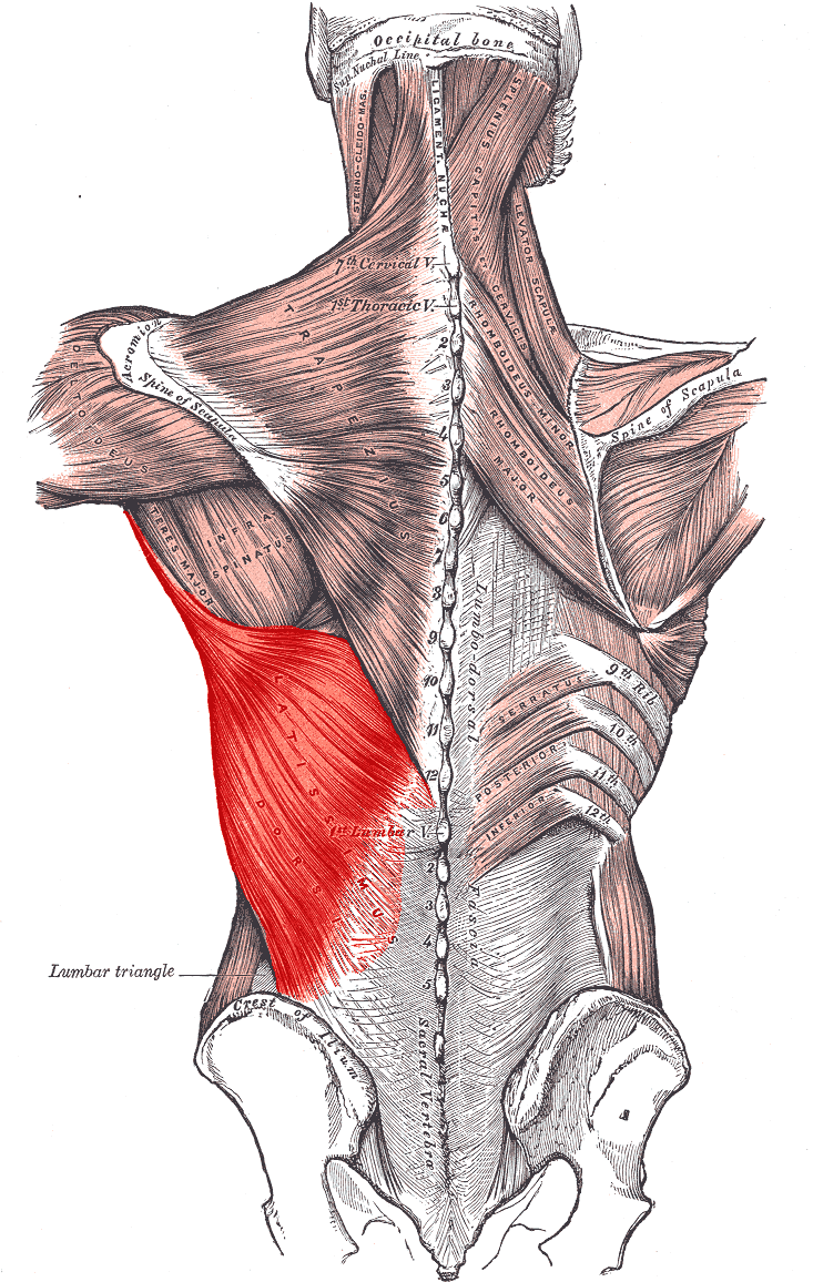
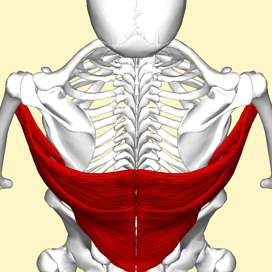
- Origin: spinous processes of T7 to T12 via the thoracolumbar fascia, the lumbar and sacral spines, the posterior third of the iliac crest, the lower three or four ribs, and variably the inferior angle of the scapula.
- Insertion: the floor of the intertubercular (bicipital) groove, by a flat tendon that spirals through 180 degrees.
- Innervation: thoracodorsal nerve (C6, C7, C8) from the posterior cord, running on the deep surface with the artery.
- Dominant pedicle: thoracodorsal artery, from the subscapular artery, with a 6 to 16 cm pedicle of 1.5 to 3 mm calibre.
- The radial nerve lies only 2 to 3 cm from the tendon insertion at the humerus — the hazard of transfer harvest.
- “Lady between two majors: latissimus inserts into the FLOOR of the groove, between pectoralis major laterally and teres major medially.
- “The 180 degree spiral of the tendon means the lowest fibres insert most superiorly, so distal muscle acts on the proximal humerus.
- “The serratus branch of the thoracodorsal artery allows retrograde perfusion — dividing the thoracodorsal proximally does not always kill the flap.
- “Thoracodorsal nerve injury is a recognised complication of axillary lymph node dissection.
Overview
The latissimus dorsi is the broadest muscle in the body, a fan of fibres converging from the lower half of the trunk onto a short flat tendon at the front of the humerus. Its functional role — adduction, extension and internal rotation — is entirely duplicated by the pectoralis major, teres major and subscapularis, which is precisely why it is the most expendable large muscle available and the most heavily used donor in reconstructive surgery.
Three surgical identities dominate examination discussion. It is the transfer of choice for an irreparable posterosuperior rotator cuff tear in a younger patient. It is the workhorse flap for reconstruction of the chest wall, breast, head and neck, and limbs, either pedicled or free. And in brachial plexus birth palsy it is one of the internal rotators whose unopposed pull produces a fixed internal rotation contracture and progressive glenoid dysplasia — the same muscle that later becomes the solution when it is released and transferred.
The latissimus tendon rotates through approximately 180 degrees on its way to the humerus.
- Fibres from the iliac crest and lowest ribs, the most inferior and posterior in origin, come to lie most superior at the insertion. Fibres from the upper thoracic origin lie most inferior.
- Consequence one — mechanics. The distal, most powerful part of the muscle acts on the proximal part of the tendon footprint, and the spiral converts a broad sheet of nearly parallel fibres into a single concentrated line of pull.
- Consequence two — surgery. When the tendon is detached and transferred, the surgeon must untwist it and orient it correctly, or the transfer will be tensioned unevenly across its width and part of it will do no work.
- Consequence three — anatomy of the groove. The insertion sits in the floor of the bicipital groove, deep to the long head of biceps tendon and between pectoralis major on the lateral lip and teres major on the medial lip. The proximity to the biceps tendon matters when a subpectoral biceps tenodesis is performed nearby.
SLIRSLatissimus Dorsi Origin
Hook:Everything below the mid-back converges on one small tendon at the front of the arm — which is why the muscle is so long and its excursion so generous.
Attachments, Innervation and Relations
Origin
- Spinous processes of T7 to T12 and their supraspinous ligaments, largely through the posterior layer of the thoracolumbar fascia.
- Lumbar and sacral spinous processes, again via the thoracolumbar fascia.
- Posterior third of the outer lip of the iliac crest.
- Lower three or four ribs, by fleshy slips interdigitating with the external oblique.
- Inferior angle of the scapula, present in roughly half of individuals as a small slip. It is the reason strong latissimus contraction can rotate the scapula and the reason a scapular slip may be encountered during flap harvest.
Insertion
- The fibres converge on a flat tendon approximately 2 to 4 cm wide and 7 to 8 cm long, which spirals through about 180 degrees.
- It inserts into the floor of the intertubercular (bicipital) groove of the humerus.
- The arrangement at the groove, lateral to medial, is: pectoralis major on the lateral lip, latissimus dorsi in the floor, teres major on the medial lip.
- A constant bursa lies between the latissimus and teres major tendons; opening it is the reliable step in separating the two during transfer harvest.
- The tendon passes anterior to teres major and the two form the posterior axillary fold together.
The transfer hazard.
- Enters the triangular interval at the lower border of teres major, within 2 to 3 cm of the tendon insertions.
- Abduct and externally rotate the arm to draw the nerve away before releasing the tendon.
- Release under direct vision, working from the humerus outward; never sweep blindly inferiorly.
- A wrist drop after shoulder surgery is a devastating and avoidable outcome.
The flap lifeline.
- Hilum on the deep surface, 8 to 12 cm from the insertion and 2 to 3 cm medial to the anterior border.
- Preserve the serratus branch where possible; it provides retrograde perfusion if the main trunk is compromised.
- Endangered during axillary node dissection and during any previous axillary surgery — always ask about it before planning a flap.
Action and Biomechanics
Actions by plane
- Adduction of the humerus, powerfully and through a long arc.
- Extension of the humerus from a flexed position.
- Internal (medial) rotation of the humerus.
- Depression of the shoulder girdle, drawing the whole shoulder downward and backward.
- Trunk elevation with a fixed arm: during climbing, crutch walking and wheelchair transfers, the latissimus pulls the trunk toward the fixed arm. This is the action that matters when counselling a wheelchair user about donor morbidity.
- Accessory respiration: it contributes to forced expiration and coughing, and the scapular slip contributes to scapular depression and downward rotation.
Mechanical characteristics that make it a good donor
- Long fibres and generous excursion. Unlike the short, stiff teres major, the latissimus has a long parallel-fibred architecture and a large excursion, which is what allows it to be routed around the humerus and still reach the greater tuberosity.
- Single long pedicle. A 6 to 16 cm pedicle of adequate calibre with a wide arc of rotation.
- Functional redundancy. Its actions are duplicated by pectoralis major, teres major and subscapularis, so most patients report no functional loss after harvest.
- The trade-off is that its excursion comes with relatively modest force per unit cross-section compared with a pennate muscle, and after transfer the tendon must be tensioned carefully to place the muscle on the correct part of its length-tension curve.
Synergists and antagonists
- Synergists: teres major and pectoralis major (adduction, extension, internal rotation), subscapularis (internal rotation), posterior deltoid (extension), long head of triceps (extension).
- Antagonists: deltoid and supraspinatus (abduction), infraspinatus and teres minor (external rotation), trapezius and serratus anterior (shoulder girdle elevation).
The transfer does not make the muscle stronger; it moves the lever.
- In its native position the latissimus inserts anterior and medial to the axis of humeral rotation, in the floor of the bicipital groove. Contraction therefore produces internal rotation and adduction.
- Detached and reattached to the posterosuperior greater tuberosity, the same contraction now produces external rotation and a head-depressing force, substituting for the absent infraspinatus and supraspinatus.
- Prerequisites, all three of which must be stated:
- An intact subscapularis, providing the anterior arm of the transverse force couple against which the transfer works. Gerber's long-term data show markedly worse results without it.
- A functioning deltoid, since the transfer is not an elevator.
- An adequate passive range and a joint without established arthritis, because a transfer moves a lever and cannot move a stiff or destroyed joint.
- The route matters: the tendon is passed deep to the deltoid and superficial to the teres minor and the long head of triceps, so that it lies in the correct plane to act as an external rotator without tethering.
Surface Anatomy and Examination
Palpation and positioning
- Ask the patient to place the hand on the hip and press inward, or to cough. The posterior axillary fold becomes prominent and the broad sheet of latissimus can be traced from the fold down and medially toward the iliac crest.
- The anterior (lateral) border is the key surgical landmark and is marked pre-operatively with the patient standing and adducting against resistance; the flap skin paddle and the descending vascular branch are planned relative to it.
- The inferior angle of the scapula is palpated to identify the scapular slip and to orient the superior extent of a flap.
Named tests and clinical assessment
- How to perform
- Hand on hip pressing inward; palpate the posterior axillary fold
- Positive finding
- Weak or absent contraction, or a palpable defect
- What it means
- Latissimus or teres major deficiency or rupture
- False positives and pitfalls
- Pectoralis major substitutes; palpate posteriorly, not anteriorly
- How to perform
- Ask the patient to cough while palpating the fold
- Positive finding
- Absent contraction
- What it means
- Denervation, since latissimus is an accessory expiratory muscle
- False positives and pitfalls
- Weak cough effort in an unwell patient
- How to perform
- Ask a seated patient to press down on the arm rests and lift the buttocks off the chair
- Positive finding
- Inability to lift or marked asymmetry
- What it means
- Functional latissimus loss, directly relevant to wheelchair transfers
- False positives and pitfalls
- Triceps and pectoralis weakness produce the same failure
- How to perform
- With the scapula stabilised, measure passive external rotation at the side and at 90 degrees abduction
- Positive finding
- Restricted passive arc
- What it means
- A contracture that must be released before any transfer
- False positives and pitfalls
- Pain and guarding in an acute presentation
- How to perform
- Scapula stabilised, passive external rotation with the arm at the side
- Positive finding
- Passive external rotation of 0 degrees or less
- What it means
- Established internal rotation contracture with a risk of glenoid dysplasia
- False positives and pitfalls
- Failure to stabilise the scapula gives a falsely reassuring arc
- How to perform
- Palpate contraction with resisted adduction on the intended donor side
- Positive finding
- Absent contraction on the side of previous axillary surgery
- What it means
- Thoracodorsal nerve injury, commonly after axillary node dissection
- False positives and pitfalls
- Muscle may still be perfused and usable as a non-functional flap even if denervated
Before planning any latissimus flap or transfer, ask specifically about previous axillary lymph node dissection, previous thoracotomy or previous axillary trauma. The thoracodorsal nerve and pedicle are both at risk in the axilla, and a denervated latissimus is useless as a functional transfer even if it remains perfused and usable as a bulk flap. A single question at the bedside prevents a wasted operation.
Complications
Radial nerve injury
- Mechanism: blind dissection inferior to the tendon insertions at harvest, or traction while routing the transfer.
- Consequence: wrist drop and loss of thumb and finger extension in a patient operated on for shoulder weakness — a catastrophic and entirely avoidable outcome.
- Avoidance: abduct and externally rotate the arm, release under direct vision, and never sweep blindly.
Axillary nerve injury
- Mechanism: proximal mobilisation into the quadrangular space, or forceful passage of the tendon beneath the deltoid.
- Consequence: deltoid loss, which removes both active elevation and the option of a subsequent reverse arthroplasty.
- Avoidance: create the subdeltoid tunnel bluntly under palpation and confirm it is free before pulling the tendon.
Pedicle compromise and flap loss
- Mechanism: injury to the thoracodorsal artery during harvest, kinking as the flap is inset, compression by a tight tunnel, or previous axillary surgery having already damaged the pedicle.
- Avoidance: dissect the pedicle to the subscapular trunk to gain length, preserve the serratus branch where possible for retrograde perfusion, avoid tension and kinking at inset, and always take a history of previous axillary surgery or irradiation.
- Recognition: monitor the flap clinically and with implantable Doppler where available; early re-exploration salvages a thrombosed anastomosis.
Donor site morbidity
- Seroma is the commonest complication of latissimus harvest, particularly with wide pedicled dissection. Quilting sutures to obliterate the dead space and prolonged drainage reduce, but do not eliminate, it.
- Functional loss is usually negligible in ambulant patients because the actions are duplicated. It is not negligible in wheelchair users, crutch users, climbers, rowers and swimmers, who depend on latissimus for transfers and propulsion. Counsel these groups explicitly and consider a split harvest.
- Loss of the posterior axillary fold contour and a long visible scar.
- Shoulder stiffness from prolonged immobilisation after a transfer.
Failure of a transfer
- Insufficient subscapularis is the dominant predictor of failure, shown clearly in long-term series.
- Tensioning error: tensioning in internal rotation leaves the transfer slack and functionless.
- Rupture of the transfer at the tuberosity, particularly if active use begins too early.
- Failure of motor re-education: the patient must learn to fire an internal rotator as an external rotator. Without a structured programme with biofeedback, a technically perfect transfer produces no functional gain.
Denervation of the graft
- A transfer whose thoracodorsal nerve is injured, or a flap harvested from a side with previous axillary node clearance, becomes a passive tenodesis rather than a functioning muscle. Confirm contraction pre-operatively.
Clinical Relevance
Pathology and clinical roles
- Typical Patient
- Professional throwers, water skiers, weightlifters
- Presentation
- Sudden posterior axillary pain during acceleration or a violent pull
- Key finding
- Loss of posterior axillary fold contour; MRI shows retraction from the groove
- Management
- Non-operative in most; repair considered in elite throwing athletes
- Typical Patient
- Infant with a C5-C6 upper trunk lesion
- Presentation
- Fixed internal rotation and adduction, loss of passive external rotation
- Key finding
- Glenoid retroversion and posterior head subluxation on imaging
- Management
- Subscapularis slide with latissimus and teres major release and transfer
- Typical Patient
- Active patient under about 60
- Presentation
- Pain and weakness of elevation and external rotation with preserved passive motion
- Key finding
- Goutallier 3 to 4 supraspinatus and infraspinatus with an intact subscapularis
- Management
- Latissimus dorsi transfer to the greater tuberosity
- Typical Patient
- After axillary lymph node dissection or posterior cord injury
- Presentation
- Weakness of adduction and extension; difficulty with wheelchair transfers and crutch use
- Key finding
- Absent latissimus contraction on resisted adduction or cough
- Management
- Usually accepted; matters most when the muscle was intended as a donor
- Typical Patient
- Any age after axillary burn
- Presentation
- Restricted abduction with a taut posterior band
- Key finding
- Clinical diagnosis
- Management
- Z-plasty or local flap release
- Typical Patient
- Anatomical variant
- Presentation
- Usually asymptomatic; occasionally axillary neurovascular compression or a palpable axillary mass
- Key finding
- A muscular slip crossing the axilla from latissimus to pectoralis major or the coracoid
- Management
- Division if symptomatic; recognition prevents confusion at axillary surgery
The muscle is first a cause of deformity and later a solution.
- Why it deforms. An upper trunk (C5-C6) lesion weakens the external rotators — infraspinatus, teres minor and deltoid — while the internal rotators, subscapularis, latissimus dorsi (C6-C8), teres major and pectoralis major, retain much more of their function. The latissimus is relatively spared precisely because its root supply extends below the level of the lesion.
- The consequence. A progressive internal rotation and adduction contracture. The head is held internally rotated and drifts posteriorly; the growing glenoid remodels into retroversion with a biconcave or false glenoid, the deformity graded on MRI by the Waters classification.
- The warning sign. Loss of passive external rotation with the scapula stabilised. It signals that the deformity is no longer purely dynamic.
- The operation. Anterior release — a subscapularis slide rather than a tenotomy, so internal rotation is not lost entirely — together with release of the latissimus dorsi and teres major from the groove, and transfer of those two muscles posterolaterally to the rotator cuff insertion (the Hoffer modification of the L'Episcopo procedure).
- The caution. Over-release leaves the child unable to reach the midline for perineal care and dressing, which patients and families find as disabling as the original deformity. Follow to skeletal maturity, since contracture can recur and dysplasia can progress.
Grading and imaging
- MRI identifies the tendon at the floor of the bicipital groove, anterior to teres major, and demonstrates rupture, retraction and fatty change. Muscle quality on MRI is assessed before transfer.
- Imaging in birth palsy: ultrasound in infants avoids anaesthesia and assesses head subluxation dynamically; MRI is used to grade glenoid version and morphology.
- Angiographic or duplex assessment of the thoracodorsal pedicle is used selectively before free flap transfer, particularly where there has been previous axillary surgery, irradiation or trauma.
- Denervation appears as diffuse muscle oedema-like signal followed by fatty replacement, uniform across the belly.
Surgical Relevance
Latissimus dorsi transfer for irreparable posterosuperior cuff tear
- Detail
- Irreparable posterosuperior tear, patient generally under about 60, pain and weakness with preserved passive motion
- Rationale
- The transfer restores the posterior arm of the transverse force couple
- Detail
- Intact subscapularis, functioning deltoid, no significant glenohumeral arthritis, adequate passive range, a patient who can be re-educated
- Rationale
- Without an anterior arm to the couple, or a fulcrum, or passive motion, the transfer fails
- Detail
- Two-incision technique: a posterior axillary incision to harvest, and a superior or deltoid-splitting incision to deliver and fix
- Rationale
- Allows tendon harvest at the humerus and passage under the deltoid
- Detail
- Open the bursa between latissimus and teres major; latissimus lies anterior and inserts in the floor of the groove, teres major posterior on the medial lip
- Rationale
- Harvesting the wrong tendon or both together destroys the vector
- Detail
- Deep to the posterior deltoid, superficial to teres minor and the long head of triceps
- Rationale
- Places the transfer in the plane of an external rotator without tethering
- Detail
- To the posterosuperior greater tuberosity with anchors or transosseous sutures, plus suture to residual infraspinatus and teres minor stumps
- Rationale
- Reconstitutes both a footprint and a soft-tissue vector
- Detail
- Tension with the arm in abduction and external rotation; immobilise in an abduction and external rotation brace for about six weeks
- Rationale
- Prevents the transfer from being tensioned in the position that would slacken it
- Detail
- Protected passive motion, then progressive re-education with biofeedback to fire an internal rotator as an external rotator
- Rationale
- Motor relearning is the rate-limiting step
The radial nerve enters the triangular interval at the lower border of teres major, within roughly 2 to 3 cm of the latissimus and teres major insertions.
- Position the arm in abduction and external rotation to draw the nerve away from the field before releasing the tendon.
- Release under direct vision, working from the humeral surface outward and taking the tendon subperiosteally where possible.
- Never sweep blindly inferior to the tendon. Blunt finger dissection in this plane is the classic mechanism of radial nerve injury during a transfer.
- Remember the axillary nerve at the other end, in the quadrangular space at the upper border of teres major, at risk during proximal mobilisation and when the tunnel beneath the deltoid is created. Make the tunnel bluntly with a finger and confirm it is free before pulling the tendon through.
- Protect the thoracodorsal pedicle on the deep surface throughout. In a transfer the muscle must remain innervated and perfused; a devascularised transfer is simply a graft.
Flap surgery
- Pedicled myocutaneous flap on the thoracodorsal pedicle: the standard reconstruction for the breast after mastectomy, for chest wall defects, for head and neck reconstruction, and for coverage of the shoulder, axilla and proximal arm. Its arc of rotation reaches the head and the mid-chest.
- Free flap: the workhorse muscle free flap. Its long pedicle, adequate calibre, large surface area and reliable anatomy make it the standard choice for large defects of the lower limb, the scalp and complex wounds anywhere.
- Functional free muscle transfer: with microvascular anastomosis and coaptation of the thoracodorsal nerve to a recipient motor nerve, the latissimus is used to restore elbow flexion in brachial plexus reconstruction, and in facial reanimation.
- Split or segmental flap: because the pedicle divides into descending and transverse branches, half the muscle can be taken while the other half remains innervated and perfused — reducing donor morbidity.
- Reverse (turnover) flap: based on the paraspinous perforators, reaching midline lumbar and lower thoracic defects. This is the practical expression of the Mathes-Nahai Type V pattern.
- Latissimus dorsi transfer for elbow flexion (bipolar transfer): the whole muscle is moved on its pedicle to the anterior arm to substitute for an absent biceps.
Other uses and landmarks
- L'Episcopo and Hoffer transfers in brachial plexus birth palsy, and the modified L'Episcopo combined with reverse arthroplasty in adults with combined loss of elevation and external rotation.
- The lumbar triangle of Petit and the auscultatory triangle are defined by its borders and appear regularly in anatomy vivas.
- The anterior border guides thoracotomy incisions and is preserved in a muscle-sparing thoracotomy specifically to keep the flap option available.
- Age under about 60 with high functional demand
- Intact, functioning subscapularis
- Functioning deltoid and axillary nerve
- Preserved passive range with no arthritis
- Motivated patient able to undertake motor re-education
- Age over about 70
- Cuff tear arthropathy with acetabularisation
- Subscapularis also irreparable
- True pseudoparalysis with anterosuperior escape
- Previous failed transfer or reconstruction
Guidelines, Registries & Global Practice
Anatomical variation across populations
- The scapular slip from the inferior angle is present in roughly half of individuals in cadaveric series, with variation between populations. Its presence matters at flap harvest and explains why strong latissimus contraction can rotate the scapula.
- The axillary arch (Langer's arch), a muscular slip crossing the axilla from latissimus to pectoralis major, the coracoid or the conjoined tendon, is described with reported prevalence differences across populations. It can compress the axillary neurovascular bundle, confuse axillary node dissection, and be mistaken for a mass.
- The branching pattern of the thoracodorsal artery varies, particularly the level at which the serratus branch and the intramuscular division arise, which is the basis for planning split flaps and must be confirmed intra-operatively rather than assumed.
- The point at which the thoracodorsal nerve enters the muscle also varies, affecting the arc of rotation achievable in a functional transfer.
Side-by-side guidance
- Position Relevant to Latissimus Dorsi
- Recognises tendon transfer as an option for irreparable posterosuperior cuff tears in younger active patients with an intact subscapularis, and reverse arthroplasty for older cuff-deficient shoulders.
- Position Relevant to Latissimus Dorsi
- Recommend early specialist referral for brachial plexus birth palsy, serial measurement of passive external rotation with the scapula stabilised, and imaging when it is lost.
- Position Relevant to Latissimus Dorsi
- Emphasises identification of the radial nerve relative to the lower border of teres major during transfer harvest and the correct plane of transfer beneath the deltoid.
- Position Relevant to Latissimus Dorsi
- Treat the latissimus as the standard workhorse free flap, with pre-operative assessment of the pedicle mandatory where there has been previous axillary surgery or irradiation.
Registry and outcome signals
- Tendon transfers about the shoulder are low-volume procedures and are not captured by national joint registries; the evidence base is long-term institutional series. The consistent signal across those series is that pain relief is the most reliable outcome, functional gain is more variable, and subscapularis integrity dominates the result.
- Breast reconstruction registries and national audits capture latissimus flap use in reasonable volume. Seroma is consistently the commonest donor complication, and total flap loss with a pedicled latissimus is rare.
- Brachial plexus birth palsy outcomes are tracked through specialist multidisciplinary networks rather than registries. The consistent message is that early recognition of the internal rotation contracture and rebalancing before fixed glenoid dysplasia develops determines long-term shoulder function.
- Reverse arthroplasty volumes in national registries continue to grow, and in many settings a reverse has replaced tendon transfer for older patients who would previously have had a latissimus transfer.
High-resource versus limited-resource practice
- Well-resourced settings: microvascular free tissue transfer, arthroscopically assisted latissimus transfer, MRI for muscle quality grading, and implantable Doppler flap monitoring.
- Limited-resource settings: the pedicled latissimus flap is one of the most valuable operations available anywhere, requiring no microsurgical equipment and covering an enormous range of defects. Similarly, latissimus and teres major transfer for birth palsy needs no implants and changes a lever rather than replacing a joint.
- The single universally applicable step costs nothing: ask about previous axillary surgery and confirm latissimus contraction clinically before planning any operation that depends on this muscle.
MCQ Practice Points
Q: Where does latissimus dorsi insert? A: The floor of the intertubercular (bicipital) groove. Pectoralis major inserts on the lateral lip and teres major on the medial lip — the Lady between two majors.
Q: What is the innervation of latissimus dorsi? A: The thoracodorsal nerve (middle subscapular nerve), C6, C7, C8, from the posterior cord, arising between the upper and lower subscapular nerves.
Q: Describe the dominant pedicle of the latissimus dorsi flap. A: The thoracodorsal artery, terminal branch of the subscapular artery from the third part of the axillary artery. Length 6 to 16 cm, calibre 1.5 to 3 mm, hilum 8 to 12 cm from the insertion and 2 to 3 cm medial to the anterior border.
Q: What Mathes-Nahai type is the latissimus dorsi and what does that permit? A: Type V — one dominant pedicle plus secondary segmental pedicles. It permits both a conventional flap on the thoracodorsal pedicle and a reverse turnover flap on the paraspinous perforators.
Q: Why does the latissimus tendon spiral through 180 degrees? A: Because the muscle originates posteriorly and inferiorly but inserts anteriorly on the humerus. The consequence is that the most inferior fibres insert most superiorly, and the tendon must be untwisted and correctly oriented when transferred.
Q: Why is the serratus branch of the thoracodorsal artery important? A: It permits retrograde perfusion of the flap through the lateral thoracic and intercostal system if the thoracodorsal trunk is divided proximally. Preserve it where possible.
Q: How close is the radial nerve to the latissimus dorsi insertion? A: Approximately 2 to 3 cm. It enters the triangular interval at the lower border of teres major. Abduct and externally rotate the arm and release the tendon under direct vision.
Q: What is the single strongest predictor of failure of a latissimus transfer for an irreparable cuff tear? A: An insufficient subscapularis. The transfer restores the posterior arm of the transverse force couple and requires a competent anterior arm to work against.
Q: Which two anatomical triangles are bounded by latissimus dorsi? A: The lumbar triangle of Petit (anterior border of latissimus, posterior border of external oblique, iliac crest) and the auscultatory triangle (superior border of latissimus, medial border of the scapula, inferolateral trapezius).
Q: Which patients notice latissimus dorsi donor morbidity most, and what is the commonest complication? A: Wheelchair users, crutch users, climbers, rowers and swimmers, who use it to lift the trunk toward a fixed arm. The commonest complication of harvest is seroma.
Exam Viva Scenarios
Practise clinical reasoning and management decisions out loud
“A 54-year-old carpenter has an irreparable posterosuperior cuff tear with Goutallier 4 supraspinatus and infraspinatus, preserved passive motion and no arthritis. He wants to keep working. Talk me through whether you would offer a latissimus dorsi transfer and what you would check first.”
“You are asked to raise a free latissimus dorsi flap for a large open tibial wound. Describe the pedicle anatomy you rely on and what would make you abandon this donor.”
“A 14-month-old with a C5-C6 brachial plexus birth palsy holds the arm internally rotated. Passive external rotation with the scapula stabilised is minus 15 degrees. Why is the latissimus dorsi part of this problem and part of the solution?”
Attachments
- Origin: T7 to T12 spines via thoracolumbar fascia, lumbar and sacral spines
- Posterior third of the iliac crest, lower three or four ribs
- Inferior angle of the scapula in about half of individuals
- Insertion: floor of the bicipital groove
- Tendon 2 to 4 cm wide, 7 to 8 cm long, spirals 180 degrees
Nerve and Pedicle
- Thoracodorsal (middle subscapular) nerve, C6-C8, posterior cord
- Thoracodorsal artery from subscapular from third part of axillary
- Pedicle 6 to 16 cm, calibre 1.5 to 3 mm
- Hilum 8 to 12 cm from insertion, 2 to 3 cm medial to the anterior border
- Descending and transverse intramuscular branches allow split flaps
- Serratus branch permits retrograde perfusion
Flap Facts
- Mathes-Nahai Type V
- Pedicled: breast, chest wall, head and neck, shoulder and proximal arm
- Free: lower limb, scalp, large complex defects
- Functional free transfer for elbow flexion and facial reanimation
- Reverse turnover flap on paraspinous perforators for lumbar defects
- Seroma is the commonest donor complication
Transfer for Cuff Tear
- Indication: irreparable posterosuperior tear, patient under about 60
- Prerequisites: intact subscapularis, functioning deltoid, passive range, no arthritis
- Route: deep to deltoid, superficial to teres minor and triceps long head
- Fix to the posterosuperior greater tuberosity
- Brace in abduction and external rotation for about six weeks
- Pain relief is the most reliable gain
Hazards and Landmarks
- Radial nerve 2 to 3 cm from the insertion at the lower border of teres major
- Axillary nerve in the quadrangular space above
- Thoracodorsal nerve at risk in axillary node dissection
- Lumbar triangle of Petit and auscultatory triangle
- Axillary arch of Langer is a recognised variant
Evidence Base
Latissimus Dorsi Transfer for the Treatment of Irreparable Rotator Cuff Tears
- 67 patients with 69 irreparable full-thickness tears of at least two complete tendons, reviewed at a mean of 53 months
- Subjective Shoulder Value rose from 28 to 66 per cent and the age and gender-matched Constant score from 55 to 73 per cent
- Flexion improved from 104 to 123 degrees, abduction from 101 to 119 degrees, external rotation from 22 to 29 degrees, and abduction strength from 0.9 to 1.8 kg
- Thirteen patients had deficient subscapularis function pre-operatively; results were better in shoulders with a negative pre-operative lift-off test
Latissimus Dorsi Transfer for the Treatment of Massive Tears of the Rotator Cuff: A Preliminary Report
- Transfer of the latissimus dorsi tendon from the humeral shaft to the superolateral humeral head, providing a large vascularised tendon to close a massive cuff defect
- The transfer exerts an external rotation and head-depressing moment, allowing more effective action of the deltoid
- Performed in 14 patients without any significant complications
- Pain relief and functional results were reported in the subgroup with adequate follow-up
Latissimus Dorsi and Teres Major Transfer to Rotator Cuff for Erb's Palsy
- Fifty-six children with Erb's palsy operated on over 20 years; 35 followed for a mean of 5 years
- Pre-operative passive external rotation averaged 5 degrees with external rotation strength below grade 2
- Post-operative active external rotation averaged 31 degrees, with only two recurrent internal rotation contractures
- Active abduction improved from a mean of 74 to 120 degrees
Classification of the Vascular Anatomy of Muscles: Experimental and Clinical Correlation
- Describes five patterns of muscle circulation based on studies of muscle vascular anatomy
- The classification is validated against arc of rotation, skin territory, distally based flaps, microvascular transplantation and muscle-delay models
- Its purpose is to predict, from vascular pattern, which muscles can be raised as reliable flaps and how they should be designed
- Within this system the latissimus dorsi is a Type V muscle — one dominant pedicle plus secondary segmental pedicles
Glenohumeral Deformity Secondary to Brachial Plexus Birth Palsy
- 94 patients with brachial plexus birth palsy entered prospectively; 42 imaged with CT or MRI for pre-operative planning
- Mean glenoscapular angle (glenoid retroversion) minus 25.7 degrees on the affected side against minus 5.5 degrees on the unaffected side
- Assessment covered glenohumeral incongruity, humeral head deformity and glenoid hypoplasia
- Function was rated using the Mallet classification on a scale of 1 to 5
Management and Outcomes of Latissimus Dorsi and Teres Major Injuries in Professional Baseball Pitchers
- Retrospective review of 16 professional baseball pitchers with MRI-confirmed latissimus dorsi or teres major tears treated between 2002 and 2008
- 6 tendon avulsions and 10 strains; all treated non-operatively with rest, rehabilitation and a graded throwing programme
- 15 of 16 (94 per cent) returned to the same or a higher level of play; mean time to throwing 35.6 days, to pitching 61.9 days
- 9 of 16 (56 per cent) were season-ending and 2 of 16 (13 per cent) recurred; 75 per cent had prior shoulder or elbow injury