The Suspensory Muscle of the Shoulder Girdle
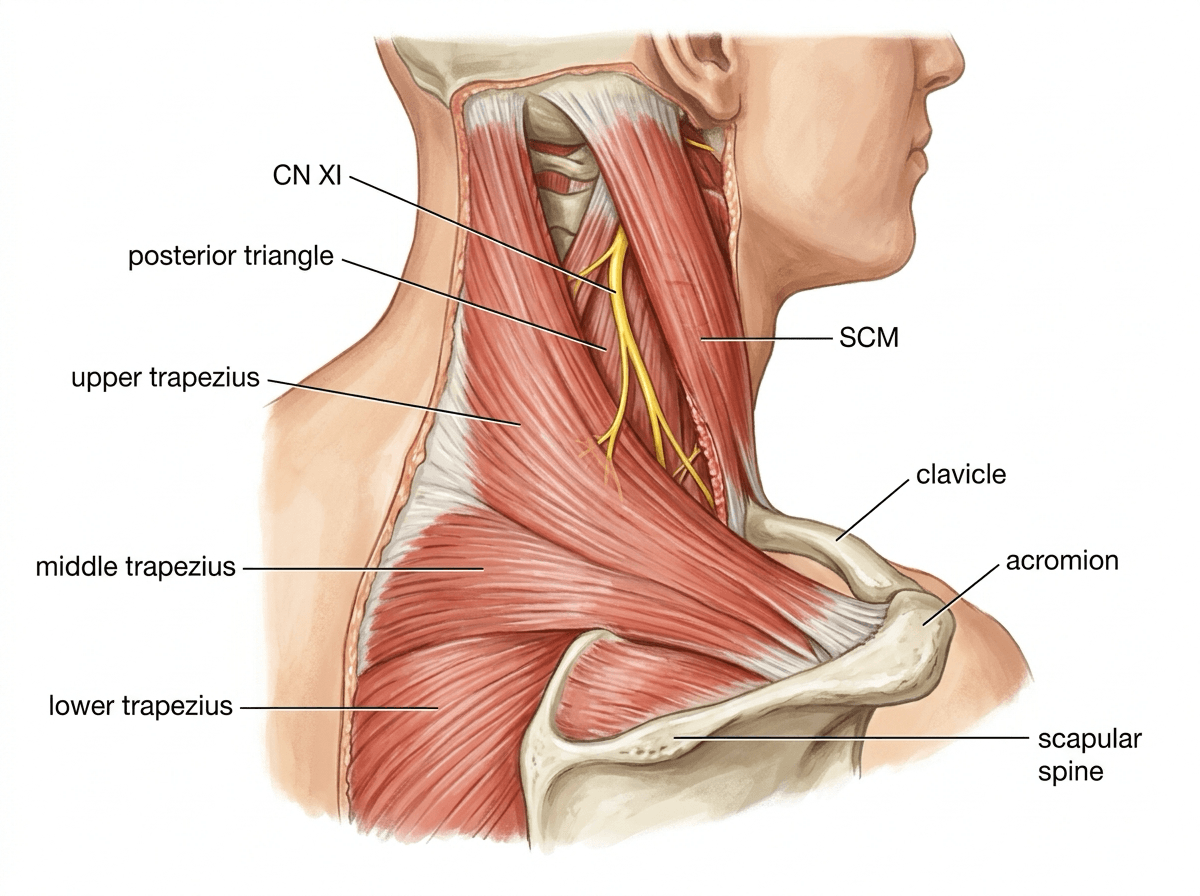
- Sole motor supply is the spinal accessory nerve (CN XI); C3 and C4 ventral rami contribute proprioceptive and some motor fibres.
- CN XI crosses the posterior triangle superficially, invested only in the investing layer of deep cervical fascia over levator scapulae, making it the most commonly injured nerve during lymph node biopsy.
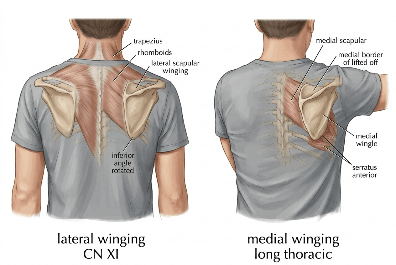
- Trapezius palsy produces LATERAL scapular winging with a drooped shoulder and asymmetric neckline; serratus palsy produces MEDIAL winging with a normal or elevated shoulder.
- The scapular upward-rotation force couple is upper trapezius plus lower trapezius plus serratus anterior; losing trapezius shifts the whole couple onto serratus.
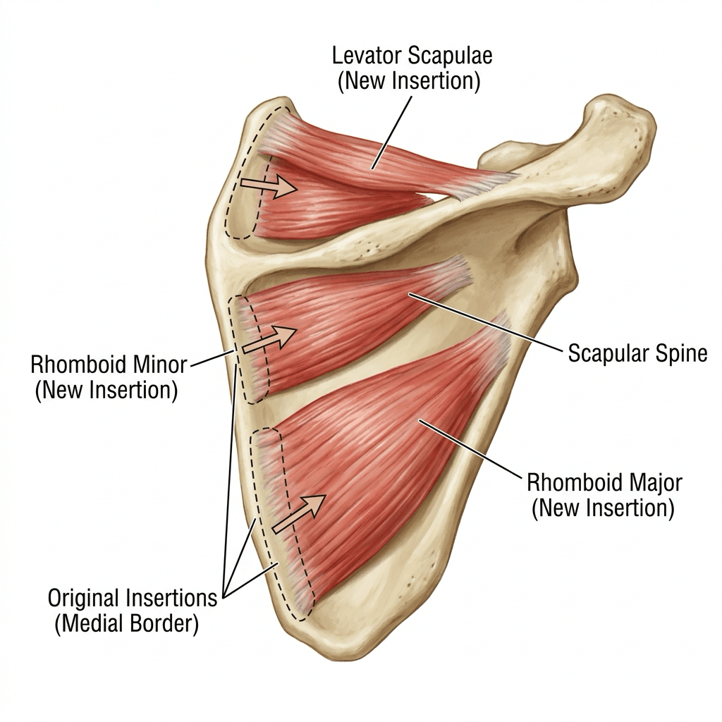
- Eden-Lange transfer moves levator scapulae, rhomboid minor and rhomboid major laterally to substitute for the three parts of the trapezius.
- “CN XI emerges from the posterior border of sternocleidomastoid at the junction of its upper and middle thirds, roughly 1 cm cranial to where the great auricular nerve wraps around the same border (Erb's point).
- “The nerve enters the deep surface of trapezius about 2 to 5 cm above the clavicle at the anterior border of the muscle - keep dissection in the posterior triangle superficial to the prevertebral fascia and use only blunt spreading.
- “Loss of the upper trapezius drops the shoulder and produces traction brachial plexopathy - the pain, not the weakness, is often what brings the patient in.
- “In spinal accessory to suprascapular nerve transfer, harvest CN XI distal to the branches to the upper trapezius so that shoulder suspension is preserved.
Overview
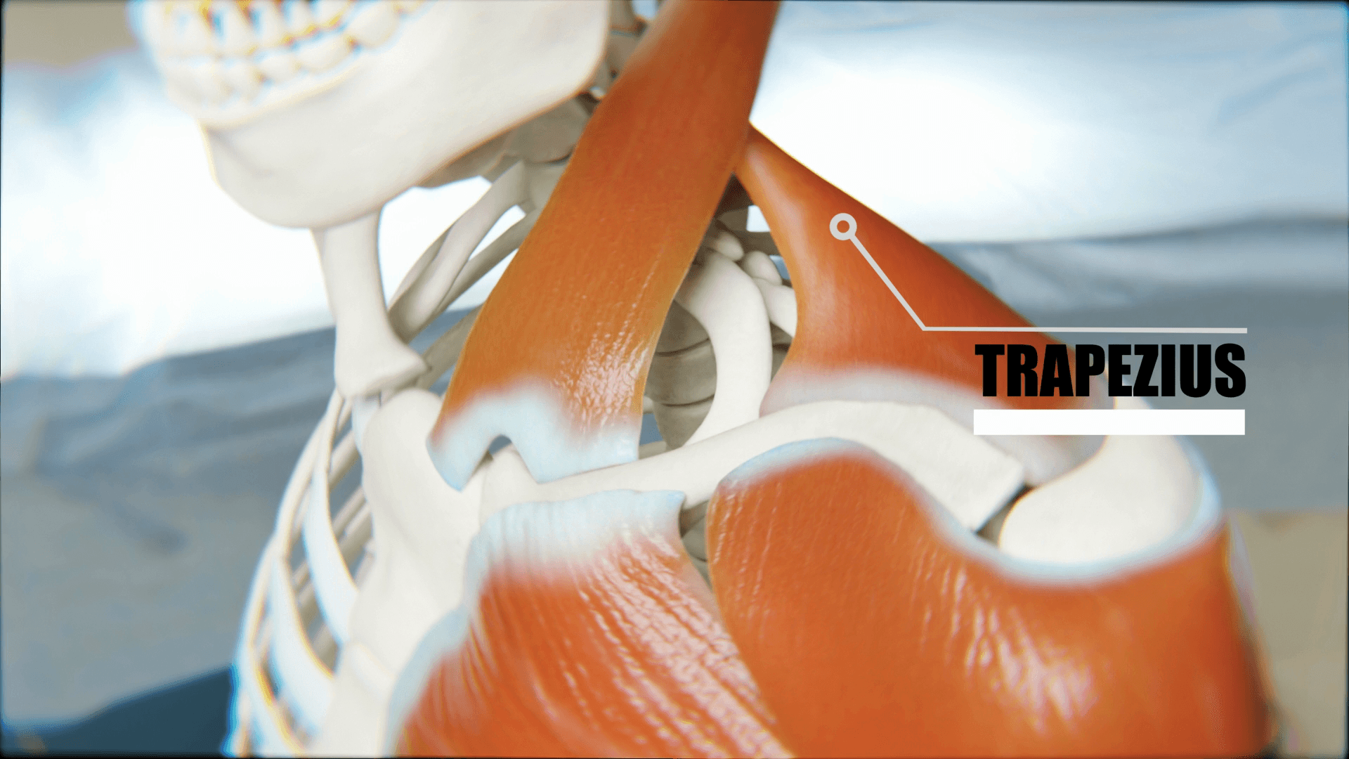
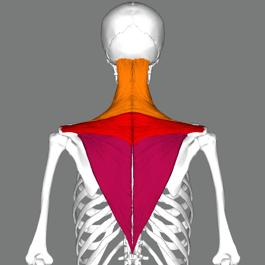
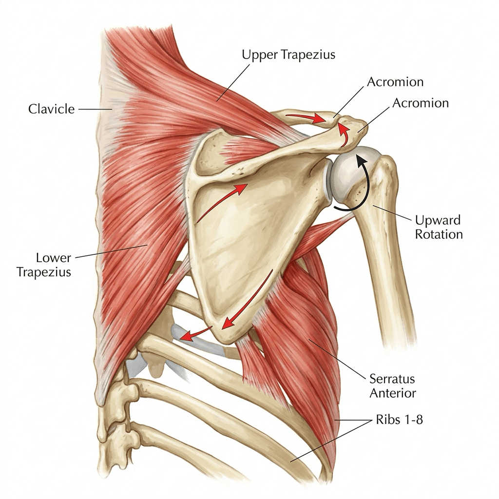
The trapezius is the largest and most superficial muscle of the posterior shoulder girdle, a broad diamond spanning from the external occipital protuberance to T12 and converging laterally onto the clavicle, acromion and scapular spine. Functionally it is not one muscle but three muscles sharing a nerve: a descending (upper) part that suspends the girdle, a transverse (middle) part that retracts it, and an ascending (lower) part that depresses the medial scapular spine and thereby upwardly rotates the glenoid.
Surgically, the trapezius matters for four reasons. It is the roof of the posterior triangle and therefore the muscle whose nerve is at risk in every neck node biopsy. Its palsy is the commonest cause of lateral scapular winging, a diagnosis routinely missed for months. It is a donor - lower trapezius to the infraspinatus footprint for irreparable posterosuperior cuff tears, upper trapezius to the proximal humerus for deltoid paralysis. And it is the muscle that must be split, elevated or reflected in almost every posterior approach to the cervicothoracic spine and scapula.
Elevation of the arm above shoulder height requires roughly 60 degrees of scapular upward rotation contributing to the 150 to 180 degrees of total elevation - the scapulohumeral rhythm. That rotation is produced by a three-muscle force couple acting about an instantaneous axis that migrates from near the root of the scapular spine early in elevation to near the acromioclavicular joint late in elevation:
- Upper trapezius pulls the lateral clavicle and acromion up and medially.
- Lower trapezius pulls the medial end of the scapular spine down and medially.
- Serratus anterior (especially the lower digitations) pulls the inferior angle laterally and forward around the chest wall.
The two trapezius vectors are opposed in the vertical plane, so their vertical components cancel and their rotatory moments add - the definition of a force couple. Take away the trapezius and the couple becomes a single muscle pulling the scapula forward: the inferior angle drifts laterally, the acromion drops, and the glenoid loses the upward tilt that keeps the deltoid vector efficient. That single mechanical statement explains lateral winging, the drooped shoulder, the loss of the last 40 degrees of abduction, and the traction pain over the brachial plexus.
ERDTrapezius Parts and Their Actions
Hook:Top to bottom: Elevate, Retract, Depress. Upper and lower together upwardly rotate.
Attachments, Innervation and Relations
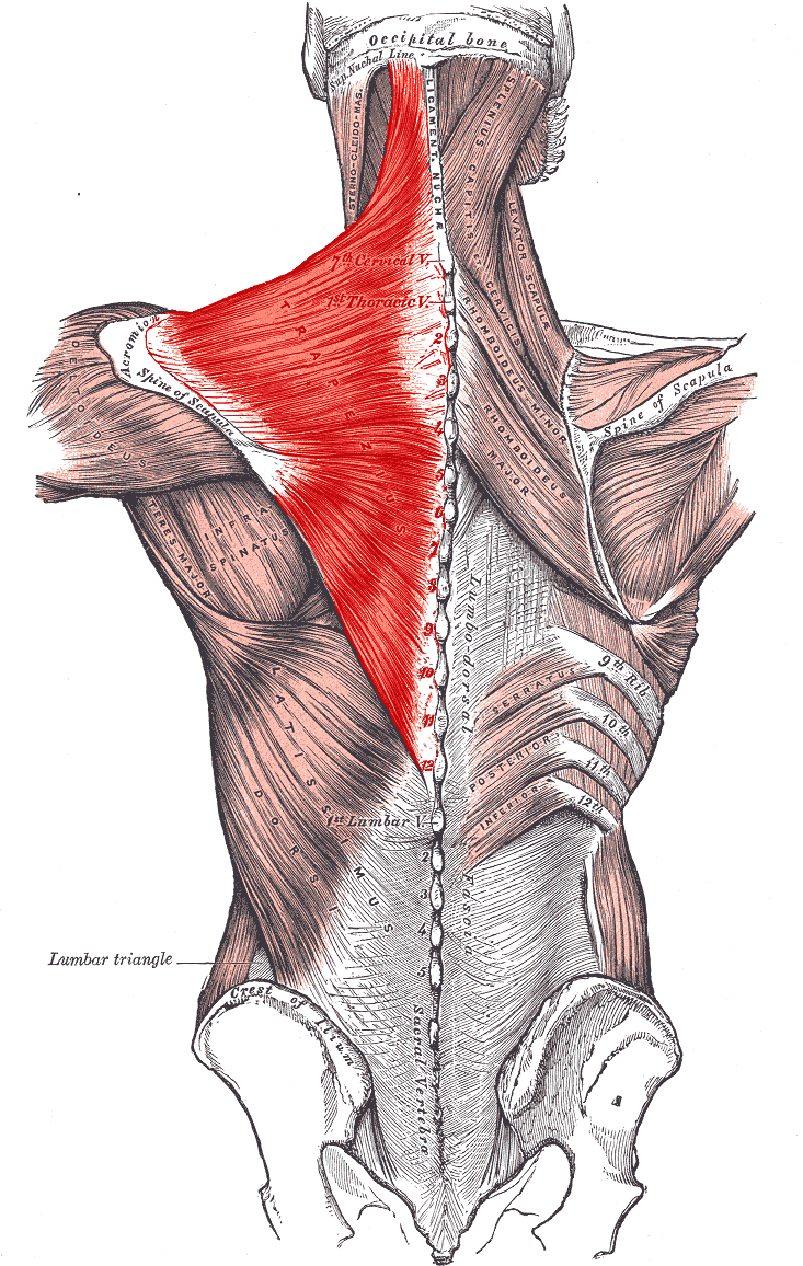
Origin (medial, continuous from occiput to T12)
- Medial third of the superior nuchal line and the external occipital protuberance.
- Ligamentum nuchae (spinous processes C1 to C6 by proxy).
- Spinous process of C7 and the spinous processes of all twelve thoracic vertebrae, plus their supraspinous ligaments.
- Around the C7-T3 level the aponeurotic origin widens into a diamond-shaped tendinous sheet shared with the opposite side - the trapezial aponeurosis. This is the thickest, most reliable tissue for suture purchase in a medial repair.
Insertion (lateral, three distinct footprints)
- Upper part: posterior border of the lateral third of the clavicle. This is the only part of the trapezius attached to the clavicle, and the reason the shoulder drops when it fails.
- Middle part: medial margin of the acromion and the superior lip of the crest of the scapular spine.
- Lower part: converges into an aponeurosis that glides over a small bursa at the medial end of the scapular spine and inserts into the tubercle at the apex of the medial (deltoid) end of the scapular spine.
Practical points
- The upper and middle parts share the acromioclavicular region with the deltoid origin; the two muscles are continuous in a common fascial sleeve, which is exploited in the trapezius transfer for deltoid paralysis.
- The inferior angle of the scapula receives no trapezius fibres - it is controlled entirely by serratus anterior and rhomboids.
- The free anterior border of the upper trapezius forms the posterior boundary of the posterior triangle of the neck and is the landmark by which the surgeon judges how far posteriorly a node biopsy may safely go.
Superficial, small, unprotected.
- CN XI has no artery running with it and no fascial tunnel to mark it.
- It sits in the same fat pad as the level V nodes being biopsied.
- It is frequently mistaken for a small vessel, a lymphatic or a fibrous band and divided with diathermy.
- Posterior triangle lymph node biopsy is the single most common cause of iatrogenic accessory nerve palsy.
Rules that actually work.
- Perform node biopsies in the posterior triangle under general anaesthesia with a nerve stimulator, not under local in clinic.
- Identify the great auricular nerve first; CN XI lies about 1 cm cranial to it on the posterior border of sternocleidomastoid.
- Use blunt spreading parallel to the nerve's oblique course; no monopolar diathermy in the fat pad.
- If a node is fixed or deep, convert to a formal exploration with proximal identification rather than blind excision.
Action and Biomechanics
Actions by part
- Line of Pull
- Supero-medial on lateral clavicle
- Primary Action
- Elevates and suspends the girdle; contributes to upward rotation via the clavicle
- Failure Consequence
- Shoulder droop, traction plexopathy, asymmetric neckline
- Line of Pull
- Horizontal, medial on acromion and spine
- Primary Action
- Retracts the scapula; stabilises against protraction
- Failure Consequence
- Protracted, forward-slumped scapula; weak horizontal abduction
- Line of Pull
- Infero-medial on medial scapular spine
- Primary Action
- Depresses the medial spine, producing upward rotation and posterior tilt
- Failure Consequence
- Loss of terminal abduction, anterior tilt, subacromial narrowing
- Line of Pull
- Combined
- Primary Action
- Upward rotation and external rotation of the scapula; posterior tilt
- Failure Consequence
- Lateral winging with the inferior angle displaced laterally
Moment arms and length-tension
- The lower trapezius has the longest upward-rotation moment arm of the three parts, because its insertion at the medial end of the scapular spine is furthest from the rotation axis. Its moment arm increases with elevation, so it is a late-range rotator.
- The upper trapezius moment arm is small in the first 30 degrees and grows once the clavicle begins to elevate and retract at the sternoclavicular joint. This is why isolated upper trapezius overactivity in a patient with lower trapezius weakness produces the classic shoulder-hitch compensation rather than true upward rotation.
- The muscle also externally rotates and posteriorly tilts the scapula. Posterior tilt is what lifts the anterior acromion away from the cuff; loss of it is a mechanical contributor to secondary subacromial impingement in the overhead athlete.
Synergists and antagonists
- Synergists for upward rotation: serratus anterior (all digitations, especially the lower four).
- Antagonists / downward rotators: levator scapulae, rhomboids, pectoralis minor, latissimus dorsi.
- Antagonist for retraction: serratus anterior and pectoralis minor.
What happens when it fails
Complete trapezius palsy produces a predictable chain: the clavicle loses its only posterior suspensory muscle, so the whole girdle drops; the scapula translates laterally and downward and rotates downward; the inferior angle swings laterally away from the midline (lateral winging); the glenoid tilts inferiorly; deltoid works at a mechanical disadvantage so abduction beyond 90 degrees fails; and the drooping girdle puts the brachial plexus on stretch, giving a deep, dragging shoulder and neck pain that is often the dominant complaint.
- Trapezius palsy (CN XI)
- Laterally
- Serratus palsy (long thoracic)
- Medially
- Trapezius palsy (CN XI)
- Depressed
- Serratus palsy (long thoracic)
- Elevated, prominent
- Trapezius palsy (CN XI)
- Drooped
- Serratus palsy (long thoracic)
- Normal or elevated
- Trapezius palsy (CN XI)
- Abduction in the coronal plane
- Serratus palsy (long thoracic)
- Forward flexion / wall push
- Trapezius palsy (CN XI)
- Neck node biopsy, neck dissection
- Serratus palsy (long thoracic)
- Viral illness, backpack, chest tube, first rib resection
- Trapezius palsy (CN XI)
- Asymmetric, hollow trapezial ridge
- Serratus palsy (long thoracic)
- Normal
The two provocative tests are complementary and both must be performed: wall push-up for serratus, resisted abduction for trapezius. Doing only the wall push and calling a normal test "no winging" is the classic examination failure.
Surface Anatomy and Examination
Palpation and positioning
- Ask the patient to shrug against resistance: the upper trapezius stands out as a ridge from the neck to the lateral clavicle. Compare the two sides from behind, with the patient standing and the shirt off to the waist.
- Middle fibres: with the arms at 90 degrees of horizontal abduction, ask for retraction; palpate the transverse band between the T1-T3 spinous processes and the scapular spine.
- Lower fibres: prone, arm elevated to 130 degrees in the plane of the scapula with the thumb up, ask the patient to lift the arm off the couch; the ascending fibres tighten as a V converging on the medial scapular spine.
The isolation tests
Isolates the upper (descending) fibres.
- Examiner stands behind; hands on the top of both shoulders.
- Patient shrugs maximally against downward resistance.
- Positive: reduced shrug height, absent trapezial ridge, flat supraspinous contour.
- Means: accessory nerve palsy until proven otherwise after neck surgery.
The trapezius winging manoeuvre.
- Patient abducts in the coronal plane against light resistance; watch the medial border throughout the arc.
- Positive: the inferior angle rotates laterally, the medial border lifts, the shoulder sits low.
- Means: lateral winging of trapezius insufficiency, not the medial winging of serratus palsy.
Grades the ascending fibres alone.
- Prone, arm elevated to about 130 degrees in the plane of the scapula, thumb up; patient lifts and holds against pressure at the elbow.
- Positive: cannot hold the arm off the couch, or substitutes by lifting the medial border with rhomboids and latissimus.
- Means: isolated lower trapezius weakness - scapular dyskinesis in the overhead athlete, or a partial CN XI lesion.
Performed to exclude the other diagnosis.
- Patient pushes against a wall with the elbows extended.
- Positive: medial border and inferior angle lift medially - serratus anterior palsy.
- Means: if the wall push is normal but abduction wings, the lesion is trapezius, not serratus.
Grading and pitfalls
- Grade with MRC power grading of the shrug, but record the height difference in centimetres as well; MRC grading of a shrug is notoriously insensitive because levator scapulae and rhomboids substitute.
- False positives: pain inhibition after acromioclavicular injury, a stiff cervical spine, and voluntary winging in generalised laxity.
- False negatives: partial CN XI lesions preserving the upper fibres (proximal C3-C4 contribution), and a well-compensated patient who has learned to hitch the shoulder. Ask the patient to hold an abducted position for 30 seconds - fatigue unmasks the winging.
- Always test both patterns. A patient sent with "winging" and a normal wall push is not a normal patient; they may have trapezius palsy, rhomboid palsy or a bony/soft-tissue cause of pseudo-winging (osteochondroma of the scapular undersurface, scapulothoracic bursitis).
Complications
Iatrogenic accessory nerve injury
- Mechanism: transection, diathermy, traction from vigorous retraction, or entrapment in a suture during closure of the posterior triangle.
- Avoidance: operate on posterior triangle nodes in theatre; identify the great auricular nerve; blunt dissection only; use a stimulator; never use monopolar diathermy in the fat pad; if the nerve is inadvertently divided and recognised, repair it immediately - a primary repair at the index operation is worth far more than any later reconstruction.
- Recognition: any patient with a new shoulder droop or shrug weakness after neck surgery has an accessory palsy until EMG says otherwise.
Donor-site morbidity after transfer
- Eden-Lange: loss of the levator and rhomboids as independent stabilisers; if the dorsal scapular nerve is subsequently injured the patient has no scapular stabilisers at all. Recurrence is uncommon where the trapezius palsy was isolated, but is the expected outcome if a coexisting long thoracic or dorsal scapular lesion was missed before surgery.
- Lower trapezius transfer: weakness of scapular retraction and depression; a seroma in the interscapular dead space is common; injury to the spinal accessory nerve during medial harvest is the catastrophic complication because it destroys both the donor and the remaining trapezius.
- Upper trapezius transfer for deltoid palsy: persistent shoulder droop, hardware prominence at the humeral bone block, and failure of the interposition graft at its junctions.
Nerve and vessel injury in trapezius-related approaches
- Suprascapular nerve at the notch during elevation of the trapezius from the scapular spine - stay lateral to a point 2 to 3 cm medial to the acromioclavicular joint along the spine, or identify and protect the nerve.
- Dorsal scapular nerve and artery 1 to 2 cm medial to the medial scapular border during any interscapular dissection - divide the rhomboids under direct vision, from lateral to medial, and control the artery before it retracts.
- Pneumothorax during scapulothoracic fusion or deep interscapular dissection at the level of ribs 3 to 7 - request a post-operative chest radiograph as routine.
Failure of scapular reconstruction
- Recurrent winging after transfer usually reflects an unrecognised second palsy (serratus anterior or rhomboid) or a fixed contracture that was never addressed. In the long-term series both poor results occurred in patients with a concomitant long thoracic and dorsal scapular nerve lesion, not from failure of the transfer itself.
- Secondary adhesive capsulitis and subacromial impingement develop in the neglected palsy because the scapula stops rotating; both must be treated (physiotherapy, occasionally arthroscopic release) before or alongside any reconstruction.
Clinical Relevance
Spinal accessory nerve palsy
Iatrogenic (the great majority)
- Posterior triangle lymph node biopsy - the commonest single cause. Typically an excision biopsy done for lymphadenopathy under local anaesthesia, without nerve monitoring, by a surgeon not expecting the nerve.
- Radical and modified radical neck dissection - classical radical neck dissection sacrifices CN XI by design; modified radical and selective dissections preserve it but still cause a traction neurapraxia in a substantial proportion ("shoulder syndrome" after neck dissection).
- Carotid endarterectomy, internal jugular vein cannulation, cervical rib resection, brachial plexus exploration.
- Facelift and parotid surgery where dissection strays into the upper posterior triangle.
Non-iatrogenic
- Blunt or penetrating trauma to the posterior triangle, including seat-belt and rugby injuries.
- Traction injury from a fall onto the shoulder with the head forced to the opposite side.
- Parsonage-Turner syndrome (neuralgic amyotrophy) - CN XI involvement is uncommon but described; look for other patchy palsies (long thoracic, suprascapular, anterior interosseous).
- Radiotherapy to the neck - late fibrotic neuropathy, often years afterwards.
- Skull base tumours and jugular foramen lesions - here CN IX, X and XI fail together (Vernet syndrome), so ask about swallowing and voice.
An accessory nerve divided at biopsy will not recover spontaneously. Early exploration with neurolysis, direct repair or interposition grafting within 6 months gives markedly better trapezius recovery than late surgery; beyond about 12 to 20 months the motor endplates and muscle are no longer salvageable and the operation becomes a tendon transfer, not a nerve operation. If EMG at three months shows no reinnervation in a patient whose nerve was in the operative field, refer immediately - do not "give it another six months".
Other trapezius pathology
- Trapezius myalgia: the commonest painful condition of the muscle. Sustained low-level activation of the upper fibres, typically occupational; treat with load modification and scapular endurance work, not injection.
- Trapezius rupture and avulsion: rare, described after a direct blow or a violent forced depression of the shoulder. Presents as an acute defect with a step in the muscle contour and can mimic palsy; ultrasound distinguishes them.
- Congenital absence or hypoplasia of part of the muscle - usually the lower fibres, and often asymptomatic. Associated with Poland syndrome and with Sprengel deformity, where the trapezius may be partly replaced by the fibrous or bony omovertebral bar.
- Klippel-Feil and Sprengel deformity: the trapezius is abnormal in both; scapular elevation and restricted abduction dominate the picture, and the Woodward procedure detaches and repositions the trapezius (with the rhomboids) caudally on the spinous processes.
Surgical Relevance
Approaches that use or divide the trapezius
- What Happens to Trapezius
- Fascial roof incised over level V fat
- Structure at Risk
- Spinal accessory nerve
- Distance from Landmark
- Emerges 1 cm cranial to great auricular nerve on the posterior border of sternocleidomastoid; enters trapezius 2-5 cm above the clavicle
- What Happens to Trapezius
- Deltoid and trapezius elevated off the scapular spine
- Structure at Risk
- Suprascapular nerve at the notch
- Distance from Landmark
- 2-3 cm medial to the base of the coracoid, under the transverse scapular ligament
- What Happens to Trapezius
- Split in the midline raphe, subperiosteal elevation
- Structure at Risk
- Dorsal scapular nerve laterally; CN XI on the deep surface
- Distance from Landmark
- Keep subperiosteal on the spinous processes; do not stray more than 4 cm lateral to the midline at C7-T1
- What Happens to Trapezius
- Lateral border of trapezius retracted medially
- Structure at Risk
- Dorsal scapular artery in the rhomboid layer
- Distance from Landmark
- 1-2 cm medial to the medial scapular border
- What Happens to Trapezius
- Trapezius split with deltoid over the acromion
- Structure at Risk
- Suprascapular nerve; CN XI terminal branches
- Distance from Landmark
- Suprascapular notch 2-3 cm medial to the acromioclavicular joint along the scapular spine
- What Happens to Trapezius
- Trapezius and rhomboids elevated from the medial border
- Structure at Risk
- Dorsal scapular vessels; pleura beneath the ribs
- Distance from Landmark
- Wires or plate around ribs 3-7; the pleura is millimetres deep to the rib
Nerve surgery for accessory palsy
- Neurolysis for a nerve in continuity with a conduction block and preserved motor units - the commonest finding at early exploration after a biopsy.
- Direct end-to-end repair where the nerve has been cleanly divided and the gap is short; the posterior triangle allows a few millimetres of mobilisation only.
- Interposition sural nerve grafting for a gap greater than about 1 cm or a dense neuroma-in-continuity. Trapezius reinnervation after grafting is good in the upper fibres and less reliable in the lower.
- Nerve transfer options where the proximal stump is unavailable, including branches of the cervical plexus.
Tendon transfer for irreparable trapezius palsy - the Eden-Lange procedure
The concept
Eden-Lange substitutes for each of the three parts of the trapezius by moving the surviving scapular stabilisers laterally, so that their line of pull becomes a stabilising and rotating vector rather than a purely medial one. All three donor muscles are innervated by the dorsal scapular nerve (levator scapulae also by C3-C4), so the transfer is only viable if the dorsal scapular nerve is intact - confirm this on EMG before committing.
The three limbs
- Levator scapulae is detached with a wafer of bone from the superior angle and transferred laterally onto the scapular spine, substituting for the upper trapezius.
- Rhomboid minor is detached from the medial border and transferred laterally onto the infraspinatus fossa, substituting for the middle trapezius.
- Rhomboid major is detached and transferred laterally onto the infraspinatus fossa, below the rhomboid minor, substituting for the lower trapezius.
Fixation
Each tendon is passed through a subperiosteal tunnel or a bone trough on the dorsal scapular surface and fixed with transosseous sutures or suture anchors through drill holes, with the arm held in about 30 degrees of abduction so the transfers are tensioned in a shortened position.
The trapezius as a donor
Indication
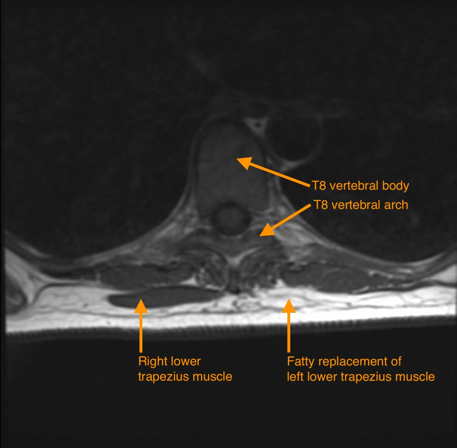
Irreparable axillary nerve palsy with a flail deltoid, in a patient with an intact cuff, a supple shoulder and a normally functioning trapezius (CN XI intact). Typical setting: a late-presenting axillary nerve injury after anterior shoulder dislocation or a proximal humeral fracture, or a failed nerve transfer, where nerve reconstruction is no longer an option (usually more than 12 months from injury or with a fatty-infiltrated deltoid on MRI).
Technique (Bateman / Saha type transfer)
- Sabre-cut or strap incision over the lateral clavicle, acromion and scapular spine.
- The trapezius insertion is mobilised together with a block of bone, classically an osteotomised segment of the lateral clavicle and acromion, preserving the continuity of the muscle with its bony insertion.
- The bone block with the attached trapezius is transferred distally onto the lateral aspect of the proximal humerus, at or just distal to the level of the deltoid insertion, and fixed with screws through the bone block.
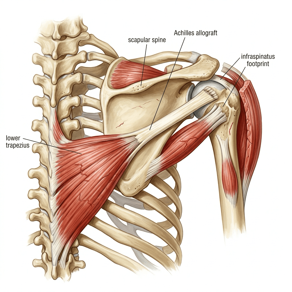
- Length is frequently insufficient, so an interposition graft (fascia lata, Achilles allograft or a tendon graft) is used to bridge the trapezius to the humerus, tensioned with the arm in 60 to 90 degrees of abduction.
- Immobilise in an abduction brace at 60 to 80 degrees for 6 weeks, then progressive weaning over 3 months.
Realistic outcome
Restores active abduction to around 60 to 90 degrees with reasonable pain relief; it is a salvage of function, not a restoration of normal deltoid. Compare with the alternatives: latissimus dorsi and teres major bipolar transfer (L'Episcopo type) to the proximal humerus, and arthrodesis in the manual labourer.
Guidelines, Registries & Global Practice
Anatomical variation
- The point at which CN XI crosses the posterior border of sternocleidomastoid varies between roughly the upper third and the midpoint of the border across cadaveric series; the constant relationship is its position cranial to the great auricular nerve, which is why the great auricular nerve, not a fixed distance from the clavicle, is the landmark taught.
- The nerve pierces sternocleidomastoid rather than passing deep to it in a minority of individuals, changing the level at which it appears in the triangle.
- A dominant dorsal scapular artery arising directly from the subclavian artery rather than as the deep branch of the transverse cervical artery is present in a substantial minority of shoulders and alters flap planning.
- Partial congenital absence of the lower trapezius is described and is usually asymptomatic; it matters if that patient is being considered as a lower trapezius transfer candidate.
Practice framing across bodies
- Position Relevant to the Trapezius and CN XI
- Posterior triangle node biopsy should be performed in theatre with formal identification of the accessory nerve, not as an office procedure under local anaesthesia.
- Position Relevant to the Trapezius and CN XI
- Recognise trapezius palsy as a distinct and frequently delayed diagnosis; recommend both winging tests in the assessment of any scapular dyskinesis.
- Position Relevant to the Trapezius and CN XI
- Support early referral of suspected iatrogenic accessory nerve palsy to a peripheral nerve or shoulder service, with EMG by three months.
- Position Relevant to the Trapezius and CN XI
- Explore lesions that fail to show reinnervation by three to six months; nerve reconstruction yields diminishing returns after about 12 months.
Resource-dependent practice
- Well-resourced settings: intra-operative nerve monitoring for neck dissection, MRI quantification of trapezius fatty infiltration, and access to Achilles allograft for extending a lower trapezius transfer.
- Limited-resource settings: clinical diagnosis remains highly reliable if both winging patterns are tested; Eden-Lange requires no implants beyond sutures and drill holes and is entirely feasible; fascia lata is a serviceable substitute for allograft in trapezius transfer.
Medicolegal dimension
Iatrogenic accessory nerve injury after node biopsy is one of the most frequently litigated peripheral nerve injuries. The two defensible practices are documented pre-operative consent naming the nerve and prompt referral once weakness is noticed - delay, not the injury itself, is what most reports criticise.
MCQ Practice Points
Q: What is the motor nerve to the trapezius, and what do C3 and C4 contribute? A: The spinal accessory nerve (CN XI). C3 and C4 ventral rami carry proprioceptive afferents and variable motor fibres, but CN XI is the clinically decisive motor supply.
Q: Where does CN XI emerge in the posterior triangle? A: From the posterior border of sternocleidomastoid at the junction of its upper and middle thirds, about 1 cm cranial to where the great auricular nerve wraps around the same border.
Q: Which way does the inferior angle move in trapezius palsy? A: Laterally - lateral winging, with a drooped shoulder, worst on resisted abduction. Serratus palsy gives medial winging on forward flexion with a normal or elevated shoulder.
Q: How far above the clavicle does CN XI enter the trapezius? A: Approximately 2 to 5 cm above the clavicle at the anterior border of the muscle.
Q: Which three muscles are transferred in the Eden-Lange procedure, and what single nerve must be intact? A: Levator scapulae, rhomboid minor and rhomboid major. All are supplied by the dorsal scapular nerve (C5), which must be confirmed intact on EMG.
Q: Why is the lower trapezius the preferred donor for restoring external rotation in an irreparable cuff tear? A: Its line of pull is almost identical to that of the infraspinatus - infero-medial in the plane of the scapula - and it is in phase with external rotation during elevation.
Q: Name the three components of the scapular upward-rotation force couple. A: Upper trapezius, lower trapezius and serratus anterior. Lower trapezius has the longest moment arm and dominates late-range rotation.
Q: What is the pedicle of the lower trapezius island flap? A: The deep branch of the transverse cervical artery (dorsal scapular artery). The upper trapezius (Conley) flap is based on the superficial branch.
Q: What forms the triangle of auscultation? A: Medial border of the scapula laterally, lateral border of trapezius medially, superior border of latissimus dorsi inferiorly, with rhomboid major in the floor.
Q: In spinal accessory to suprascapular nerve transfer, where should the accessory nerve be divided? A: Distal to the branches supplying the upper trapezius, so that shoulder suspension by the descending fibres is preserved.
Exam Viva Scenarios
Practise clinical reasoning and management decisions out loud
“A 34-year-old woman is referred with a painful right shoulder four months after excision of a lump in the right side of her neck. She cannot lift the arm above her head and the shoulder looks lower on that side. How do you assess her?”
“A 41-year-old builder has a complete trapezius palsy three years after a neck dissection. He has a painful, drooping shoulder and cannot abduct beyond 80 degrees. Passive movement is full. What can you offer him?”
“A 28-year-old man has a flail deltoid 14 months after an anterior shoulder dislocation with an axillary nerve injury. The rotator cuff is intact, the joint is supple, and EMG shows no deltoid reinnervation. He asks whether anything can be done. What are the options and where does the trapezius fit?”
Anatomy
- Origin: occiput, ligamentum nuchae, C7-T12 spinous processes
- Insert: lateral third clavicle (upper), acromion and spine (middle), medial spine tubercle (lower)
- Nerve: spinal accessory (CN XI) motor; C3-C4 proprioceptive
- Blood: transverse cervical artery - superficial branch upper, deep branch lower
Nerve Course
- Jugular foramen, deep to sternocleidomastoid, supplies it
- Emerges at junction of upper and middle thirds of SCM posterior border
- About 1 cm cranial to great auricular nerve
- Enters trapezius 2-5 cm above clavicle at its anterior border
Palsy
- Cause: posterior triangle node biopsy (commonest)
- Lateral winging - inferior angle moves laterally
- Drooped shoulder, weak shrug, loss of abduction past 90 degrees
- EMG at 3 months; explore by 6 months
Reconstruction
- Eden-Lange: levator to spine, rhomboid minor and major to infraspinatus fossa
- Modified: lateralise the levator for a longer moment arm
- Bateman upper trapezius transfer for deltoid paralysis
- Lower trapezius plus Achilles allograft to greater tuberosity for irreparable cuff
Evidence Base
Surgical Outcomes of 111 Spinal Accessory Nerve Injuries
- Retrospective review of 111 surgical repairs of the spinal accessory nerve over 23 years, mean follow-up 25.6 months
- 103 of 111 injuries (93 per cent) were iatrogenic, and 82 of those (80 per cent) followed lymph node biopsy
- Graft repair was the commonest procedure (58 patients), with end-to-end repair in 26 and neurolysis in 19
- More than 95 per cent of patients treated by neurolysis supported by positive nerve action potential recordings improved to Grade 4 or higher
- Of 84 patients repaired by graft or suture, most achieved favourable functional recovery
Transfer of the Levator Scapulae, Rhomboid Major and Rhomboid Minor for Paralysis of the Trapezius
- 22 patients with trapezius paralysis from spinal accessory nerve injury underwent transfer of levator scapulae and both rhomboids
- Aetiology was cervical node biopsy in 13, trauma in 7 and radical neck dissection in 2
- All had pain, neckline asymmetry, a drooping girdle with lateral displacement of the scapula and weak active elevation
- 14 patients had been given an incorrect clinical diagnosis and 12 had had an inaccurate or incomplete electromyographic examination
- EVERY PATIENT HAD ALREADY FAILED either physical therapy or an operative attempt at neurolysis or nerve reconstruction, so this is a salvage cohort and its results should not be read as first-line outcomes
- A LONG THORACIC NERVE PALSY DEVELOPED IN THREE PATIENTS after the transfer - a complication that matters particularly here, because Romero and Gerber found concomitant long thoracic lesions to be the strongest predictor of a poor Eden-Lange result
- At a mean of 7.5 years (range 2 to 14) the result was excellent in 13, satisfactory in 6 and unsatisfactory in 3, with adequate pain relief and functional improvement in all but 3
Levator Scapulae and Rhomboid Transfer for Paralysis of Trapezius - The Eden-Lange Procedure
- 16 patients with chronic trapezius paralysis reviewed; 4 treated conservatively regained no satisfactory function, although 2 became pain-free
- 12 underwent Eden-Lange transfer, with the levator scapulae taken to the ACROMION and the rhomboids to the infraspinatus fossa
- At a mean follow-up of 32 years the Constant-score outcome was excellent in 9, fair in 2 and poor in 1; pain was adequately relieved in 11 and overhead function restored in 9
- Both poor outcomes had a concomitant long thoracic and dorsal scapular nerve lesion
- The authors concluded the procedure gives very satisfactory long-term results for ISOLATED trapezius paralysis, and is less successful where there is additional serratus anterior palsy or weak rhomboids
Outcome of Triple-Tendon Transfer, an Eden-Lange Variant, to Reconstruct Trapezius Paralysis
- 22 patients with persistent trapezius paralysis after spinal accessory nerve injury underwent a triple-tendon transfer
- The levator scapulae was taken to the LATERAL aspect of the scapular spine, rhomboid minor just medial to it, and rhomboid major to the medial spine, each with its bony insertion
- At a mean of 35 months winging was corrected in every patient with improved shoulder asymmetry
- Active abduction improved from a mean of 71 to 118 degrees and flexion from 102 to 150 degrees, with significant gains in Constant score, subjective shoulder value and DASH
Outcome of Lower Trapezius Transfer to Reconstruct Massive Irreparable Posterosuperior Rotator Cuff Tear
- 33 patients, mean age 53 years, with massive irreparable posterosuperior cuff tears reconstructed by lower trapezius transfer prolonged with Achilles tendon allograft
- At a mean of 47 months, 32 patients had significant improvement in pain, subjective shoulder value, DASH and range of movement
- Mean postoperative flexion was 120 degrees, abduction 90 degrees and external rotation 50 degrees
- Patients with more than 60 degrees of preoperative flexion made the greatest gains, while external rotation improved in ALL patients regardless of preoperative loss
Shoulder Disability after Different Selective Neck Dissections (Levels II-IV versus Levels II-V)
- 40 patients matched into two groups of 20, comparing selective neck dissection of levels II-IV with levels II-V, with the accessory nerve anatomically preserved
- Shoulder droop was present in 30 per cent of the level II-V group versus 5 per cent of the level II-IV group, and half the level II-V group had abnormal abduction with contralateral head rotation
- Electromyographic abnormalities were far commoner after level II-V dissection (85 versus 40 per cent), with upper trapezius abnormalities in 85 versus 20 per cent
- The authors concluded that clearance of the posterior triangle increases shoulder morbidity, but that subclinical nerve impairment occurs even after level II-IV dissection if the submuscular recess is routinely dissected