Calcium Homeostasis & Metabolic Bone Disease
Overview
Serum calcium is held in a very narrow range (about 2.2 to 2.6 mmol/L) because it is essential for nerve and muscle function, clotting, and as a building block of bone. The body achieves this with three hormones acting on three organs: parathyroid hormone, active vitamin D (calcitriol) and calcitonin acting on bone, gut and kidney.
Bone is both the largest calcium store and a tissue under constant remodelling, so calcium balance and skeletal health are inseparable. Metabolic bone disease is what appears when this system goes wrong or when bone turnover itself is disordered. This topic builds the calcium axis first, then uses it to make sense of each disease and its biochemistry.
Physiology of the Calcium Control System
Where the calcium is. About 99 percent of body calcium is stored in bone as hydroxyapatite. The small fraction in blood exists in three forms: ionised (the active, regulated fraction, about half), protein-bound (mostly to albumin), and complexed with anions.
Why we correct for albumin. Only ionised calcium is biologically active, so a low albumin lowers total calcium without making the patient hypocalcaemic. Either correct calcium for albumin or measure the ionised fraction directly.
- Source
- Parathyroid chief cells
- Main actions
- Resorbs bone, increases renal calcium reabsorption, loses phosphate, activates vitamin D in kidney
- Net effect on calcium
- Raises calcium
- Source
- Kidney (final step)
- Main actions
- Increases gut absorption of calcium and phosphate, supports bone mineralisation
- Net effect on calcium
- Raises calcium and phosphate
- Source
- Thyroid C cells
- Main actions
- Inhibits osteoclasts
- Net effect on calcium
- Lowers calcium (minor role in humans)
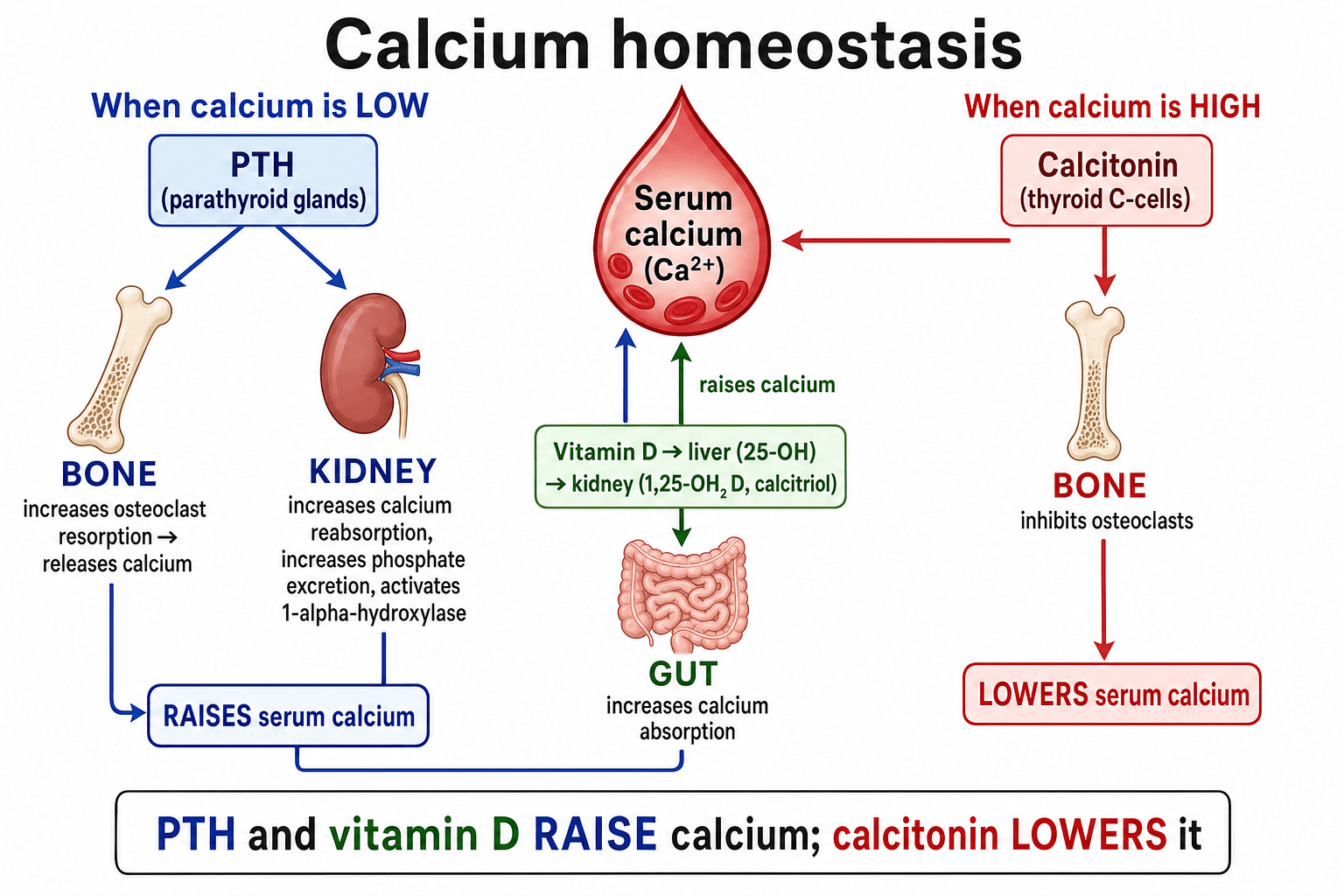
The feedback loop. A fall in ionised calcium is sensed by the calcium-sensing receptor on parathyroid cells, which release PTH. PTH stimulates osteoclastic resorption in bone (indirectly, through RANKL on osteoblasts), makes the kidney reabsorb calcium while excreting phosphate, and switches on renal 1-alpha-hydroxylase to convert 25-OH-D to calcitriol, which increases gut absorption of calcium. As calcium rises, PTH secretion is suppressed - a classic negative feedback loop.
Phosphate runs the other way. PTH is phosphaturic, so a high PTH tends to give a high calcium and a low phosphate, while calcitriol raises both. Tracking calcium, phosphate, PTH and alkaline phosphatase together is what lets you read any metabolic bone picture.
The hormone the classic teaching leaves out. "Three hormones" handles calcium well but understates phosphate, which is governed chiefly by fibroblast growth factor 23 (FGF23) — a hormone examiners increasingly ask about. It is secreted by osteocytes and osteoblasts in response to a phosphate load and to calcitriol, and acts on the kidney using the obligatory co-receptor Klotho.
What it does. Its two actions both lower phosphate and lower calcitriol: it is strongly phosphaturic (it down-regulates the proximal-tubule sodium-phosphate cotransporters NaPi-IIa and NaPi-IIc) and it suppresses renal 1-alpha-hydroxylase while up-regulating 24-hydroxylase. So it opposes PTH on vitamin D activation but shares PTH's phosphaturic effect.
Where it shows up clinically. FGF23 rises very early in chronic kidney disease, often the first measurable abnormality of CKD-MBD before phosphate or PTH change. Excess FGF23 drives the hereditary and tumour-induced phosphate-wasting forms of rickets and osteomalacia, while loss of FGF23 activity instead causes hyperphosphataemic tumoral calcinosis.
The Vitamin D Pathway
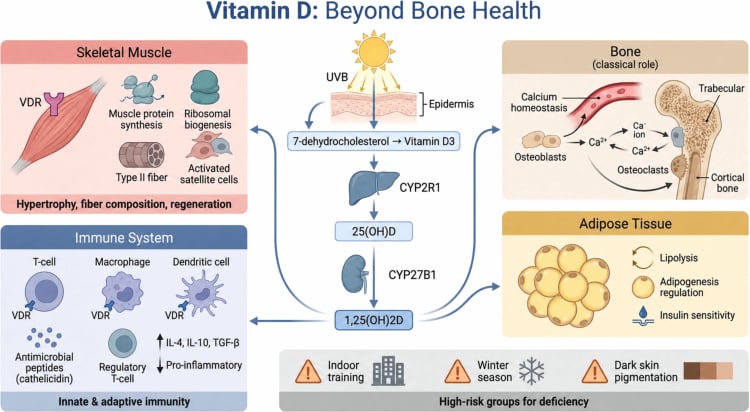
The precursor. Vitamin D begins as an inactive precursor: cholecalciferol (D3) made in skin under UVB light or taken in the diet, and ergocalciferol (D2) from plants. It then passes through two hydroxylation steps, in two different organs.
- Liver: adds a hydroxyl at carbon 25 to make 25-hydroxyvitamin D (25-OH-D). This is the stable, abundant storage form and is the one measured to assess vitamin D status.
- Kidney: adds a hydroxyl at carbon 1 (by 1-alpha-hydroxylase, stimulated by PTH and low phosphate) to make active 1,25-dihydroxyvitamin D (calcitriol).
What calcitriol then does. It increases intestinal absorption of calcium and phosphate, supports normal bone mineralisation, and feeds back to suppress PTH.
Where the pathway breaks. Two clinical consequences follow directly from the two-organ activation: liver disease impairs the first step, and kidney disease impairs the activating second step, so chronic kidney disease produces low calcitriol, low calcium and secondary hyperparathyroidism (renal osteodystrophy).
Measure 25-OH-D to judge vitamin D status, not calcitriol. Calcitriol has a short half-life and can be kept normal by a high PTH even when stores are low, so it is a poor marker of deficiency.
The Metabolic Bone Diseases
Each pattern below has a page of its own, and they are best learnt as a set because the biochemistry is what separates them: osteoporosis is all-normal chemistry with low bone density; osteomalacia and its childhood form rickets are a mineralisation defect with low phosphate, high alkaline phosphatase and secondary hyperparathyroidism, usually from vitamin D deficiency; primary hyperparathyroidism is a high calcium with an inappropriately unsuppressed PTH; and Paget disease is a very high alkaline phosphatase with normal calcium and phosphate. Renal osteodystrophy is the one that breaks the pattern, because the kidney is both the organ that activates vitamin D and the organ that excretes phosphate. The cellular machinery all of this regulates is bone remodeling.
Reading the biochemistry
The panel to read is calcium, phosphate, PTH and alkaline phosphatase, and each disease has a signature on it.
- Calcium
- Normal
- Phosphate
- Normal
- PTH
- Normal
- Alk phos
- Normal
- Calcium
- Low or low-normal
- Phosphate
- Low
- PTH
- High (secondary)
- Alk phos
- High
- Calcium
- High
- Phosphate
- Low
- PTH
- High or inappropriately normal
- Alk phos
- High or normal
- Calcium
- Low or normal
- Phosphate
- High
- PTH
- High
- Alk phos
- High
- Calcium
- Normal
- Phosphate
- Normal
- PTH
- Normal
- Alk phos
- Very high (isolated)
Osteoporosis
What it is. Osteoporosis is reduced bone mass with deteriorated microarchitecture, so bone is normal in composition but there is simply less of it, raising fragility-fracture risk. Mineralisation and routine biochemistry are normal, which is the key distinction from osteomalacia.
How it is diagnosed. By DXA (a T-score of -2.5 or lower at hip or spine) or by a low-trauma fragility fracture.
How it is treated. Lifestyle measures, calcium and vitamin D sufficiency, and bone-active drugs: anti-resorptives such as bisphosphonates and denosumab, or anabolic agents such as teriparatide and romosozumab in high-risk patients.
Osteomalacia and Rickets
The defect. Osteomalacia is defective mineralisation of osteoid in the adult skeleton, so bone is normal in amount but poorly mineralised; in the growing child the same defect at the growth plate is called rickets.
The causes. The usual cause is vitamin D deficiency (low sunlight, poor intake, malabsorption), but it also follows chronic kidney disease, certain anticonvulsants, and rare phosphate-wasting disorders.
How it presents. Adults have bone pain, proximal muscle weakness and fractures. Children develop bowing, metaphyseal widening and growth failure.
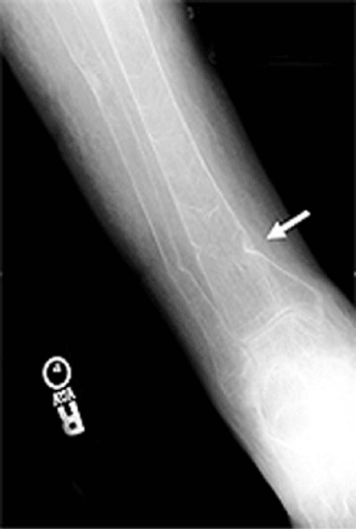
The radiographic hallmark. The Looser zone (pseudofracture) is a transverse lucent band where unmineralised osteoid has accumulated, and these sit on the concave side of the bone. Treatment is vitamin D and calcium replacement, correcting the underlying cause.
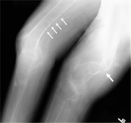
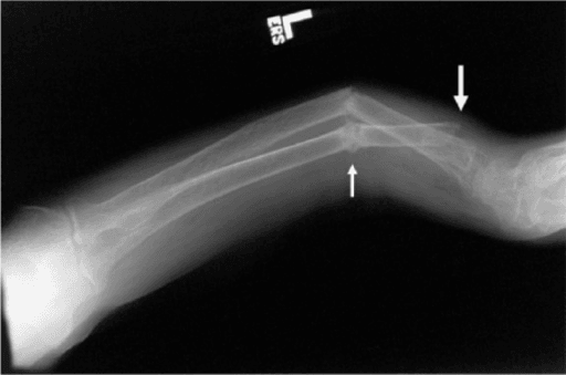
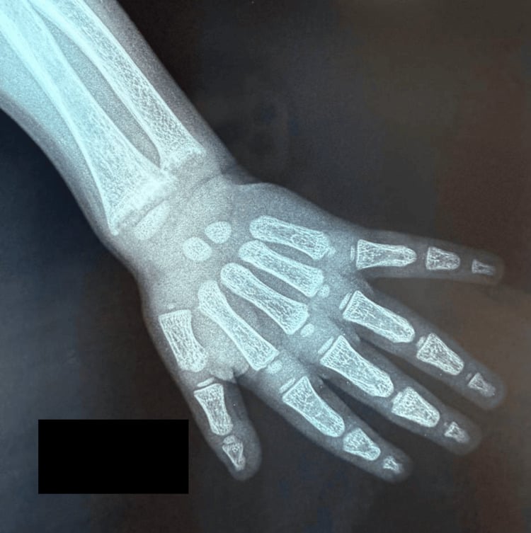
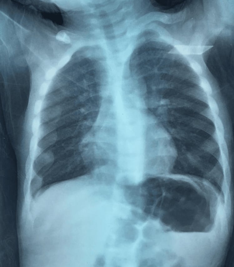
Two aetiological groups. Rickets and osteomalacia are a final common pathway of defective mineralisation, and examiners expect the aetiological split into a calcium/vitamin D group and a phosphate-wasting group.
Calcipenic, the common group. These share a biochemistry of low or low-normal calcium with a secondary high PTH and low phosphate.
- Nutritional vitamin D (or dietary calcium) deficiency
- Vitamin-D-dependent rickets type 1 — 1-alpha-hydroxylase deficiency, so 25-OH-D is normal but calcitriol is low, and it responds to calcitriol
- Vitamin-D-dependent rickets type 2 — end-organ resistance from a vitamin D receptor mutation, so calcitriol is high, classically with alopecia
Phosphopenic, from renal phosphate loss. Usually FGF23-mediated, these show normal calcium with a normal or only mildly raised PTH and an isolated low phosphate — the key discriminator from the calcipenic group.
- X-linked hypophosphataemic rickets — the commonest heritable form, a PHEX mutation raising FGF23, treated with phosphate plus calcitriol, or the anti-FGF23 antibody burosumab
- Tumour-induced (oncogenic) osteomalacia — an FGF23-secreting mesenchymal tumour, curable by excision
- Renal tubular disorders such as Fanconi syndrome and renal tubular acidosis
Hyperparathyroidism
- Mechanism
- Autonomous adenoma (most), hyperplasia or carcinoma
- Calcium
- High
- PTH
- High or inappropriately normal
- Mechanism
- Gland responds to low calcium (often renal failure or vitamin D deficiency)
- Calcium
- Low or normal
- PTH
- High
- Mechanism
- Long-standing secondary becomes autonomous (often after years of renal disease)
- Calcium
- High
- PTH
- Very high
Primary disease. This is the classic cause of high calcium in the well outpatient, usually from a single adenoma, and it may be silent or cause stones, bone disease and the wider hypercalcaemia symptoms.
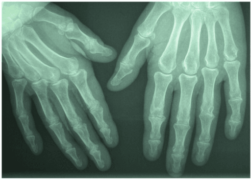
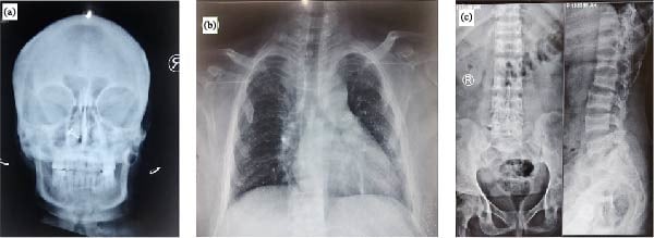
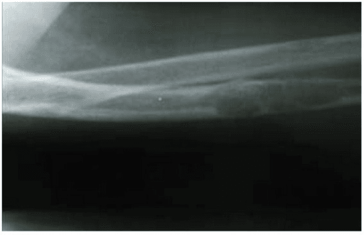
When it is left to run. Severe disease produces osteitis fibrosa cystica: brown tumours, subperiosteal resorption and a salt-and-pepper skull. Parathyroidectomy is the definitive treatment when criteria are met.
Paget Disease of Bone
What it is. Paget disease is disordered, accelerated remodelling producing structurally weak, enlarged bone. It often affects the pelvis, spine, skull and long bones, and may be silent or cause bone pain, deformity (bowing), deafness and rarely high-output cardiac failure or sarcomatous change.
How it reads. Biochemistry shows an isolated, often very high alkaline phosphatase with normal calcium and phosphate.
How it is treated. Bisphosphonates, especially a single zoledronic acid infusion, suppress the high turnover and relieve pain.
Hypercalcaemia: Finding the Cause
What a high calcium does to the patient. The classic picture is stones, bones, abdominal moans and psychic groans, with thirst and polyuria on top.
- Bones — bone pain and metabolic bone disease
- Stones — kidney stones and nephrocalcinosis
- Abdominal moans — constipation, nausea, pancreatitis, ulcers
- Psychic groans — confusion, depression, fatigue
- Thirst and polyuria — polydipsia and polyuria from impaired concentration
The long list of causes. Beyond the parathyroids and malignancy, the causes worth carrying are drugs, granulomatous disease, vitamin excess, excessive intake and the endocrine associations.
CHIMPANZEESCauses of Hypercalcaemia
Hook:CHIMPANZEES covers the causes of high calcium - the big two are hyperParaThyroidism and Malignancy.
A high or inappropriately normal PTH points to the parathyroids. A low PTH points elsewhere: in the unwell or known-cancer patient that means malignancy, where PTH is suppressed.
Calcium Emergencies
Both ends of the range are dangerous, and the table below is the recognition list.
- Typical features
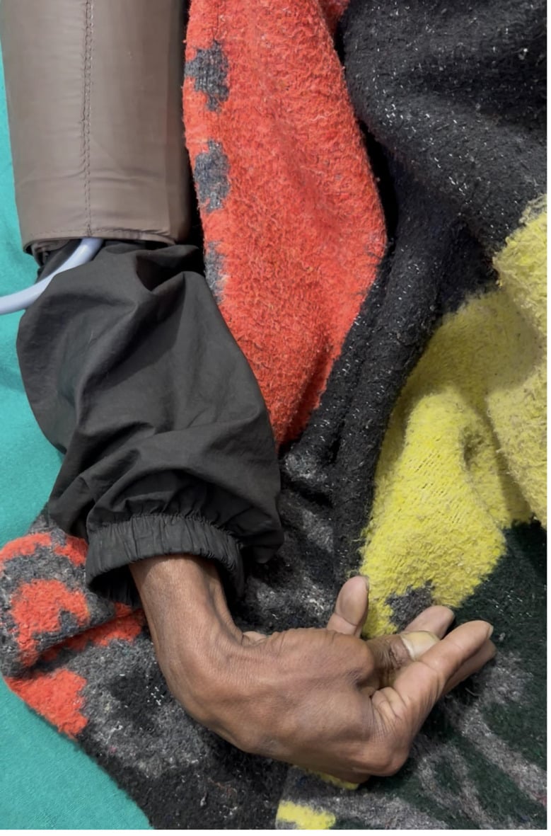
- Confusion, polyuria, dehydration, arrhythmia (short QT)
- Immediate management
- Aggressive IV fluids, then IV bisphosphonate; treat the cause
- Typical features
- Tetany, perioral and digital paraesthesia, Chvostek and Trousseau signs, long QT, seizures
- Immediate management
- IV calcium gluconate with cardiac monitoring, then correct cause and magnesium
- Typical features
- Profound hypocalcaemia after parathyroidectomy as bone rapidly remineralises
- Immediate management
- Aggressive calcium and active vitamin D replacement
Calcium above about 3.5 mmol/L, or any patient with confusion, marked dehydration or arrhythmia, needs urgent treatment. Fluid first: generous intravenous rehydration restores volume and promotes calcium excretion, and only then does an intravenous bisphosphonate follow, alongside treatment of the underlying cause. Loop diuretics are only added once the patient is fully rehydrated.
Clinical Relevance
Calcium homeostasis underpins much of orthopaedic and exam practice. In fracture clinic and fracture liaison services, recognising and correcting vitamin D deficiency and osteoporosis is central to secondary prevention. In basic-science vivas, the PTH-vitamin D axis and the biochemical patterns of metabolic bone disease are classic asks.
In trauma and elective surgery, poorly mineralised or low-density bone changes fixation strategy and healing, and in perioperative care both severe hypercalcaemia and symptomatic hypocalcaemia are emergencies. Being able to read the panel and act on it is the practical core that examiners reward.
Guidelines, Registries and Global Practice
- Osteoporosis and bone-health guidance worldwide (for example AAOS/AOA bone-health initiatives, NICE/NOGG in the UK, and endocrine society guidance) recommends ensuring calcium and vitamin D sufficiency as the foundation on which bone-active drug therapy is added.
- Vitamin D supplementation evidence supports targeted replacement of those who are deficient or at high risk rather than blanket high-dose use in healthy, replete older adults.
- Primary hyperparathyroidism consensus guidelines list bone protection (low bone density or fragility fracture), high calcium, young age and renal involvement among the recognised indications for parathyroidectomy.
- Renal bone disease (CKD-MBD) guidance frames management around controlling phosphate, providing active vitamin D analogues, and managing secondary hyperparathyroidism, with the aim of protecting both bone and the cardiovascular system.
Exam Viva Scenarios
Practise clinical reasoning and management decisions out loud
“The examiner shows you a normal serum calcium and asks how the body keeps calcium in such a narrow range. Take me through the control system.”
“A 62-year-old woman has widespread bone pain and proximal muscle weakness. Calcium is low-normal, phosphate is low, alkaline phosphatase is high and PTH is elevated. What is the diagnosis and how would you manage her?”
The Axis
- PTH raises calcium, lowers phosphate (phosphaturic)
- Calcitriol raises gut absorption of calcium and phosphate
- Calcitonin lowers calcium (minor role in humans)
- Organs: bone (resorption), kidney (reabsorb + activate vit D), gut (absorb)
Vitamin D
- Liver makes 25-OH-D (storage form, measured)
- Kidney makes active 1,25-(OH)2-D (calcitriol)
- Renal failure: low calcitriol, low calcium, secondary hyperparathyroidism
Read the Biochemistry
- Osteoporosis: all normal, low bone density
- Osteomalacia: low phosphate, high alk phos, secondary high PTH
- Primary hyperparathyroidism: high calcium, low phosphate, high PTH
- Paget: isolated very high alkaline phosphatase
Red Flags
- Severe hypercalcaemia - fluids first, then IV bisphosphonate
- Tetany / long QT - symptomatic hypocalcaemia needs IV calcium
- Looser zones - radiographic hallmark of osteomalacia
- Correct calcium for albumin or measure ionised calcium
Evidence for Calcium and Vitamin D
The pairing is deliberate: calcium PLUS vitamin D reduces fractures (Weaver/NOF), but vitamin D ALONE in already-replete healthy elders does not (DO-HEALTH) — so the message is targeted replacement, not blanket high-dose supplementation. The parathyroidectomy data show surgery also protects the skeleton.
Calcium plus Vitamin D Reduces Fractures (NOF Meta-analysis)
- Meta-analysis of 8 randomised controlled trials, 30,970 participants
- Calcium plus vitamin D supplementation cut total fractures by about 15 percent (relative risk 0.85)
- Hip fractures were reduced by about 30 percent (relative risk 0.70)
- Benefit seen in both community-dwelling and institutionalised middle-aged to older adults
Vitamin D Alone Does Not Improve Outcomes in Healthy Elderly (DO-HEALTH)
- Double-blind RCT, 2157 generally healthy adults aged 70 or older, 3-year follow-up
- Vitamin D3 2000 IU/day, omega-3, and exercise tested alone and in combination
- No significant benefit on nonvertebral fractures, blood pressure, physical performance or cognition
- Suggests routine high-dose vitamin D does not help robust, replete older adults
Parathyroidectomy for Normocalcemic Primary Hyperparathyroidism is Associated with Improved Bone Mineral Density Regardless of Postoperative PTH
- Single-centre cohort of 661 parathyroidectomies for primary hyperparathyroidism, of whom 68 had the normocalcaemic variant (28 with paired bone-density data)
- The normocalcaemic patients gained bone density at the lumbar spine (about 1.8 percent) and total hip (about 1.6 percent) at 18 months
- Bone density improved even when postoperative PTH remained persistently elevated
- Supports surgery for bone protection in selected (including normocalcaemic) primary hyperparathyroidism