Osteoid-Producing Soft-Tissue Sarcoma
- EXTRASKELETAL OSTEOSARCOMA is a RARE, usually HIGH-GRADE soft-tissue SARCOMA that produces an OSTEOID or bone matrix but, by definition, DEVELOPS IN SOFT TISSUE with NO attachment to bone or periosteum - which distinguishes it from intramedullary (conventional) and from surface (parosteal/periosteal) osteosarcomas.
- It typically occurs in OLDER ADULTS - older than the peak age of conventional osteosarcoma - presenting with a DEEP soft-tissue MASS, most commonly in the THIGH/lower limb (also the retroperitoneum and upper limb); a minority arise in previously irradiated tissue or after trauma.
- It is a HIGH-GRADE sarcoma with a POOR PROGNOSIS and a HIGH rate of (haematogenous) METASTASIS - particularly to the LUNG - and of local recurrence, so it must be taken seriously and staged accordingly (chest imaging) despite arising in soft tissue.
- The CRITICAL DIFFERENTIAL is MYOSITIS OSSIFICANS, a BENIGN reactive heterotopic ossification that characteristically shows ZONAL maturation - MATURE, well-organised bone at the PERIPHERY (rim) with an immature/cellular CENTRE - which is the OPPOSITE of the disorganised, centrally-dense ossification of extraskeletal osteosarcoma; mistaking one for the other is a classic and dangerous error in both directions.
- DIAGNOSIS follows soft-tissue-sarcoma principles - MRI of the primary, plain radiograph/CT for the matrix mineralisation, and a properly PLANNED BIOPSY at the treating sarcoma unit; a needle biopsy can be misleading (in one report it was called a giant cell tumour of soft tissue) and the diagnosis may only be confirmed on the resection specimen, so caution and specialist review are needed.
- MANAGEMENT is WIDE surgical RESECTION with clear margins, the mainstay; unlike conventional osteosarcoma, CHEMOTHERAPY has NOT been shown to help, and the evidence is now quantified rather than merely 'debated'. A systematic review of 12 retrospective studies and 761 patients with LOCALISED disease found 5-year disease-free survival of 47.9 PERCENT (187 of 390) with surgery plus (neo)adjuvant chemotherapy against 40.4 PERCENT (150 of 371) with surgery alone - a pooled ODDS RATIO of 1.23 (95% CI 0.69-2.19, p=0.479, I-squared 37 percent). The authors' conclusion is stronger than 'uncertain': ROUTINE use of adjuvant chemotherapy should be AVOIDED, pending randomised trials. None of the included studies was randomised, so confounding by indication runs in both directions.
- Given the poor prognosis, CAREFUL long-term FOLLOW-UP for recurrence and metastasis is recommended - all within a specialist sarcoma multidisciplinary team.
- “Extraskeletal osteosarcoma = rare HIGH-GRADE soft-tissue sarcoma producing osteoid/bone but NOT attached to bone/periosteum; OLDER adults; deep mass (often thigh). Poor prognosis, high lung metastasis.
- “CRITICAL differential = MYOSITIS OSSIFICANS (benign, ZONAL: mature bone at the PERIPHERY, immature centre) - the OPPOSITE of the disorganised central ossification of extraskeletal OS. Also distinguish from surface/conventional OS (attached to bone).
- “Diagnosis by MRI + radiograph/CT + planned biopsy (needle biopsy can mislead - may confirm only on resection). Wide resection is the mainstay; long-term surveillance; sarcoma-centre care.
- “Quantify the chemotherapy answer rather than calling it 'debated': across 12 retrospective studies and 761 LOCALISED cases, 5-year disease-free survival was 47.9% with (neo)adjuvant chemotherapy vs 40.4% with surgery alone - pooled OR 1.23 (95% CI 0.69-2.19, ns). The reviewers advise AVOIDING routine adjuvant chemotherapy. No randomised trial exists.
High-grade soft-tissue sarcoma making osteoid, not attached to bone; older adults; deep mass; disorganised, centrally-dense ossification; poor prognosis.
Zonal maturation - mature bone at the periphery (rim), immature centre (the opposite zoning); often post-traumatic; matures over time. Don't excise it as a sarcoma (or vice versa).
Epidemiology, Prognosis and the Fuller Differential
- Epidemiology. It is rare - roughly 1-2% of soft-tissue sarcomas and a few percent of all osteosarcomas - peaking in the fifth-to-seventh decade (older than conventional osteosarcoma); a recognised subset is radiation-associated, arising in a prior radiotherapy field after a latency of years.
- Prognosis. Five-year survival is poor, commonly quoted around 25-50% and generally worse than most soft-tissue sarcomas; adverse factors are large size (over 5 cm), high grade, positive margins and metastasis at presentation, with frequent local recurrence.
- The fuller differential. Beyond myositis ossificans, a bone-forming soft-tissue mass may be a dedifferentiated liposarcoma with osteosarcomatous differentiation (which carries MDM2/CDK4 amplification - absent in primary extraskeletal osteosarcoma), a synovial sarcoma with calcification (SS18 fusion), or another sarcoma with divergent bone formation - so a primary extraskeletal osteosarcoma is diagnosed only after excluding an osteosarcomatous component of another tumour.
Q: What is the epidemiology and prognosis of extraskeletal osteosarcoma, and what is the fuller differential?
A: Rare (~1-2% of soft-tissue sarcomas), peak fifth-to-seventh decade (older than conventional OS); a radiation-associated subset arises in a prior RT field after years' latency. Five-year survival poor (~25-50%, worse than most STS); adverse = size over 5 cm, high grade, positive margins, metastasis. The fuller differential of a bone-forming soft-tissue mass: dedifferentiated liposarcoma with osteosarcomatous differentiation (MDM2/CDK4-amplified - absent in primary EOS), synovial sarcoma with calcification (SS18), other sarcomas with divergent bone - so primary EOS is a diagnosis of exclusion.
The Chemotherapy Question, Answered With Numbers
Saying the benefit of chemotherapy is "uncertain" is a weaker answer than the evidence allows. A systematic review pooled 12 retrospective studies and 761 patients with localised extraskeletal osteosarcoma, comparing surgery plus (neo)adjuvant chemotherapy against surgery alone. Five-year disease-free survival was 47.9 percent (187 of 390) with chemotherapy and 40.4 percent (150 of 371) without - a pooled odds ratio of 1.23 with a confidence interval crossing 1 (0.69 to 2.19, p=0.479) and modest heterogeneity (I-squared 37 percent). The authors conclude that the effect appears limited and that routine use of adjuvant chemotherapy should be avoided, while calling for randomised trials. This is the distinction worth making: the evidence is not silent, it is negative-but-imprecise. None of the 12 studies was randomised, so patients selected for chemotherapy probably differed systematically from those who were not, and the confidence interval remains wide enough that a real benefit has not been excluded.
In conventional intramedullary osteosarcoma, multi-agent chemotherapy transformed survival and is standard. Extraskeletal osteosarcoma arises in an older population and behaves like a high-grade soft-tissue sarcoma, and the pooled data above do not reproduce that benefit. Carrying the conventional-osteosarcoma protocol across on the strength of a shared name is the error the numbers guard against.
Note the control arm: 40.4 percent five-year disease-free survival after surgery alone for localised disease. That is the denominator behind "poor prognosis" - and it applies to patients without metastases at presentation, so it is the optimistic end of the range rather than the average case.
Pathophysiology & Molecular Basis
- Lineage. A malignant mesenchymal tumour whose cells produce neoplastic osteoid/bone without any contact with bone or periosteum; the cell of origin is presumed to be a primitive mesenchymal progenitor with osteoblastic differentiation. The WHO places it among soft-tissue osteosarcomas, distinct from conventional intramedullary and surface osteosarcoma.
- Molecular profile. A complex (aneuploid) karyotype with no defining translocation - unlike the fusion-driven sarcomas in its differential (synovial sarcoma, myxoid liposarcoma). SATB2 immunostaining marks osteoblastic differentiation but is non-specific.
- Diagnosis of exclusion. A dedifferentiated liposarcoma with osteosarcomatous differentiation carries MDM2/CDK4 amplification (absent in primary ESO), and a calcifying synovial sarcoma carries the SS18 fusion - both must be excluded before calling a tumour primary ESO.
- Radiation-associated subset. A minority (about 6 percent in the Chung-Enzinger series) arise in previously irradiated tissue after a latency of years to decades (median 13.2 years in the Finnish postirradiation sarcoma registry analysis, where four of the ten osteosarcomas were extraskeletal).
Investigations
- Plain radiograph and CT show the matrix: central, amorphous "cloud-like", disorganised mineralisation with a non-mineralised periphery - the reverse of myositis ossificans' mature peripheral rim. CT also confirms the absence of bone attachment.
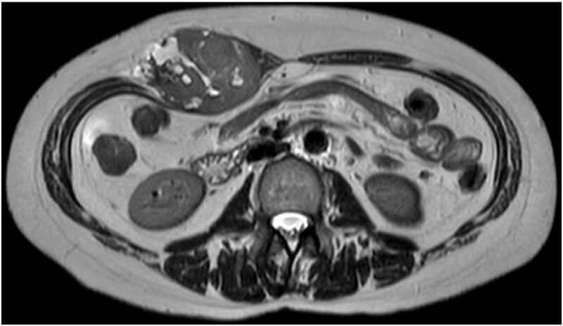
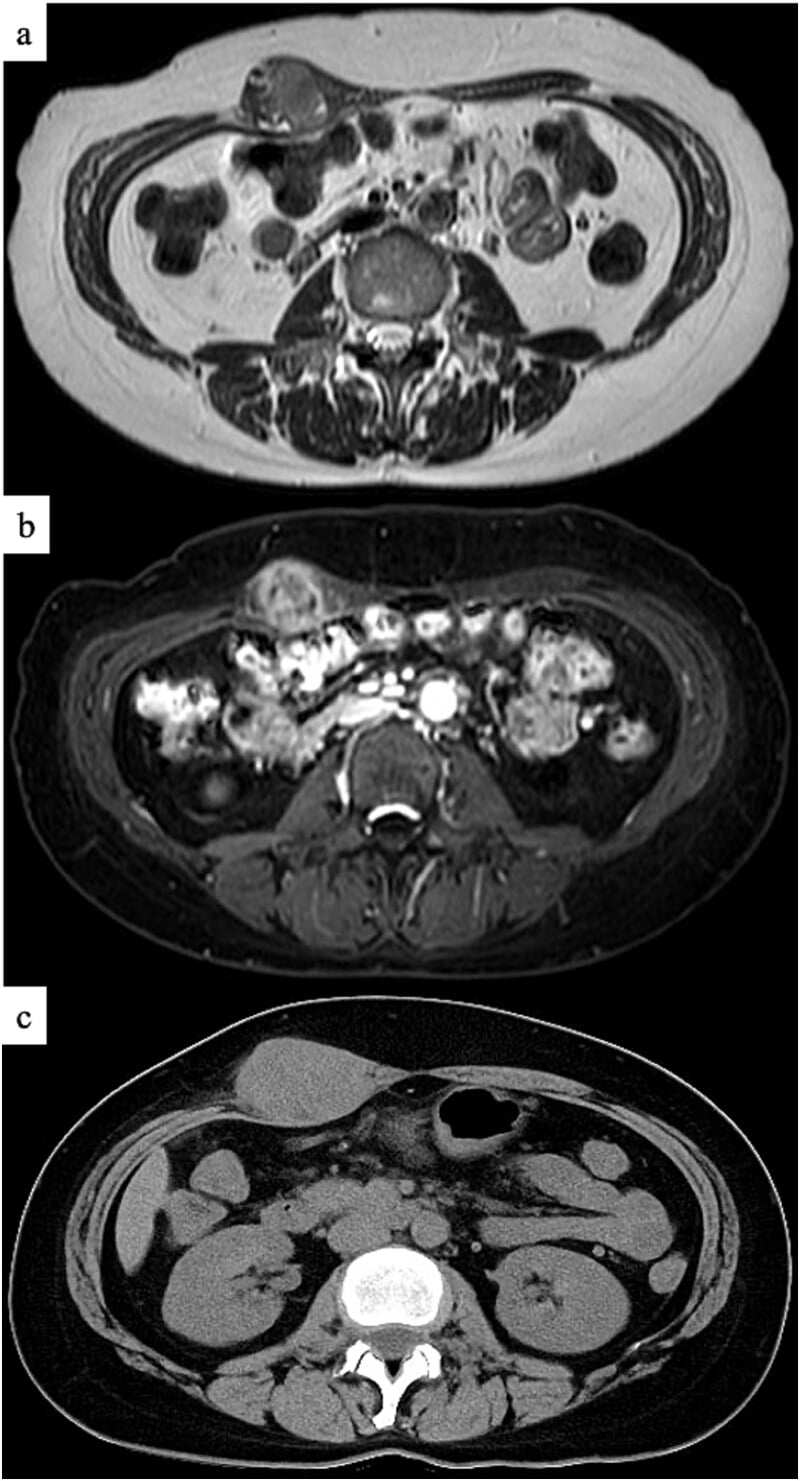
- MRI defines the soft-tissue mass (heterogeneous, with necrosis and haemorrhage) for surgical planning.
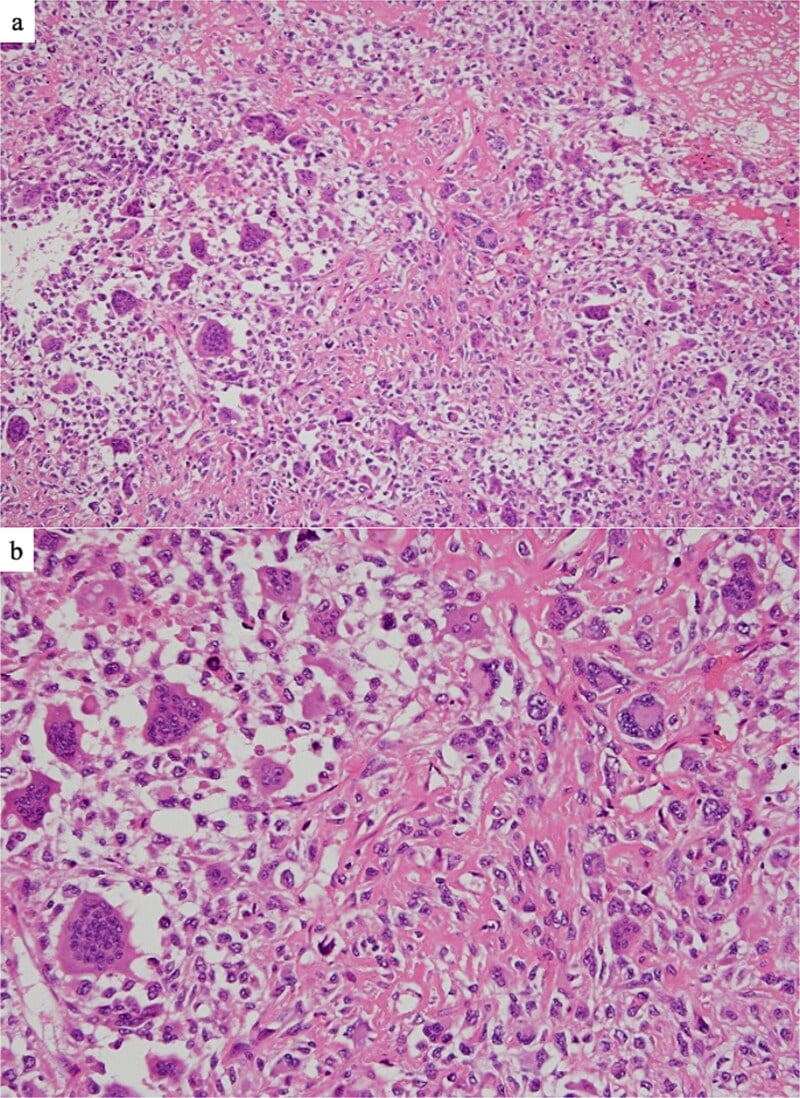
- Planned biopsy at the treating sarcoma unit - with the explicit warning that needle biopsy can mislead (reported as giant cell tumour of soft tissue in the Nakayama case, delaying diagnosis); specialist review of imaging zoning and histology together, with re-biopsy or resection-specimen confirmation when the picture does not fit.
- Staging: chest imaging (lung is the dominant metastatic site), plus consideration of nodal assessment - the Chung-Enzinger series found metastases in regional lymph nodes as well as lung and bone.
- Molecular work-up where the differential demands it: MDM2/CDK4 to exclude dedifferentiated liposarcoma, SS18 for synovial sarcoma.
Subtypes, Imaging Pattern and Molecular Profile
- The histological subtypes. Like conventional osteosarcoma, extraskeletal osteosarcoma is subclassified by its predominant matrix into osteoblastic (commonest), chondroblastic and fibroblastic, with less common telangiectatic, small-cell and rare well-differentiated/low-grade variants; the diagnostic requirement is malignant cells producing neoplastic osteoid, and most are high-grade.
- The imaging pattern. On radiograph/CT the mineralisation is typically central, amorphous ("cloud-like") and disorganised, often with a non-mineralised soft-tissue periphery - mirroring the histology and the reverse of myositis ossificans' peripheral rim. MRI shows a heterogeneous mass with mineralisation, necrosis and haemorrhage, and no attachment to bone or periosteum (confirming the extraskeletal origin).
- The molecular profile. Extraskeletal osteosarcoma has a complex (aneuploid) karyotype - like other high-grade pleomorphic sarcomas and unlike the fusion-driven sarcomas (e.g. synovial sarcoma) - so there is no defining translocation; SATB2 marks osteoblastic differentiation but is non-specific, and the diagnosis rests on identifying malignant osteoid after exclusion.
Q: What are the histological subtypes and molecular features of extraskeletal osteosarcoma?
A: Subclassified (like conventional OS) into osteoblastic (commonest), chondroblastic, fibroblastic, ± telangiectatic/small-cell/rare low-grade; the requirement is malignant neoplastic osteoid, mostly high-grade. On CT the mineralisation is central/amorphous/disorganised with a non-mineralised periphery (mirroring the histology, the reverse of myositis ossificans). Molecularly it has a complex (aneuploid) karyotype - no defining fusion (unlike synovial sarcoma); SATB2 is a non-specific osteoblastic marker.
Features & The Critical Differential
Extraskeletal osteosarcoma is a rare, high-grade soft-tissue sarcoma that makes osteoid/bone but arises in soft tissue with no attachment to bone or periosteum, typically in older adults as a deep mass (often the thigh). It is high-grade with a poor prognosis and high lung-metastatic potential. The critical differential is myositis ossificans, a benign reactive ossification with zonal maturation (mature bone at the periphery/rim, immature centre) - the opposite of the disorganised, centrally-dense ossification of extraskeletal osteosarcoma; it is also distinguished from the surface/conventional osteosarcomas, which are attached to bone. Needle biopsy can mislead, so diagnosis may rest on the resection specimen with specialist review.
- Extraskeletal osteosarcoma
- Malignant (high-grade sarcoma)
- Myositis ossificans
- Benign reactive heterotopic ossification
- Extraskeletal osteosarcoma
- Disorganised; denser centrally
- Myositis ossificans
- Zonal: mature bone at periphery (rim), immature centre
- Extraskeletal osteosarcoma
- Progresses/enlarges
- Myositis ossificans
- Matures and may stabilise/shrink
- Extraskeletal osteosarcoma
- Older adults; +/- prior radiation
- Myositis ossificans
- Often younger; post-traumatic
- Extraskeletal osteosarcoma
- Poor (metastasis/recurrence)
- Myositis ossificans
- Benign (self-limiting)
Management
- Diagnosis: MRI of the primary; radiograph/CT for matrix mineralisation; planned biopsy at the treating sarcoma unit (needle biopsy can mislead - confirm on resection if needed; specialist review).
- Staging: chest imaging (high lung-metastatic risk).
- Surgery: wide resection with clear margins - the mainstay.
- Surveillance: careful long-term follow-up (recurrence/metastasis); specialist sarcoma MDT throughout.
The lesions this must be separated from all have their own pages, and the separation matters in both directions. The benign reactive mimic is myositis ossificans, part of the wider spectrum of heterotopic ossification; the bone-based tumour whose name it shares is osteosarcoma, with the surface forms covered under high-grade surface osteosarcoma. Among the malignant mimics, a calcifying synovial sarcoma and a dedifferentiated liposarcoma with osteosarcomatous differentiation both need excluding, the latter by its MDM2 and CDK4 amplification. The referral pathway for the undiagnosed deep mass is soft-tissue masses and sarcoma referral.
The central diagnostic safety point in extraskeletal osteosarcoma is its differentiation from myositis ossificans, because the consequences of getting it wrong are serious both ways. Myositis ossificans is a benign reactive heterotopic ossification with characteristic zonal maturation - mature, well-organised bone at the periphery (the rim) and an immature, cellular centre - which matures over time and needs no aggressive treatment; mistaking it for a sarcoma can lead to unnecessary radical surgery. Extraskeletal osteosarcoma is a high-grade malignancy that produces disorganised osteoid, denser centrally, arises in soft tissue unattached to bone, occurs in older adults and carries a poor prognosis with high lung-metastatic potential; mistaking it for myositis ossificans (or for a benign lesion on a misleading needle biopsy) under-treats a dangerous cancer. Because needle biopsy can be misleading, the imaging zoning pattern, the clinical context and specialist pathological review - sometimes only confirmed on the resection specimen - are essential, and any osteoid-producing soft-tissue mass in an older adult should be worked up and managed at a specialist sarcoma centre, with wide resection the mainstay of treatment.
Complications & Prognosis
- Local recurrence - 43 percent in the Chung-Enzinger AFIP series (28 of 65 with follow-up); many patients recurred more than once.
- Metastasis - 63 percent in that series (39 of 62 assessed), prevailing sites lung, regional lymph nodes and bone; 61.5 percent of patients with follow-up had died (36 from tumour).
- Prognosis - five-year survival is poor, commonly quoted around 25-50 percent (a figure repeated across the literature rather than from a single defined cohort); even for localised disease the surgery-alone arm of the Tsukamoto review achieved only 40.4 percent five-year disease-free survival. Adverse factors: size over 5 cm, high grade, positive margins, metastasis at presentation.
- Treatment morbidity - wide resection of a deep extremity or retroperitoneal mass carries the morbidity of compartment or neurovascular sacrifice; where the wrong diagnosis was made on needle biopsy, inappropriate therapy (denosumab in the Nakayama case) wastes time while the tumour enlarges.
Mnemonics & Memory Aids
OSTEOID
Hook:OSTEOID: Osteoid in soft tissue (not attached), Soft-tissue sarcoma, Thigh/radiation, Exclude myositis ossificans, Outcome poor, Imaging+biopsy, Do wide resection.
RIM
Hook:RIM - the zoning rule: mature RIM (periphery) is benign myositis ossificans; central disorganised ossification is the sarcoma. The single most examinable discriminator.
AVOID
Hook:AVOID routine adjuvant chemotherapy in localised extraskeletal osteosarcoma - the pooled data are negative-but-imprecise, and the reviewers say so explicitly.
Clinical Decision Scenarios
Practise clinical reasoning and management decisions out loud
“An older adult has a deep, ossifying soft-tissue mass in the thigh. How do you distinguish extraskeletal osteosarcoma from myositis ossificans, and how would you manage it?”
What it is
- Rare high-grade soft-tissue sarcoma producing osteoid/bone
- Arises in soft tissue, NOT attached to bone or periosteum
- Older adults; deep mass (often thigh); +/- prior radiation
Behaviour & differential
- Poor prognosis; high lung-metastatic and local-recurrence risk
- Critical differential: myositis ossificans (zonal - mature peripheral rim, immature centre)
- Distinguish from surface/conventional osteosarcoma (attached to bone)
Diagnosis & management
- MRI + radiograph/CT (matrix/zoning); planned biopsy (needle biopsy can mislead)
- Stage chest; wide resection with clear margins = mainstay
- Avoid ROUTINE adjuvant chemotherapy: 5-yr DFS 47.9 vs 40.4 percent, OR 1.23 (0.69-2.19), n=761 localised
- Manage like a high-grade STS; long-term surveillance; sarcoma-centre care
Evidence & Key Studies
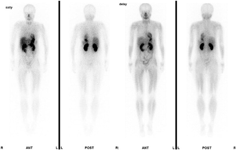
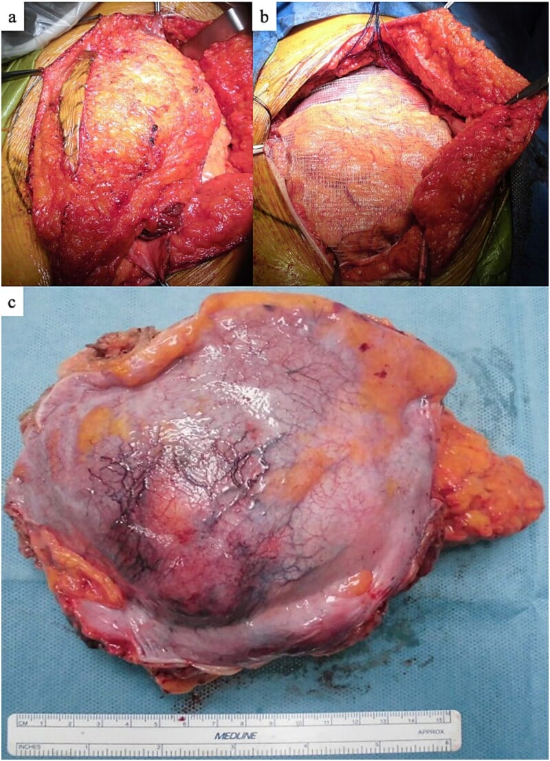
Extraskeletal osteosarcoma in the rectus abdominis muscle: a case report
- Extraskeletal osteosarcoma is a rare malignant tumour with an osteoid or bone matrix that develops in soft tissues not in contact with bone or periosteum.
- In this case (a 47-year-old woman with an abdominal-wall mass) the needle biopsy was misleading - reported as a giant cell tumour of soft tissue, for which denosumab was given while the tumour enlarged - and the diagnosis of extraskeletal osteosarcoma was confirmed only on the wide-resection specimen.
- Because the tumour has a poor prognosis, wide tumour resection was performed and careful long-term follow-up recommended; the patient was alive and disease-free one year after surgery.
The definition of extraskeletal osteosarcoma (an osteoid/bone-producing malignant tumour arising in soft tissue not in contact with bone or periosteum), the diagnostic difficulty (a misleading needle biopsy confirmed only on resection), the poor prognosis, and management by wide resection with careful long-term follow-up come from the cited Nakayama report. The older-adult demographic, the high lung-metastatic risk, the critical differential from myositis ossificans (zonal peripheral maturation vs disorganised central ossification) and from surface/conventional osteosarcoma are standard, well-established teaching. The chemotherapy figures - the 5-year disease-free survival in each arm, the pooled odds ratio and the recommendation against routine adjuvant use - come from the Tsukamoto systematic review, whose 12 constituent studies are all retrospective. No randomised trial of chemotherapy in this disease exists. The 25 to 50 percent five-year survival range quoted for the disease overall is a figure repeated across the literature rather than one drawn from a single defined cohort, and no validated staging system or surveillance schedule specific to extraskeletal osteosarcoma has been published.