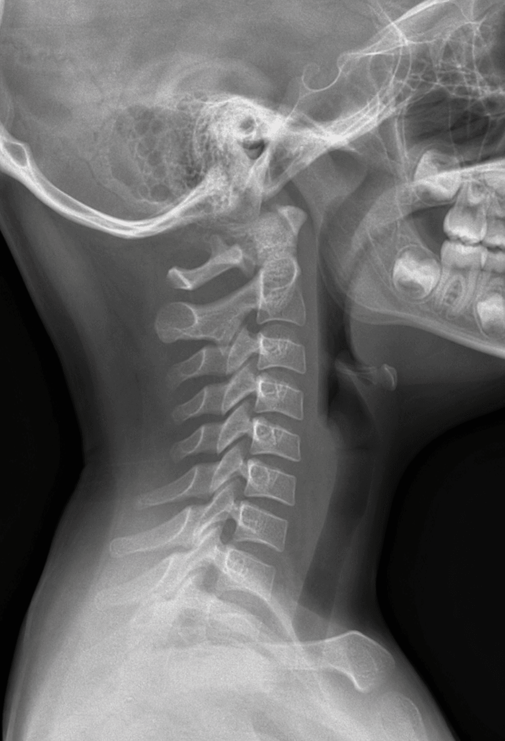
Physiological C2-C3 Displacement in Children
- Pseudosubluxation is a PHYSIOLOGICAL (normal) anterior displacement of C2 on C3 (and sometimes C3 on C4) of up to about 2-3 mm seen in young children - it is a NORMAL VARIANT and NOT an injury, and it occurs because the immature cervical spine has ligamentous laxity, relatively horizontal facet joints, and a high fulcrum of flexion-extension motion (around C2-C3) together with a large head-to-body ratio.
- It is common and age-related: C2-C3 pseudosubluxation has been reported in around 9% of children younger than 7 years, and it generally resolves as the spine matures; it is the single most important paediatric cervical normal variant to recognise so as not to misdiagnose a normal child as having a fracture-dislocation.
- The key tool to distinguish pseudosubluxation from a true injury is the SWISCHUK POSTERIOR CERVICAL (spinolaminar) LINE: a line drawn along the anterior cortex of the spinous processes (the spinolaminar junctions) of C1, C2 and C3 should pass within about 2 mm of the anterior cortex of the C2 spinous process - in physiological pseudosubluxation the C2 spinolaminar point lies on or close to this line, whereas in a true C2 injury (e.g. a hangman's fracture) the posterior arch of C2 is displaced and the line is disrupted.
- Further reassuring features are that pseudosubluxation REDUCES on an EXTENSION (or neutral) view, the alignment is smooth without a focal kyphotic angulation, there is no prevertebral soft-tissue swelling beyond the normal range, and there is no fracture - so correlating the radiograph with the child's AGE, the MECHANISM of injury and the clinical examination is essential.
- Pseudosubluxation sits among a family of paediatric cervical NORMAL VARIANTS that can each be mistaken for trauma: hypermobility and pseudospread of the atlas on the axis, absence of the normal cervical lordosis, anterior WEDGING of the (still partly cartilaginous) vertebral bodies, prevertebral soft-tissue PSEUDOWIDENING (worse on expiration/neck flexion), and incomplete ossification with open synchondroses - knowing these prevents over-diagnosis.
- The practical consequence is to AVOID OVER-TREATING a normal variant: recognise physiological pseudosubluxation with the Swischuk line and clinical correlation rather than immobilising or investigating a normal child unnecessarily - while still remembering that children (especially under 8) are prone to upper cervical injuries and SCIWORA, so genuine clinical concern warrants appropriate assessment.
- “Pseudosubluxation = NORMAL anterior displacement of C2 on C3 (and C3 on C4) up to ~2-3 mm in young children (~9% under age 7) - a variant, not an injury.
- “Use the SWISCHUK posterior cervical line: within ~2 mm of the C2 spinolaminar cortex = normal; disrupted = true C2 injury. It reduces on extension.
- “Other paediatric variants mimic trauma (pseudospread, anterior wedging, prevertebral pseudowidening); correlate with age/mechanism - don't over-treat.
Anterior displacement of C2 on C3 (up to ~2-3 mm) in a young child - a normal variant from laxity, horizontal facets and a high motion fulcrum.
The Swischuk posterior cervical line is intact (within ~2 mm of the C2 spinolaminar cortex), and it reduces on extension. Correlate with age/mechanism.
What It Is & How to Tell It From Injury
Pseudosubluxation is the physiological anterior displacement of C2 on C3 (and sometimes C3 on C4) of up to about 2-3 mm in young children - a normal variant, not an injury - caused by ligamentous laxity, horizontal facet joints and a high (C2-C3) fulcrum of motion with a large head-to-body ratio. It is common, reported in around 9% of children under 7, and resolves with maturity. The key way to confirm it is normal is the Swischuk posterior cervical (spinolaminar) line: drawn along the anterior cortex of the spinous processes of C1, C2 and C3, it should pass within about 2 mm of the anterior cortex of the C2 spinous process - intact in pseudosubluxation, disrupted in a true C2 injury (e.g. hangman's fracture). Pseudosubluxation also reduces on extension, has smooth alignment, and shows no fracture or abnormal prevertebral swelling.
Other Variants & Practical Approach
- Know the family of normal variants. Besides pseudosubluxation: hypermobility and pseudospread of the atlas on the axis, absence of lordosis, anterior wedging of the partly-cartilaginous vertebral bodies, prevertebral soft-tissue pseudowidening (worse on expiration/flexion), and incomplete ossification/open synchondroses.
- Use the Swischuk line and an extension view. An intact posterior cervical line and reduction on extension confirm a normal variant.
- Correlate with age, mechanism and examination. Interpret the paediatric cervical radiograph in light of the child's age, the injury mechanism and the clinical picture - not in isolation.
- Don't over-treat - but don't be complacent. Avoid unnecessary immobilisation/investigation of a normal variant, while remembering young children are prone to upper cervical injuries and SCIWORA, so genuine clinical concern still warrants proper assessment (and advanced imaging where indicated).
The single most useful skill with paediatric pseudosubluxation is using the Swischuk posterior cervical line to separate a normal variant from a true injury: if the line drawn along the spinolaminar cortices of C1-C2-C3 passes within about 2 mm of the C2 spinous process and the displacement reduces on extension, the C2-on-C3 'subluxation' is physiological; if the line is disrupted, suspect a true C2 injury such as a hangman's fracture. Equally important is the converse caution - children under 8 are prone to upper cervical injuries and to SCIWORA (spinal cord injury without radiographic abnormality), so a normal-looking radiograph does not exclude injury when there is real clinical concern, and the radiograph must always be read with the child's age, mechanism and examination in mind.
Evidence & Key Studies
Pediatric cervical spine in emergency: normal anatomy, variants and pitfalls
- The paediatric cervical spine has distinct radiographic features - hypermobility between C2 and C3, pseudospread of the atlas on the axis, pseudosubluxation, absence of lordosis, anterior wedging of vertebral bodies, prevertebral soft-tissue pseudowidening and incomplete ossification/synchondroses - that can be mistaken for traumatic injuries.
- Younger children (under 8 years) tend to sustain upper cervical injuries because of the anatomy and biomechanics of the immature spine.
- Interpretation must take into account the child's age, the location of any injury and the mechanism of trauma; comprehensive knowledge of normal variants prevents over-diagnosis.
Acute evaluation and management of pediatric spinal cord injury (C2-C3 pseudosubluxation)
- Radiographic evaluation of the child's cervical spine is challenging because of normal variants such as C2-C3 pseudosubluxation, which occurs in about 9% of children younger than 7 years.
- SCIWORA (spinal cord injury without radiographic abnormality) is common in children under 10 and is associated with more complete neurological injuries.
- Children cannot be evaluated and treated as small adults; immobilisation and imaging must account for the larger head-to-torso ratio and the upper-cervical injury pattern.
According to PubMed, the catalogue of paediatric cervical normal variants that mimic trauma (pseudosubluxation, pseudospread, anterior wedging, prevertebral pseudowidening, incomplete ossification) and the principle of correlating with age/location/mechanism come from the cited Adib review; the prevalence of C2-C3 pseudosubluxation (about 9% under age 7) and the importance of SCIWORA in young children from the cited Betz review. The Swischuk posterior cervical line and the reduction on extension are standard, well-established teaching. (See also our Paediatric Cervical Spine Injuries and Hangman's Fracture topics.)
Clinical Decision Scenarios
Practise clinical reasoning and management decisions out loud
“A young child's lateral cervical radiograph after a fall shows anterior displacement of C2 on C3. How do you decide if this is a normal variant or an injury?”
“What other paediatric cervical radiographic variants can mimic trauma?”
Mnemonics & Memory Aids
SWISCHUK
Hook:SWISCHUK: the line Within ~2 mm, Intact/smooth, Subluxation reduces on extension, C2-C3, Horizontal facets, Under 8, Keep clinical context (SCIWORA).
What it is
- Physiological anterior displacement of C2 on C3 (+/- C3 on C4) up to ~2-3 mm
- Normal variant in young children (~9% under age 7); resolves with maturity
- Cause: ligamentous laxity, horizontal facets, high motion fulcrum, big head
Confirm it's normal
- Swischuk posterior cervical line within ~2 mm of the C2 spinolaminar cortex
- Reduces on extension; smooth alignment; no fracture
- No abnormal prevertebral soft-tissue swelling
Other variants
- Pseudospread of atlas on axis; absence of lordosis
- Anterior wedging of vertebral bodies; prevertebral pseudowidening
- Incomplete ossification / open synchondroses
Practical approach
- Correlate with age, mechanism and examination
- Don't over-treat a normal variant
- Remember upper-cervical injuries and SCIWORA in the young