When the Sixth-Compartment Tendon Leaves Its Groove
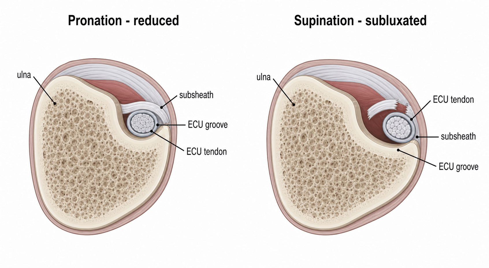
- The extensor carpi ulnaris (ECU) runs in the SIXTH dorsal compartment within its OWN fibro-osseous SUBSHEATH, in a bony GROOVE on the dorsal aspect of the ulnar head; this subsheath (distinct from the overlying extensor retinaculum) is what keeps the ECU in its groove during forearm rotation.
- When the SUBSHEATH is TORN or attenuated, the ECU SUBLUXATES or DISLOCATES out of its groove - typically VOLARLY/ULNARLY - during forearm SUPINATION, ulnar deviation and wrist flexion, and reduces back into the groove on pronation; this produces a painful, often visible/palpable SNAPPING at the dorsoulnar wrist.
- It is classically a sporting injury from a forceful or repetitive SUPINATION-ULNAR DEVIATION-FLEXION load (tennis, golf, racquet and bat sports), but also occurs degeneratively and in rheumatoid disease; it is an important and under-recognised cause of ULNAR-SIDED WRIST PAIN.
- DIAGNOSIS is largely clinical: reproduce the snapping with the provocative manoeuvre (active SUPINATION with ULNAR DEVIATION and flexion, watching/palpating the ECU jump out of its groove), and use the ECU SYNERGY TEST (resisted radial abduction of the thumb with the forearm supinated reproduces dorsoulnar pain) to localise the ECU; DYNAMIC ULTRASOUND (during supination/pronation) best demonstrates the subluxation, and MRI shows subsheath disruption and tendinopathy.
- It must be distinguished from the other causes of ULNAR-SIDED WRIST PAIN - TFCC tears, DRUJ instability/arthritis, lunotriquetral instability, ulnar impaction and pisotriquetral arthritis.
- MANAGEMENT: acute cases can be treated CONSERVATIVELY with IMMOBILISATION in a long-arm cast/splint in PRONATION and slight wrist extension/radial deviation (the position that reduces and rests the ECU) for several weeks, plus activity modification and NSAIDs; SURGERY is indicated for chronic, recurrent or refractory instability - REPAIR of the subsheath (acute) or RECONSTRUCTION (chronic) using a slip of EXTENSOR RETINACULUM, a periosteal flap or DRUJ capsule, sometimes with ulnar GROOVE-DEEPENING - while corticosteroid injection is reserved for tendinopathy, not for instability.
- “ECU = 6th compartment in its own SUBSHEATH on the dorsal ulnar groove; subsheath tear -> subluxation.
- “Snapping/subluxation provoked by SUPINATION + ulnar deviation + flexion; reduces on pronation. Dynamic ultrasound is the key test.
- “Acute: immobilise in PRONATION/slight extension. Chronic/recurrent: subsheath repair or reconstruction (retinaculum/periosteum/DRUJ capsule) +/- groove deepening.
A torn subsheath lets the ECU jump out of its ulnar groove on supination/ulnar deviation/ flexion and snap back on pronation - painful, often visible/palpable.
Reproduce the snap with the provocative manoeuvre; dynamic ultrasound during supination/ pronation shows the subluxation; MRI shows subsheath disruption/tendinopathy.
Anatomy & Mechanism
The ECU occupies the sixth dorsal compartment, where it sits in a bony groove on the dorsal ulnar head held by a dedicated fibro-osseous subsheath - a structure separate from the overlying extensor retinaculum. During forearm rotation the ECU must stay in this groove; the subsheath is the key restraint. A forceful or repetitive load in SUPINATION, ULNAR DEVIATION and wrist FLEXION (as in racquet/bat sports) can tear or stretch the subsheath, after which the ECU subluxates/dislocates out of the groove - usually toward the volar-ulnar side - during supination and reduces on pronation, snapping painfully each time. Chronic/degenerative attenuation and rheumatoid disease can do the same.
Assessment & Diagnosis
The diagnosis is mainly clinical. Reproduce the instability with the provocative manoeuvre - active supination with ulnar deviation and wrist flexion - watching and palpating the ECU jump out of its groove, then relocate on pronation. The ECU SYNERGY TEST (with the elbow flexed and forearm supinated, the patient radially abducts the thumb against resistance, which co-contracts the ECU and reproduces dorsoulnar pain) helps localise the ECU as the pain source. DYNAMIC ULTRASOUND through supination and pronation is the best test to demonstrate the subluxation in real time; MRI shows the disrupted subsheath, ECU position and any tendinopathy. Consider and exclude the other causes of ulnar-sided wrist pain - TFCC tears, DRUJ instability/arthritis, lunotriquetral instability, ulnar impaction and pisotriquetral arthritis.
Management
- Acute / first presentation: CONSERVATIVE treatment - immobilise in a long-arm cast or splint in PRONATION with slight wrist extension/radial deviation (the position that reduces and rests the ECU) for several weeks, with NSAIDs and activity modification; many acute cases settle.
- Tendinopathy/tenosynovitis (no instability): rest, splinting and a corticosteroid injection within the sheath can help - but a steroid injection does NOT treat instability.
- Chronic / recurrent / refractory instability: SURGERY -
- Subsheath REPAIR for an acute/repairable subsheath.
- Subsheath RECONSTRUCTION for chronic cases, using a slip of extensor retinaculum, a periosteal flap (Schlesinger), or the DRUJ dorsal capsule (preserving the extensor retinaculum).
- Ulnar groove deepening may be added when the groove is shallow. Reinforcement of the repair/reconstruction reduces recurrence and, in athletes, allows return to sport at a few months.
Evidence & Key Studies
Recurrent dislocation of the extensor carpi ulnaris tendon in a water-polo athlete
- ECU dislocation/subluxation is rare in the general population but common in athletes whose wrists undergo forceful rotational movements.
- Pain and a snapping sensation at the dorsoulnar wrist, especially during supination, are the predominant symptoms and often need surgery.
- Direct subsheath repair reinforced with an extensor retinaculum graft resolved recurrent dislocation and allowed return to high-level sport at four months.
A novel technique using the dorsal capsule of the DRUJ for ECU subsheath reconstruction
- Symptomatic recurrent ECU subluxation is an increasingly recognised cause of ulnar-sided wrist pain that usually requires subsheath reconstruction.
- Reconstruction using the dorsal DRUJ capsule (preserving the extensor retinaculum) gave mostly satisfactory short-term outcomes (Modified Mayo Wrist Score) in 7 patients.
- It is offered for patients failing conservative treatment or primary repair, and can salvage failed procedures while protecting extensor tendon function.
According to PubMed, the athletic mechanism, the supination-provoked dorsoulnar snapping and the repair-plus-retinaculum-graft technique come from the cited Stathopoulos case, and the recognition of recurrent ECU subluxation as a cause of ulnar-sided wrist pain and the need for subsheath reconstruction (with options preserving the retinaculum) from the cited Png series. The 6th-compartment/subsheath anatomy, the provocative test and the conservative-then-surgical ladder are standard hand-surgery teaching. (See also our Dorsal Wrist Extensor Compartments, TFCC Injuries, DRUJ and Ulnar-Sided Wrist Pain topics.)
Clinical Decision Scenarios
Practise clinical reasoning and management decisions out loud
“A tennis player has painful snapping on the ulnar side of the wrist when they supinate. What is the likely diagnosis, and how would you confirm it?”
“How would you manage ECU instability, conservatively and surgically?”
Mnemonics & Memory Aids
SNAP
Hook:The ECU SNAPs out in Supination and relocates in Pronation.
6th
Hook:6th compartment, Subsheath, Ultrasound, Repair/Reconstruct.
Anatomy & mechanism
- ECU in 6th dorsal compartment in its OWN subsheath, in a dorsal ulnar groove
- Subsheath tear -> ECU subluxates (volarly) on supination/ulnar deviation/flexion
- Athletic (racquet/bat) load; also degenerative/rheumatoid
Diagnosis
- Provocative supination + ulnar deviation/flexion reproduces the snap
- ECU synergy test reproduces dorsoulnar pain
- Dynamic ultrasound (best) + MRI; exclude TFCC/DRUJ/LT/ulnar impaction
Conservative
- Acute: long-arm immobilisation in PRONATION + slight extension/radial deviation
- Activity modification, NSAIDs
- Steroid injection for tendinopathy only (not instability)
Surgical
- Acute: subsheath repair
- Chronic/recurrent: subsheath reconstruction (retinaculum/periosteum/DRUJ capsule)
- +/- ulnar groove deepening; reinforce to reduce recurrence